It is possible to eliminate a mole without a trace only when it is small in size. However, removal is often resorted to for large and voluminous nevi that cause aesthetic discomfort or cause certain concerns. Therefore, spots after mole removal are a common phenomenon that can form when using various manipulations, whether laser or surgical removal.
After the procedure to remove nevi, there will be a spot on the skin for some time that needs to be treated for better healing.
Possible complications after mole removal
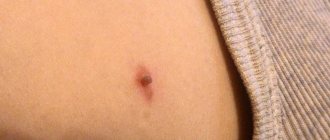
Traditionally, 2-3 days after a successful operation to remove a mole, a dark, dry crust forms in its place , visually resembling a wound from a cut. The size of the crust corresponds to the removed nevus. Over time, it disappears on its own, and in place of the mole there remains a depression, the color is slightly darker than the natural shade of the skin. This process takes from 2 to 4 weeks. And after about 100 days, practically no traces remain on the skin .
If the specialist does not have a sufficient level of qualifications or the patient has a weakened immune system, some complications , including:
- Swelling, suppuration or edema at the site of the removed mole;
- The appearance of itching or other unpleasant sensations;
- Change in skin color at the site of the former nevus (white, red or maroon spots);
- A case of relapse, in which a new mole forms at the site of the removed nevus;
- Intensive growth of a malignant tumor.
It is important! The greatest danger is posed by complications after operations before which a histological examination was not performed. In this case, the possibility of the presence of malignant cells in the affected area cannot be excluded.
What to do if redness and inflammation appear
If you notice that the skin around one of your moles is red and inflamed, you should contact a dermatologist or oncologist for advice without panic. There is no need to be afraid of this, because in most cases the examination results can be obtained almost immediately. If the fears are confirmed and the redness turns out to be a symptom of malignancy, the necessary tests and subsequent surgery will be prescribed. If there is no cause for concern, the specialist will prescribe a pharmaceutical remedy that relieves inflammation.
YOUR DOCTOR WILL BE ABLE TO FIND OUT WHETHER CERTAIN FOLK RECIPES ARE SUITABLE FOR YOU, IT IS NOT RECOMMENDED TO USE THEM WITHOUT THE CONSENT OF A SPECIALIST TO AVOID COMPLICATIONS AND SIDE EFFECTS.
If the initial visual examination of the nevus is insufficient to make an accurate diagnosis, the doctor may prescribe additional tests - dermatoscopy and siascopy. These are absolutely painless procedures that are performed non-invasively, that is, without damaging the skin. Dermatoscopy allows you to look at the tumor under magnification using a device that operates on the principle of a microscope. Siascopy is performed with a special scanner, which allows you to study the composition of the mole in more detail. In particularly difficult cases, a biopsy is prescribed, which involves taking a certain amount of nevus tissue for subsequent analysis of the nature of its occurrence.
Why do spots appear?
The spots remaining at the site of the removed mole are the result of manipulations on the skin performed by a specialist, regardless of the removal technique. They may vary in color.
Red spots after mole removal
The patient may accidentally tear off the crust until it falls off on its own. In this case, a red dry spot will remain under the crust. Sometimes redness indicates a wound infection . In this case, the skin area becomes inflamed, body temperature rises and the risk of complications increases.
White spot after removal
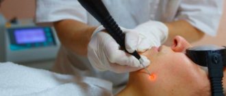
White spots appear after a mole is burned out with a laser. With regular exposure to ultraviolet rays (for example, while tanning in a solarium or in direct sunlight), the removal site becomes noticeably lighter. health hazard from the appearance of white spots . Typically, such spots disappear on their own within 3-6 months, completely merging with the color of the skin. In very rare cases, the process of natural pigmentation takes 1 to 2 years.
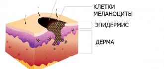
Also, white spots appear on the skin if the body of the mole was located in the deep layers of the epidermis and melanocytes, which form the pigment, were removed along with it during the operation . As they grow, the white spot will gradually acquire a natural, normal color.
Dark spots after removal
Dark spots that appear at the site of the removed nevus require careful attention, as they may indicate complications after surgery:
- Black . A dry, dark brown or black crust is not dangerous. Essentially, it is clotted blood . When the wound is completely healed, the crust will fall off on its own. Ideally, there will be a pink depression underneath (young skin);
- Brown . The presence of such a stain indicates that removal operation was not completed completely . Nevus cells remained in the layers of the skin. In this case, a visit to the clinic will be required. Most often, the specialist will refer the patient for a second operation;
- Violet . The most difficult case . The appearance of a burgundy or purple spot is an alarming symptom. Thus, the body signals inflammation as a result of an infection or that the mole was not completely removed, and the remaining nevus cells in the body began to degenerate into a malignant form. The sooner the patient goes to the hospital, the higher the chances of avoiding serious consequences.
How does healing occur?
After removing the pigment spot, a red mark remains, which is gradually covered with a thin crust. For the first month after surgery, it is advisable to protect the place where the procedure was performed. The crust that appears instead of the nevus should fall off on its own; tearing it off is prohibited. It protects the wound until new skin forms.
When the crust falls off, a tender area of skin appears that needs protection from the sun. Rarely does a hole appear after removal of a mole or new formations in the form of tubercles of a dense structure.
The duration of wound healing after surgery cannot be determined; it depends on the characteristics of the body.
On average, the wound healing time is 4 weeks; the newly formed skin will hurt and cause discomfort.
Should you be concerned about pigmentation changes?
After surgery to remove a mole, traces of the procedure may be visible on the skin. Light dry spots do not pose a danger to the body. This also applies to the black, dense crust. Over time, the pigmentation of the affected area will be restored.
You should only be wary of dark red and purple spots that cause discomfort. If the operation site is inflamed, moist and hot, this indicates the presence of an inflammatory process. In this case, you should not self-medicate and urgently need to go to the clinic.
Deep face resurfacing under general anesthesia and its consequences
General anesthesia can be used for deep resurfacing of the face and eyelid area. This helps to avoid pain and guarantees complete immobility, which improves the result of the procedure. However, it is important to know the consequences of general anesthesia:
- pressure drop,
- disturbances of blood circulation, heart rhythm, breathing, kidney and liver function,
- increased intracranial pressure - nausea, vomiting, severe headache,
- dry mouth,
- allergic reactions.
Most of them can be avoided with a preliminary examination. Therefore, when choosing an operation under general anesthesia, be sure to consult a therapist in advance and obtain an opinion on the possibility of such anesthesia. The choice of the clinic, the specialist who will carry out the resurfacing, and the anesthesiologist is also important.
Watch this video about how laser facial resurfacing is performed under anesthesia and what is done to quickly heal the skin:
Ways to remove stains
Pink and white spots do not require treatment. Natural pigmentation of cells occurs gradually. If personal hygiene rules are not followed and the wound is not properly cared for, a permanent white pigment spot may form on the skin.
To avoid possible complications, do not subject the removal site to mechanical damage and do not get it wet . Peeling off the crust and being in a humid environment can contribute to infection of the wound or increase the time of skin regeneration.
Limit your time in direct sunlight . If possible, protect the surgical site with clothing or tape for 3-5 months.
Photo 2. The crust formed at the site of the mole should be preserved for as long as possible. Source: Flickr (Haw Yu)
If the mole was not completely removed or, after accidentally removing the crust, obvious signs of suppuration appeared, the wound bleeds and does not heal for a long time, be sure to contact a specialized specialist .
The doctor will determine the cause of the pathology and prescribe appropriate treatment. In some cases, repeat surgery will be required.
Hyperpigmentation after laser and spots on the face after resurfacing
Most often, hyperpigmentation after laser and spots on the face during grinding are temporary consequences, their appearance is explained by the activation of pigment-producing cells - melanocytes. This reaction usually disappears on its own within 3-4 months. Excessive pigmentation can be provoked by sunlight during the period until the skin has fully recovered. Therefore, it is strictly forbidden to go outside in the first 2 months without cream with a 50 SPF filter.
Classification of hemangiomas
Doctors divide this type according to its structure and manifestation:
- Is there a red spot on the body or a blue one? Does it turn pale when pressed? This class is the simplest - flat. The color directly depends on the vessels or capillaries from which it grows.
- The bulge and appearance of the purple node is a cavernous or cavernous type of hemangioma.
- If the tumor pulsates, and small vessels spread out from the center of the mole, it is a branched tumor. If it is damaged or removed independently, bleeding is inevitable.
- A noticeable red, raised mole is called a bump-shaped or spider-shaped mole.
Self-medication methods
Sometimes it happens that incomprehensible changes occur with a tumor, but it is not possible to see a doctor in the near future, for example, if the patient is on vacation. In this case, you can use some pharmaceutical drugs that will not cause harm in any case. However, before using them, it is advisable to consult at least with a nurse if you are prone to allergies.
The most common way to quickly get rid of redness and inflammation of the skin around the tumor is to wipe it with medical alcohol. This must be done several times a day until the problem is completely eliminated or until you can make an appointment with a specialist. It is necessary to moisten cotton wool or a cotton pad with alcohol and apply it to the redness without pressure; there is no need to secure the compress with a bandage or plaster, just wiping is enough. If the pharmacy does not have medical alcohol, you can replace it with Septil.
One of the readily available ways to eliminate redness is streptocide powder. It is sold in pharmacies in finished form or in tablets that must first be crushed. Sprinkle the powder on the reddened mole, without wrapping it with a bandage or securing it with a band-aid, and leave it on for several minutes. This method is not suitable if you have previously used any cream to relieve inflammation, since streptocide will react with it.
Another remedy that allows you to quickly relieve redness and inflammation of the skin is calendula tincture - an anti-inflammatory, antiseptic, healing and regenerating agent. You can buy it at the pharmacy or prepare it yourself, for this you will need to pour 2 tablespoons of dried calendula flowers with 100 ml of alcohol or vodka, and then leave in a dark place. It is necessary to wipe the inflamed mole with the tincture several times a day until the problem is completely eliminated.
Preoperative examination of the patient
Professional preoperative diagnosis is the main condition for the prevention of melanoma. The doctor first talks with the patient, then examines him: assesses the total number of birthmarks on the body, their size, contours, consistency of the neoplasm, color. Even an experienced specialist cannot always accurately determine the degree of danger of a neoplasm without additional methods.
Dermatoscopy is an examination of a nevus using 10-40x magnification, which makes it possible to examine changes on the surface of the mole with high accuracy.
If doubt remains about the benignity of the nevus, a more serious examination is prescribed, including the following methods:
- echography of the affected area to determine the depth of germination,
- radiography,
- local thermometry (malignant areas have a higher temperature),
- indication by radioactive phosphorus isotopes,
- puncture biopsy.
Such an examination is necessary to select the optimal removal method.
What affects the outcome of the operation
The consequences of mole removal are affected by:
- Features of the neoplasm itself - the deeper the mole is located, and the larger it is, the greater the likelihood of scar formation.
- Removal method - each of them has its own indications, features, advantages and disadvantages. Scars rarely form after laser coagulation, but always after surgical excision. Benign moles are removed using gentle methods. If malignant changes are suspected, the method of choice is surgery.
- Qualification of a doctor - a specialist must know how to determine the nature of the neoplasm, exclude the possibility of malignant degeneration, master the appropriate diagnostic techniques, based on this, choose the optimal method for eliminating the birthmark, and have experience in carrying out such manipulations.
- Congenital characteristics of the human body - there is a category of people prone to scar formation.
- The patient’s compliance with the doctor’s recommendations regarding the wound care regimen during the postoperative period.


