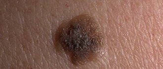
It is impossible to find at least one person who does not have a single unique spot - a mole. The appearance of these marks can be due to many reasons, including hormonal changes and heredity. Moles sometimes occur due to prolonged exposure to ultraviolet rays, most often on areas of the skin that have been injured. Since moles are a common phenomenon, many people see no reason to worry if their appearance does not change, does not increase, and does not hurt or itch. But not everyone understands what to do if a pimple forms on a mole. Not everyone knows what is the best thing to do in this case: visit a doctor or wait until the pimple goes away on its own. It is necessary to understand what causes the occurrence of foci of inflammation around the mole.
Allergic processes
Such skin reactions are increasingly common among modern people.
Allergic pimples usually do not hurt, they are numerous and itchy.
May be accompanied by a burning sensation.
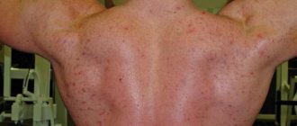
Extensive rashes may affect pigmented areas of the skin.
As a result, everything looks as if a person suddenly developed a lot of pimples in the form of moles.
If the allergy is quite active, then after the process subsides and resolves, peeling and crusts remain.
You cannot rip it off, as it can easily cause infection.
How to remove inflammation
Purulent inflammation has dangerous consequences. Accompanied by pain without touch or exposure to the sun. The site of inflammation is itchy, painful and constantly hot.
You should act according to the recommendations of an oncologist or dermatologist. For instant pain relief, turn to universal folk and classical methods.
| Means | Mode of application | How many times a day | How does it work |
| Alcohol or alcohol tincture | Moisten the area of the hanging mole | 1 to 3 times | Disinfects the wound, eliminates the symptoms of inflammation |
| Tincture of homemade herbs (yarrow, ginger, etc.) | Wipe or apply lotion | 3 times a day | Knits, eliminates inflammation |
| Ointment with zinc, salicylic acid | Lubricate boils and scratches | 1 time | Kills harmful bacteria |
| Hydrogen peroxide | For open boils, soak with cotton wool. | 1-2 times for 7-10 minutes | Disinfects and cleanses the wound from harmful bacteria |
| Antiseptic | Grind into powder, lubricate (if tablet) | 1 time for wound post-treatment | Kills germs |
A scratch, injury, stye, scrape or abrasion can cause bleeding that is difficult to stop.
Immediately fix the wound and apply a sterile bandage moistened with hydrogen peroxide or alcohol.
What not to do if a boil appears under a nevus:
- try to release the pus on your own;
- remove inflammation yourself. Performed by a surgeon without any health consequences;
- try to hide with cosmetics;
- scratch the itchy area;
- contact with long-haired animals to avoid infection.
Herpes
This also applies to relatively common causes of acne.
The rash appears on the face in the lip area or on the skin of the groin.
In severe cases, it sprinkles the side surfaces of the body.
Therefore, moles can easily enter the affected area.
Especially if there are a lot of them.
Signs that a pimple in a mole on the face is of herpetic origin:
- Pain and burning several hours before the rash appears.
- A characteristic cluster of pimples in groups.
- Rapid formation of bubbles.
- Transformation into an ulcer-like surface.
- Drying with crust formation.
Perhaps herpes is the only type of inflammation that can cause skin cancer.
But only if the rash occurs often in the same place.
And herpes never leads to melanoma, no matter how many moles fall into the area of the rash.
Treatment options
The most effective treatment for nevus is removal. The operations are carried out in a few minutes, and recovery takes no more than four days. But the period depends on the quality, depth and size of education. Modern medicine distinguishes several methods of surgical intervention:
- laser;
- radio waves;
- electrocoagulation;
- cryodestruction;
- surgery.
Laser therapy is suitable for removing moles on the head; it allows you to control the depth of penetration of the rays. There is no pain during the operation; the procedure lasts about 10 minutes. After healing there are no scars left. But the quality of the operation directly depends on the power of the medical equipment.
Radio wave therapy is a quick and painless way to eliminate moles on the back and other parts of the body. The doctor may take tissue for examination and then perform the procedure. The operation takes about 15 minutes, and the patient recovers within a few days. No scars remain. Only convex nevi can be removed; it is not suitable for flat ones.
Electrocoagulation is considered a painless procedure. The doctor independently controls the depth of current penetration. But it will not be possible to remove large moles or those that have grown into the lower layers of the epithelium in this way.
Cryodestruction is the use of nitrogen. The substance can eliminate nevi of any size and depth in just a few minutes. A dense crust forms at this site, protecting against infections; it will fall off over time. But the method has some disadvantages: after the wound heals, scars may remain, and a repeat procedure will be required, since during the operation it is impossible to control the depth of action of nitrogen . Sometimes healthy tissue near the mole is affected, which will only worsen the situation.
Malignant tumors, regardless of where they may grow, are removed only by surgery. Surgeries allow you to remove nevi from the deep layers of the skin, this eliminates the recurrence of inflammation. The patient must be given local anesthesia or anesthesia. After the procedure, stitches are placed on the area of removal; in almost all cases, a scar remains.
All methods, except the last one, are suitable for eliminating benign formations. Only surgeons remove melanoma; after the operation, they prescribe chemical or radiation therapy, which will prevent the patient from getting sick a second time.
Pimple on a mole: danger signs
According to the descriptions proposed above, it is possible with a fairly high degree of probability to exclude the formation of melanoma or another type of skin cancer.
However, sometimes even an experienced doctor finds it difficult to distinguish a pimple from a mole, which threatens to degenerate into a malignant tumor.
It is necessary to urgently go to a dermatologist for examination in the following cases:
- Swelling in any part of the mole progresses relatively slowly, over several weeks or months.
- For a long time the process is painless, it can only cause moral discomfort, and sometimes causes itching.
- A mole that looks like a pimple begins to bleed and is easily injured.
- Pigment asymmetry occurs and color heterogeneity appears.
- The color of the formation darkened or, on the contrary, became lighter.
- A pimple appeared on a mole and did not change in dynamics for several days.
All these are individual signs of the degeneration of a stable pigment formation into a malignant process.
If a person has at least one of them, consultation with a dermatologist is necessary as soon as possible.
Types of nevus
A mole can be acquired or congenital. The latter are usually inherited from parents. Their shape, color and size are different. But throughout life, nevi can appear due to various factors:
- high levels of radiation;
- hormonal imbalance in the body - during pregnancy, puberty and serious illnesses;
- genetic predisposition;
- excessive dose of ultraviolet radiation.
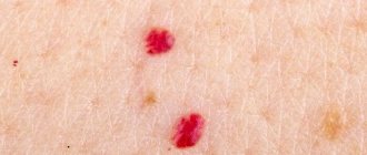
There are two classes of moles - nonvascular and hemangiomas.
The latter appear due to damage to blood vessels and are pink or red in color. Capillary nevi are located on the surface of the skin, and cavernous nevi grow under the upper layers of the epithelium. Non-vascular formations have any shape, they can be flat or hanging. Sizes range from microscopic dots to huge spots. Some moles may appear without any reason; they do not pose any danger.
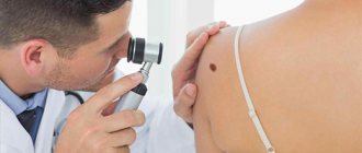
Dermatoscopy of a pimple on a mole
Dermatoscopy allows one to establish the nature of the process with sufficiently high accuracy.
This is a non-invasive technique.
Its essence is to study a suspicious area of skin at high magnification and under bright light.
This way you can look at the signs of inflammation in the area of the mole - an expanded capillary network, swelling of cells and intercellular space, accumulation of immune tissue macrophages.
The formation of new vessels, atypical cell shape, growth of melanocytes beyond the pigment layer, and infiltration of the basal epithelium speak in favor of the neoplasm.
Causes
- Mechanical damage. If a mole is injured as a result of friction against clothing, involuntary tearing, or scratching, then its integrity is compromised and thus an entry point for infection is formed. When there are a lot of microbes, the skin structures become inflamed and can fester.
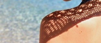
- Prolonged sunbathing is dangerous not only due to increased production of melanin on the skin (and there is already an excess of it in nevi), but also due to increased sweating and overheating of the dermis. Excessive sweating creates favorable conditions on the skin for the growth of pathogenic microflora.
- Skin pathologies. With them, inflammatory processes can develop in the epidermis, often leading to suppuration in acne, eczema, dermatitis, scabies, and furunculosis.
- Immunodeficiencies. With them, the skin partially loses its protective functions and becomes easily infected.
- Exchange disorders. Diseases such as hyperthyroidism, diabetes mellitus and obesity are accompanied by the development of skin pathologies and the appearance of various rashes on the human body; such a rash can also form under moles.
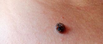
- Degeneration of nevi. It is manifested by the formation of ulcers, elevations, tubercles on it, a change in its previous color, regular bleeding, and hair loss.
- Dysfunctions in the sebaceous glands. In such cases, as a result of the accumulation of excess fat, the skin pores and omentums are poorly cleaned and become inflamed when bacteria or fungi enter.
- Folliculitis. A disease in which hair follicles are affected and suppurated.
Thus, the mechanism of formation of pus inside a mole is due to the penetration of microbes into its superficial or deep layers, as well as into the areas of the hair follicle and sebaceous glands, which cause their inflammation.
Histology of the mole
A biopsy with histological examination of the material allows a more accurate and correct diagnosis to be made.
First, the mole is removed, and then the removed tissue is carefully examined under a microscope.
In this case, a number of conditions must be met:
- If a mole becomes inflamed like a pimple, first eliminate the active process.
- Pre-conduct dermatoscopy to determine the extent of removal.
- Use only a traditional scalpel to maximize the preservation of the original mole tissue.
- The procedure is carried out in a specialized medical institution in compliance with all the rules of asepsis and antisepsis.
Doctors themselves know how to properly prepare and send the sample for histological examination.
Results are usually ready after 5-8 days.
If the conclusion states that no signs of malignancy or atypia were found, then there is nothing to be afraid of.
A pimple and a mole just turned out to be a harmless combination.
Otherwise, if a suspicion of a tumor process is detected, a consultation with an oncologist is required.