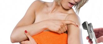
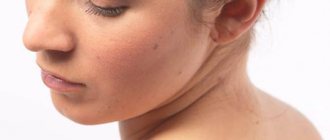
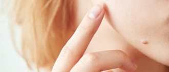
Moles on the face and body do not always fall into the category of harmless “decorations”. Often, nevi are located in places that are constantly injured - on the neck, in the armpits, in the groin, under the hair and on the bra line. There are often cases when, after intense tanning in a solarium or hormonal imbalance, a mole begins to degenerate - it increases in size, becomes inflamed, begins to hurt or itch, and rashes appear around it. In these situations, it is necessary to urgently make an appointment with an oncodermatologist in order not to miss the appearance of skin cancer - melanoma.
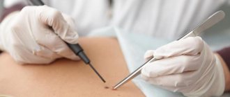
One of the most common methods of removing a mole is excision of the neoplasm with a scalpel. Typically the procedure can take up to 1 hour. To relieve pain, the doctor makes several injections into the extraction area. During the operation, in addition to the affected tissue, part of the healthy skin is captured, because otherwise suturing is impossible. When surgically removing a mole, the patient must be tested for syphilis, HIV, hepatitis B and C and other infections that are transmitted through the bloodstream.
What is a nevus (mole)
A mole or nevus is an acquired or congenital defect on the skin or mucous membranes of a person. A mole may be characterized by a high content of pigment cells, which is why it stands out against the background of the characteristic skin color.
There are no differences between congenital and acquired moles, and the process of the appearance of new growths of this type is considered normal. Moles are benign skin formations. In rare cases, cell mutation occurs and the mole transforms into melanoma.
Removal of nevi (moles) Kyiv takes place after a histological examination of structural cells. The method of removal and further actions of the surgeon depend on the pathology and condition of the mole.
Types of moles
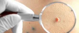
- Lentigo. The safest type of moles are dark brown or light brown in color. They are a small flat formation on the skin of uniform color. They rarely cause discomfort to their owner and are often perceived as freckles.
- Hemangioma. Vascular moles resemble nodules or flat formations on the skin that are red or pink in color. These formations on the skin are somewhat reminiscent of warts or significant age spots. The size of hemangioma varies from miniature (a few millimeters) to significant (up to a centimeter).
- Giant pigmented nevus. The congenital formation on the skin is quite impressive in size. These moles have a flat surface and a brownish color. Due to its unaesthetic appearance and large size, a giant pigmented nevus is removed at an early age.
- Epidermal-dermal nevi. Flat, undistinguished moles or slightly raised moles above the skin. The size of the formation can vary from 1 mm to 1 cm, and the color range from light brown to dark brown. This formation is localized in the buttocks and groin area, on the palms and soles of the feet.
- Blue nevi. Most often, blue nevi cover areas of skin on the arms, legs, buttocks, and face. Due to their biological activity, such formations are considered melanoma-hazardous. The color of blue nevus varies from blue to bluish-brown. It can reach a size of several centimeters or is almost invisible on the skin. Often the formation is dense, lumpy, without hair.
- Intradermal nevi. This formation has a convex shape and rises quite significantly above the skin. They have a smooth or warty surface that is black/flesh in color to the touch. Often such a formation penetrates the hair and causes a lot of discomfort due to its unattractive appearance. The growth of hair through a mole indicates the malano-mono-dangerous nature of the formation. After all, cancer cells destroy hair follicles and prevent hair growth.
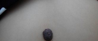
- Dysplastic nevi. Skin formations that rise above the skin and are irregularly shaped. There is a genetic predisposition to the formation of this type of mole. Quite often, such formations reach a size of several centimeters and do not have a clear outline.
Normal recovery after surgery
If the intervention is performed by an experienced specialist, then the wound healing process proceeds normally. All you need is time and proper care.
- In the first week, a crust forms. The most important thing is not to touch her. In the first few hours, the wound may bleed a little, especially if it was removed using a scalpel. Avoid getting it wet or accidentally damaging it. In some cases, it is recommended to rinse with a light solution of potassium permanganate and apply an antibacterial ointment prescribed by a doctor.
- Second week - the crust on a small growth often falls off. In larger formations, it may still persist, and the skin underneath may itch. The exposed layer of thin epidermis requires protection from sunlight, and visiting a solarium is prohibited at this time.
- By the third week, the operation site should no longer hurt or bleed, even when affecting a large area. The redness goes away. If the laser method was used, then everything should be fine by this time. The application site is no different from normal skin.
- The fourth week is considered the last. At this moment, the patient forgets that he was once bothered by a nevus or birthmark. And the wound heals completely.
What is the chance of melanoma?
Not all moles can turn into cancer.
Melanomic moles account for 95% of all cases of surgical or hardware excision of moles. Don't be upset if your body is generously covered with moles. Melanoma precautions and prevention are quite simple. Expert opinion Sakania Luiza Ruslanovna
Dermatovenerologist, cosmetologist, trichologist
Ask a Question
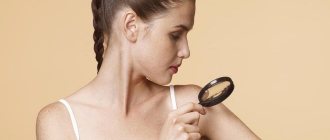
It is worth limiting your exposure to direct sunlight, not exposing moles to injury, eating right and leading a healthy lifestyle. Deformation of a mole: change in color, shape, size, increased sensitivity - indicates a disruption in the vital activity of the mole’s cells.
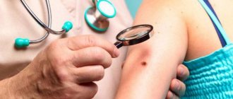
Only an experienced dermatologist during the examination will be able to predict the transformation of the mole and prescribe the necessary laboratory tests. After deciphering the tests, complete and effective treatment/removal of the mole (nevus) is prescribed.
Scar treatment
Scars and scars may appear at the site of a removed mole; this happens when the body of the nevus is located deep in the subcutaneous layer, as well as when the rules for treating fresh wounds are violated. If the wound is not properly cared for and is not kept clean and sterile, bacterial infection occurs, which leads to tissue scarring. When moles are removed with a laser, the risk of complications developing is much less, but this does not mean that the area of the resulting wounds does not require special care.
To ensure that there are no scars left at the site of the removed mole, special silicone-based patches are used. Also, the elimination area can be smeared with medicinal ointment, which resolves scars. But if a lump has formed at the site of the scar, you need to see a doctor, since this is a pathology that requires special treatment.
Is it necessary to remove the nevus?
The nevus must be removed in several cases:
- There is a suspicion of melanoma or other malignant tumors, which cannot be excluded without removal. Some nevi will need to be completely removed surgically for detailed examination under a microscope (histology). Otherwise, based on external signs, it will be impossible to exclude melanoma. If the nevus is large, and only one focus is suspicious, you can take only this piece with a scalpel or radio wave apparatus. Laser and electric knife are not suitable for these purposes. In addition to melanocytic nevi, there are sebaceous, warty and others. They will also need to be removed if skin cancer, basal cell carcinoma or skin sarcoma is suspected.
- High risk of nevus developing into melanoma. The doctor assesses the risk of each specific neoplasm turning into melanoma and, based on the degree of risk, decides whether the nevus needs to be removed. The doctor is helped in making a decision by deviations in external signs: violation of uniformity of color, clarity of boundaries, increase in size, symmetry. Damage and trauma to the nevus in the past, sunburn, and family predisposition are important. It should be noted that any melanocytic nevus has a small percentage of chance of developing into melanoma. So, if the nevus is completely removed, this risk will be significantly reduced. Sebaceous nevus has an increased likelihood of transformation into basal cell carcinoma; for prevention purposes, it also needs to be removed.
- The external repulsive appearance of a nevus causes psychological problems in humans. If a nevus causes psychological problems or interferes with a person’s daily work and personal relationships, it must be removed. It is not only important here how a person perceives himself. Some people will still avoid communication, subconsciously considering such formations to be contagious.
How is surgical removal of moles performed?
There are two methods for surgical removal of a mole: excision with cauterization (burning out using a special device) and excision followed by suturing.
The operation lasts from 40 to 60 minutes. The duration of the manipulation will depend on the individual characteristics and condition of the patient, as well as the size and location of the mole.
Excision of nevi without cosmetic sutures consists of the following steps:
- Treatment of the operated area with antiseptic drugs;
- Carrying out local anesthesia;
- Excision of the nevus using a scalpel at the skin level;
- Cauterizing the wound with a special device for cauterization or applying a drug for local use;
- Treatment of the wound with a local antibacterial agent;
- Applying a sterile dressing.
Upon completion of the operation, the doctor provides comprehensive recommendations for caring for the postoperative area and sends the resulting material for histological examination.
Excision of a mole with suturing is carried out in several stages:
- Treatment of the operated area with antiseptics;
- Use of local anesthesia;
- Excision of the nevus with the capture of surrounding healthy tissue;
- Treatment of the wound with antiseptic drugs;
- Cosmetic stitches. Depending on the depth of the formation, superficial, self-priming and deep sutures can be used;
- Treatment of the wound with a local antibiotic;
- Applying a patch and sterile dressing.
The removed material is also sent for histology to confirm the benignity of the neoplasm. If the nevus turns out to be malignant, then another intervention is performed. A week after the operation, the sutures are removed (if superficial sutures are used).
Recently, surgeons have been using special silicone absorbable patches, which make the scar left after excision less noticeable.
They are glued to the scar and worn for 15-20 days. In addition, if the patient is not satisfied with his appearance, plastic correction can be performed in the future.
Removal or treatment?
As usual, each of us has power over our own body, but not in the case of nevi.
Not only we, but also a huge number of doctors recommend not to decide the fate of tumors on the body without permission. You, of course, can decide that you will remove a mole, but this process must take place not only under the supervision of a doctor, but also with his direct participation. You should never cut or rip off a nevus, or try to burn it out with drugs that are not intended for this.
Expert opinion
Sakania Luiza Ruslanovna
Dermatovenerologist, cosmetologist, trichologist
Ask a Question
Any outside interference in the life of a nevus can lead to irreparable consequences that no person would want to experience on himself.
Therefore, for the umpteenth time, we warn every person and advise seeing a doctor if a mole begins to cause some concern and inconvenience, or you simply decide to remove it for personal reasons. Contact a qualified specialist, and he will definitely tell you how to get rid of an annoying or interfering mole.
Treatment of nevi
The laser method of mole removal is especially indicated if the formation is located on the face, neck, open areas - where the skin is sensitive. It is possible to treat nevus, but many refuse to do so. There are enough reasons for this, one of them is the simplest removal of a nevus with a laser, which completely eliminates the reappearance of the formation.
So why make unnecessary attempts at treatment? But other people reject deletion and have every right to do so. They order special ointments and tonics and try to get rid of the nevus in this way.
Pharmacy medications act almost the same:
- destructuring of external tissues;
- anti-inflammatory effect;
- anesthetic effect;
- tissue regenerating and antimicrobial effect;
- absorbable;
- normalizing and controlling skin texture.
Advantages of the method
There are various methods for removing nevus. But excision with a scalpel is the safest. Advantages of the method:
- The ability to completely get rid of education in one session.
- Possibility of removing cancer cells located near the mole, formations in the deep layers of the epidermis.
- The operation is performed under the supervision of a doctor.
- For excision, local anesthesia is used.
Attention! It is forbidden to remove a mole yourself, even if it has been injured. This can lead to infection or the development of cancer. For this procedure, you must consult a doctor.
Removal of nevi
As we have repeatedly said, removal of a pigmented nevus or any other type of nevus from the face or other part of the body is possible, but only with the permission of a qualified professional. This professional should be a dermatologist or oncologist.
It is advisable to carry out the procedure after passing tests, laboratory tests and the doctor’s resolution for removal.
It can occur in many of the previously described ways, and whether it is a papillomatous nevus, removal of a pigmented nevus, or a slight disturbance of a mole, we will try to describe the processes as broadly as possible in order to provide you with complete information.
Rehabilitation period after mole removal
Surgical excision of nevi is associated with the longest period of rehabilitation. Depending on the structure, location and size of the nevus, the recovery period can last several weeks.
Throughout the rehabilitation period, the patient should adhere to the following recommendations:
- Do not scratch or tear off the crust that covers the postoperative wound;
- Do not sunbathe on the beach or in the solarium;
- Avoid getting water on the wound for a week after the intervention;
- When excising plantar nevi, do not subject the operated area to excessive stress. This will prevent possible bleeding;
- Carry out independent replacement of postoperative dressings only with the permission of the doctor;
- Follow hygienic skin care rules.
Also, rehabilitation after excision of a nevus involves cosmetic surgery to eliminate visible scars.
Types of therapy
Treatment methods are selected depending on the location and type of nevus. Sometimes it is left untouched, but observation by a dermatologist is recommended. If the formation is benign, then hospitalization is not required, but all procedures must take place in a medical facility.
Laser method
This type is carried out in most cases using local anesthesia. The laser is effective for benign formations:
- intradermal,
- epidermal.
The good thing about this method is that it is done very quickly. On average, the procedure takes about five minutes. The doctor cuts off the nevus in layers using a device that has directed laser radiation. It is possible to adjust the depth and intensity of beam penetration.
Expert opinion
Sakania Luiza Ruslanovna
Dermatovenerologist, cosmetologist, trichologist
Ask a Question
Laser nevus removal is non-contact and one of the safest. It allows you to remove nevi on the face or other areas where the skin is especially sensitive.
With laser there is no blood loss, pain, or risk of infection. Most moles, when completely removed, never appear in that place again. The wound heals in a few days, in rare cases it takes two weeks.
There are also some negative sides. After removing large birthmarks, a white spot remains on the skin. Since the laser evaporates the formation cells, it is not possible to send them for histological examination. Therefore, such therapy is used when the nevus is carefully examined by a doctor and tests are taken.
Surgical excision
The use of a surgical scalpel is a classic method in which a bordering incision is made. It is done in such a way as to touch healthy skin. After the manipulation, the wound is sutured, and the formation itself is sent for examination.
The method has proven itself well when the nevus:
- penetrated very deeply into the layers of the skin
- has several broken parts
- suspected of transforming into melanoma
Doctors do not carry out such treatment for herpes, infectious or inflammatory diseases. A wait-and-see approach is chosen until complete recovery. The procedure is carried out under anesthesia and guarantees the removal of the entire mole.
Expert opinion
Sakania Luiza Ruslanovna
Dermatovenerologist, cosmetologist, trichologist
Ask a Question
The disadvantage of removing a nevus surgically is the long healing period of the wound. Often a scar remains in this place. In addition, after surgical excision, you should not stay in the sun for a long time.
The procedure itself takes 30 minutes. The time depends on the location of the nevus and its size. In modern clinics, after surgery, silicone absorbable patches are used, which make it possible to make the scar after surgery less noticeable.
Cryodestruction
This method makes it possible to cope with the formation of cold. To do this, very low temperatures are used to freeze and immediately destroy organic formations.
In most cases, the procedure is prescribed when there is no need to send a mole for histology; it is also used to remove formations in a precancerous state.
The indications are:
- change in nevus size
- painful sensations
- bleeding
- formation of precancerous cells.
Cold is used on tumors located in the upper layers of the dermis. Under its influence, water crystallizes in cells, and the level of electrolytes and metabolites reaches a critical threshold. This destroys the cell membrane. The procedure is carried out using liquid nitrogen or a cryodestructor.
In both cases, the removal itself takes 10 seconds. The procedure is painless, so there is no need for pain relief. The disadvantage of the method is the lack of ability to control the depth of exposure, so in inept hands, burns and tissue scarring are possible.
Electrocoagulation
The method allows you to deal with a mole using thermal effects obtained by electric current. During the process, the treated area is cauterized, allowing:
- get rid of nevus quickly
- after healing, get a smooth area of skin without a scar
- explore cells of formation
- eliminate even the smallest formations.
This method is used on small nevi or if there is a base. Contraindications include: the appearance of malignant tumors, herpes, skin coagulation disorders, the presence of foci of chronic infection.
Before the procedure, local anesthesia is administered. The mole is then removed using a metal loop that carries a current. A small number of healthy skin cells are also cauterized to “seal” the vessels and prevent blood loss.
After the procedure, high-quality education care will be required throughout your recovery.
Radio knife
Using a radio knife is one of the most gentle techniques. High-frequency waves generate energy, which is used to cut tissue and evaporate cells. This allows you to get an almost invisible mark on the work site.
The main advantage is that healthy tissues are not damaged. The procedure itself takes no more than 20 minutes and is painless. The radioknife is used on all parts of the body, and formations can be examined.
Such removal cannot be carried out if skin formations begin to turn into malignant tumors, there are herpes or inflammatory diseases. It is also contraindicated for those who have heart rate sensors in their bodies.
The device itself removes moles during operation, disinfects and stops bleeding. It is possible to use a radioknife to work with precancerous formations.
Surgitron device
The technique allows you to remove a nevus using a cold radio knife. The method does not lead to complications and has a low level of trauma. The impact on the mole is carried out using high frequency radio waves. Energy leads to rapid destruction of cellular structures. The method can be used for single and multiple nevi.
However, one of the contraindications is pregnancy. The remaining restrictions are the same as for the radio knife. A crust remains at the treatment site, which falls off within a week, leaving a mark like an abrasion. A variety of medications are used to heal it. At the same time, solariums and rest in the open sun are prohibited.
What is the best way to remove a nevus on the face?
- It is better to remove nevus on the face with a laser or radio wave method. These methods can remove the vast majority of nevi of any type. After the procedures, inflammation will be minimal, the wounds will look neat, and healing will be fast.
- Using liquid nitrogen, you can only remove seborrheic or warty nevus on the face. After the procedure, tissue swelling in the area of the nevus is observed for several days, and the wound becomes weeping.
- The surgical method is irreplaceable if it is necessary to remove a large nevus and if it is very similar to melanoma or other malignant tumor. The downside is that scars appear that are 3-4 times longer than the diameter of the nevus. They try to orient the scars along wrinkles and natural folds, however, this does not always work.
- It is better not to use electrocoagulation at all to avoid scarring.
Advantages and disadvantages of the procedure
Like any medical intervention, surgical removal of a mole has advantages and disadvantages. The benefits include:
- The cost of the operation, in comparison with other methods, is lower.
- After surgery, it is possible to send the pigment formation for histological analysis. The study will determine whether the growth is malignant or benign.
- A scalpel can be used to excise large nevi.
- Mole removal surgery involves excision of a healthy area of skin that is located around the nevus. This is necessary to ensure that the malignancy does not reappear.
- Even if the nevus is located deep in the dermis, when using a scalpel, the surgeon is able to completely remove the growth. This distinguishes the surgical technique from burning or freezing.
- During removal of a nevus, there is no pain for the person, as local anesthesia is administered.
- Minimal number of contraindications, unlike other methods.
In addition to the advantages, surgical removal of moles has disadvantages. These include:
- The recovery period is longer than with other excision methods.
- A scar or scar remains after the operation. However, the scar is not always present. If the nevus was small, a scar can be avoided. If the nevus is located on the nose, lip, neck, you should choose a different method.
- A ban on visiting the beach and solarium until the wound heals.
How to remove a nevus at home?
Extreme types of impact (cutting, tearing off) should be completely excluded, as this is fraught with the development of cancer.
For removal at home use:
- garlic
- iodine
- ointment with celandine
- vinegar, apple juice
In all these cases, a cotton pad is moistened and used to lubricate the nevus. All folk remedies require a huge amount of time, and the consequences can be unpredictable.
Thus, nevi are neoplasms that can cause harm if handled incorrectly. Therefore, the treatment method is chosen by the doctor who decides on the need for a histological examination.