Ophthalmology
Share
- Xanthomas and xanthomatosis
- Types of xanthoma
- Xanthoma of the stomach
- Treatment of xanthomas
- Conservative treatment of xanthoma
- Treatment of xanthoma in children
Xanthoma is a disease resulting from a disorder of lipid (fat) metabolism. It is a focal skin neoplasm with peculiarly altered cells that contain fatty inclusions inside. It has been proven that the manifestation of xanthomas is associated with excess cholesterol in the blood of people suffering from them.
Causes of the disease
Skin plaques occur due to metabolic disorders and fat deposition on the surface.
The formation of xanthomas is caused by the following pathologies and conditions of the body:
- endocrine diseases;
- hereditary disorder of fat metabolism;
- atherosclerotic heart disease;
- chronic arterial hypertension;
- metabolic syndrome;
- oncological diseases;
- last months of pregnancy;
- diseases of the gastrointestinal tract;
- diseases of the hematopoietic system;
- pathologies of the kidneys and bile ducts.
The formation of xanthomas on the body is caused by a number of negative factors, including a sedentary lifestyle and obesity.
Factors contributing to the appearance of xanthoma on the skin:
- binge eating;
- age-related changes and aging of the body;
- obesity;
- sedentary lifestyle;
- insufficient supply of nutrients;
- long-term use or side effects of medications;
- psychological stress;
- smoking, drinking alcohol;
- consequences of surgery.
Classification of pathology
Cholesterol plaques on the skin vary in appearance, location and reasons for their appearance. The main types of xanthomas are presented in the table:
| Name | Options | |||
| Color | What do they look like? | Size, cm | Location | |
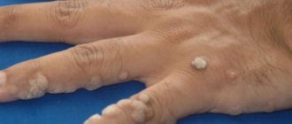
| Flat | Yellow | Flat, shapeless plaques | From 0.1 to 2.5 | Palms, fingers, feet |
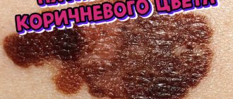
| Tuberose | Red-yellow, brown | Nodules, plaques | 1—5 | Elbows, buttocks, knees, fingers |
| Eruptive | Yellow, reddish | Multiple confluent nodules | 0,1—0,4 | Buttocks, shoulders, thighs |
| Tendon | Flesh, yellow | Subcutaneous tumors | 2—7 | Elbows, ankles, knees, tendons of the upper and lower extremities |
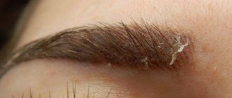
| Xanthelasma of the eyelids | Yellow | Small plaques | From 0.2 to 4 | Eye area |
| Xanthogranulomas | Minor rashes | From 0.1 to 2 | Face, head, upper body | |
Types of xanthoma
There are five clinical types of xanthoma:
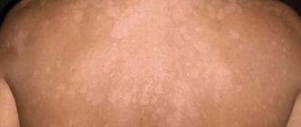
- Flat xanthoma. This type of xanthoma appears as soft, flat or slightly raised soft papules. These papules are yellow in color and round in shape. These skin formations are multiple in nature and are located in the area of skin folds, especially on the palms and soles. Flat xanthoma often affects older people suffering from atherosclerosis, obesity, and liver disease. There are two types of flat xanthomas: intertriginous and diffuse. The former are characterized by yellow-red lesions with clearly defined boundaries. Also, diffuse flat xanthomas can appear as groups of papules and multiple yellowish plaques, almost unlimited from each other. This type of xanthoma occurs in people suffering from diseases such as normolipidemia, paraproteinemia, lymphoma, myeloma, and leukemia. Sometimes xanthomas appear earlier than the first signs of underlying diseases. Intertriginous flat xanthomas can develop with hereditary hypercholesterolemia.
- Xanthelasma of the eyelids. This species is characterized by the appearance of multiple yellowish soft plaques around the eyes (from the inner corner). Xanthoma of the eyelids is the most common type of xanthoma, but not the most specific, since it is sometimes found in people who have normal blood lipid levels.
- Multiple nodular (eruptive) xanthoma. Its peculiarity is manifested in the sudden appearance of small flat or semicircular yellow papules, soft to the touch, with clear boundaries. In this case, the location of neoplasms on the body is chaotic. When papules coalesce, plaques appear. The sites of the most numerous rashes are the thighs (back surface) and buttocks.
- Tuberous xanthoma. With this type of xanthoma, plaques and nodes of a yellowish or brown color, ranging in size from 1 to 5 cm, form on the body. Laboratory studies of tuberous xanthoma have shown that the main content of these plaques is cholesterol (and not triglycerides, as in small xanthomas). Foci of tuberous xanthoma are the result of the fusion of the above-described eruptive xanthoma. On the body they are located on the skin of the buttocks, knees, elbows, and fingers. Patients with tuberous xanthoma are at high risk of developing coronary artery disease, since in this type of xanthoma the level of cholesterol in the blood is very high.
- Tendon xanthomas. As the name suggests, these xanthomas most often form in the area of the tendons (most commonly the extensor tendons of the fingers and the Achilles tendons). They look like dense, tumor formations the color of normal skin or slightly yellowish, moving along with the tendon when the joint is extended.
Symptoms of the disease
Yellowish formations can occur in different parts of the body and are prone to proliferation.
Xanthomas on the skin appear in the form of growths and plaques without pain. Main manifestations of skin defects:
- location on the face, buttocks, fingers, joints or eyelids;
- no pain;
- slow growth;
- yellow or flesh-colored, eruptive xanthomas may have a red rim around them;
- soft or dense consistency;
- thinning and dry skin at the sites of formations;
- tendency to gradually grow and merge with each other (eruptive xanthomatosis).
Features of the disease in a child
Plaques on the skin at an early age occur due to congenital disorders of lipid metabolism. As a child grows up, xanthoma develops under the influence of the following reasons:
- diseases of the gastrointestinal tract;
- diabetes;
- obesity;
- poor nutrition.
Treatment of xanthoma in childhood is as follows:
- dairy-vegetable diet;
- drug therapy;
- surgical removal.
Getting rid of unwanted manifestations is carried out using low temperatures - cryodestruction.
Xanthomas in children, after curing the underlying disease and eliminating negative factors, resolve and disappear on their own. If xanthomas remain on the face, removal is carried out using one of the following methods:
- Cryodestruction - freezing at low temperatures.
- Electrocoagulation - removal by electric current.
- Radio wave surgery is the destruction of pathological tissues by high-frequency directed waves.
Xanthomas and xanthomatosis
Xanthomas can be local (that is, concentrated on one area of the skin) or generalized. Generalized xanthomas - in case of weakening of the general immunity of the body, injuries, or the presence of chronic diseases (for example, diabetes), sometimes develop into xanthomatosis. With xanthomatosis, fatty tumors appear randomly throughout the body. This disease is especially dangerous for children, since it is often accompanied by damage to blood vessels and some internal organs.
Therefore, not only dermatologists, but also endocrinologists and therapists treat xanthoma and xanthomatosis.
A person with primary manifestations of xanthoma should know that their main cause is an oversaturation of the blood with the remains of neutral fats processed by the body.
Diagnostics
To determine the causes of defects, you must consult a dermatologist. Initially, a history of the disease is collected and a visual examination of the patient’s skin is performed. The following examinations are prescribed:
- Diascopy is an assessment of the nature of a neoplasm using a glass slide.
- General tests (blood, urine) to determine the condition of the urinary system and the functioning of the body.
- Biochemical blood test - study of disorders in internal organs.
- Lipidogram - detection of high cholesterol in the blood.
- Body mass index - determination of the patient’s weight.
An additional procedure in diagnosing pathology is magnetic resonance imaging.
Additionally, the following studies are carried out:
- Ultrasound of internal organs.
- Biopsy with histological examination - examination of a fragment of the affected tissue to determine the nature of the pathological process.
- Blood test for thyroid hormones. An assessment of hormonal levels and diabetic predisposition are determined.
- Magnetic resonance imaging (MRI). Layer-by-layer scanning of the selected area.
- Positron emission tomography (PET). It is the identification of initial metabolic disorders in the body.
Prevention
Prevention of xanthomatosis means preventing the development of hyperlipidemia. Since there are many reasons for this condition, preventive methods will be extensive. First of all this:
- limiting consumption of large amounts of animal fats;
- prevention of pathologies that can contribute to the development of hyperlipidemia, and if they have already arisen, their identification and treatment;
- timely preventive examinations by a therapist and cardiologist with regular instrumental and laboratory examinations.
All methods of preventing hyperlipidemia (and therefore xatomatosis) are valuable. But it should be remembered that in most cases it is not the “miracle” pill that plays a role, but the nutritional culture that should be instilled from childhood. Such a simple preventive measure as a proper diet and avoidance of fatty jellied meats, cutlets, and kebabs has saved more than one person from lipid disorders. This is a more sound method than experiments with “folk” remedies for removing fat from the body, as well as all sorts of lotions and rubs, with the help of which they try to reduce fat deposits (without taking into account that the same deposits can form in tendons, meninges and other structures of the human body).
Treatment of defects
Methods of influencing cholesterol plaque on the body depend on the causes of formation and the severity of the pathological process. Therapy includes:
- changing your diet to reduce fat;
- maintaining body weight;
- performing light physical exercise.
If there is a disease that causes xanthomas, the attending physician selects drug treatment individually for each patient. The main drugs are presented in the table:
| Group of drugs | Impact | Name |
| Statins | Reduce cholesterol | "Zokor" |
| Prevents cardiovascular complications | "Rosucard" | |
| Fibrates | Increases the amount of healthy cholesterol | "Traykor" |
| Bile acid sequestrants | Stimulates the production and elimination of fats | "Questran" |
| Antioxidants | Restores and heals skin cells | "Lycopene" |
| Vitamins | Saturate the body with nutrients | "Polivit Geriatric" |
| Cholesterol absorption inhibitors | Prevents fat absorption | "Ezetrol" |
| Nicotinic acid preparations | Regulate lipid levels | "Niacin" |
| Hormonal | Normalizes the production of thyroid hormones | "Euthirox" |
Cholesterol plaque can be removed by surgical intervention, one type of which is the use of a laser.
The following types of surgery help get rid of cholesterol plaque:
- Chemotherapy is the use of trichloroacetic acid to break down abnormal cells.
- Electrocoagulation is the cauterization of defects with a directed electric current.
- Laser removal - heating and destruction of xanthomas with a laser beam.
- Cryodestruction - freezing and further cauterization of growths (not used for plaques on the face).
- Radio wave excision - heating and evaporation of tumors.
- Surgical intervention - cutting out xanthomas with a scalpel.
Forecast
The prognosis for xanthomatosis is different. It depends on the causes of hyperlipidemia.
Xanthomas themselves do not pose a threat to the health and life of the patient. The prognosis for the complete elimination of fatty plaques in tissues is generally favorable - thanks to adequate treatment, new xanthomas do not appear, and those that have arisen can be eliminated surgically or cosmetically. But if hyperlipidemia occurs again, then often a relapse cannot be avoided.
On the other hand, xanthomatosis should be perceived not only as a pathology, but also as a signal about the possible development of other diseases caused by hyperlipidemia - these are:
- atherosclerosis. A particularly threatening condition is atherosclerotic damage to the coronary vessels of the heart, against which myocardial infarction (death) occurs, often resulting in death. No less dangerous is atherosclerosis of cerebral vessels, which can lead to stroke - a violation of cerebral circulation;
- heart defects
and some other pathologies.
Kovtonyuk Oksana Vladimirovna, medical observer, surgeon, consultant doctor
7, total, today
( 55 votes, average: 4.07 out of 5)
Sun allergy – photodermatitis
Thrush on the nipples during feeding: how to treat?