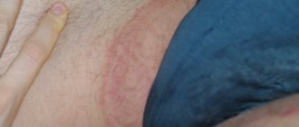
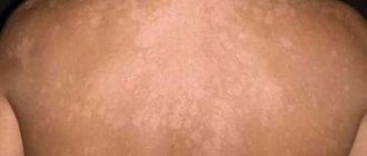
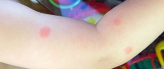
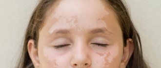
Scientists and doctors have repeatedly studied the issues of pigmentation. Brown spots on the skin appear due to an excess of melanocytes in the epidermis. They can have different shapes and sizes; the spots can be convex or flat. Defects are removed if they threaten health or cause psychological discomfort.
Causes of the disease
Diseases of high severity are quite common today. Chronic forms of these diseases often lead to disorders associated with melanin synthesis. The list of these diseases includes the following pathologies:
- Liver dysfunction due to cirrhosis and other diseases of this organ. It is this type of disease that most often leads to the appearance of small areas on the surface of the skin, colored in dark tones.
- Disturbances in the functioning of the endocrine system . Disruptions of the endocrine system have a huge impact on the quality of the metabolic system. Hormonal disorders, pathological processes in the functioning of the sweat glands, diabetes mellitus are only a small part of the reasons leading to the appearance of spots of non-standard color.
- The cachectic form of melanosis quite often accompanies a disease such as tuberculosis.
- Kidney dysfunction can also be expressed by the appearance of areas with an unnatural color. This form of the disease is called uremic type melanosis.
The above reasons are the main factors leading to the appearance of melanosis. However, this disease may also have a different nature, not associated with disturbances in the functioning of internal organs.
Treatment of dark spots on the skin
When treating dark spots, an integrated approach is used, which includes the use of tablets and medications for external use, surgery, and cosmetic procedures.
Drug therapy
The doctor selects medications based on the test results; the goal of therapy is to eliminate the manifestations of the disease and the root causes of the pathology.
Skinoren cream is prescribed as an effective anti-inflammatory agent in the fight against dark spots
What medications are used in treatment:
- anti-inflammatory drugs - Skinoren cream;
- antibacterial drugs – Rifampicin, Nystatin;
- antifungal agents – Clotrimazole;
- antihistamines - Loratadine, Claritin;
- steroid ointments – Triderm, Pimafucort;
- keratolytic agents - salicylic ointment, Resorcinol;
- vitamin complexes with riboflavin, folic acid.
As additional methods of treatment, the doctor may prescribe a diet, therapeutic exercises, and physiotherapy.
Important!
You should immediately consult a doctor if a spot appears on the skin of a child, grows, bleeds, hurts, or blood relatives have similar rashes.
Other treatments
If the appearance of age spots is not associated with serious diseases, you can get rid of them using traditional methods.
How to remove dark spots at home:
- Add 10 drops of hydrogen peroxide and ammonia to 30 g of homemade cottage cheese, spread the mixture evenly over the skin, and leave for a quarter of an hour. The procedures are carried out 2-3 times a week, a total of 10-12 sessions will be required.
- Mix 5 g of vinegar, honey and rice flour, lubricate pigment spots with the mixture, leave for 20 minutes. Apply masks once every 3 days for a month.
- The recipe for a soft scrub with an antiseptic effect is to combine 10 g of iodized salt with 5 drops of orange oil and 5 g of any cream. Apply the mixture onto the skin with light massage movements, leave for 2-3 minutes, rinse with warm water. Carry out the procedure 2-3 times a week.
At home, you can lighten age spots and age spots using retinoids, creams with AHA acids, serums with arbutin and hydroquinone.
Toxic type melanosis
This form of pathology is observed in people who are constantly exposed to various aggressive chemicals due to their profession. Prolonged contact with fuels and lubricants (oil, coal, oils) is one of the most common causes of pathology.
Prolonged contact with this category of products leads to acute toxic poisoning. Lack of attention to this problem can cause not only spots on the body, but also a chronic form of the disease. Against this background, various pathological processes are observed in the work of many body systems and a severe deterioration in health.
Becker's nevus
This type of mole looks like a small spot colored yellow-brown. Such neoplasms most often have uneven borders. Becker's nevus in most cases occurs in adolescents between the ages of ten and fifteen years. According to statistics, this disease is more common among males.
Becker's nevus is most often localized in the lower extremities and upper torso. At the initial stages of formation, the spots have a small diameter, but the development of the disease leads to a significant increase in their size. The average diameter of neoplasms can exceed ten centimeters.
Becker's nevus is a disease of unknown etiology. Experts associate the appearance of this disease with hormonal disorders.
Pigment spots, especially multiple ones, are a cosmetic defect
Dubreuil melanosis
This disease is oncological in nature. The appearance of small spots that are dark in color and irregular in shape often indicates the development of skin cancer. This form of neoplasm is most often localized in the upper body. Brown growths at the initial stage of development are very similar to moles and rise slightly above the surface of the skin.
At the initial stage of development, the spots have a small diameter, but over a short period of time their diameter increases several times. The color of the new growths can vary from light yellow to dark brown. The outline of the growths can be compared to a geographical map. The development of the disease leads to the formation of nodules and papules on the surface of the affected tissues and tumors themselves. The stain changes its structure, becoming denser.
The disease is accompanied by severe itching and redness of those areas of the skin that are nearby. The brown spot on the skin begins to peel off towards the end of the development stage. At the same stage, small spots similar to freckles begin to form on healthy areas of the skin. The appearance of these symptoms indicates the beginning of the degeneration of the spot into a malignant tumor.
Diagnostics
If it was not possible to make a diagnosis during the initial examination, a number of additional tests are prescribed to create a more accurate clinical picture.
Diagnostic methods:
- clinical, biochemical blood test;
- tumor marker test;
- analysis for STIs, allergens;
- general urine analysis;
- immunological studies;
- scraping, culture;
- biopsy of tissue samples from the affected area.
A biopsy of the affected areas of the skin is performed to accurately determine the cause of pigmentation.
Additional research methods are prescribed taking into account the general clinical picture, medical history, and age of the patient.
Acanthosis nigricans
Dermatologists are often asked the question: dark spots on the skin have appeared, what does this mean? Experts say that the appearance of black spots may be associated with the development of acanthosis nigricans. This disease is considered quite rare and has several forms, malignant and benign. Symptoms of acanthosis nigricans are most often localized in those areas of the body where there are folds of skin. These areas include the neck, buttocks, armpits and groin area.
The rapid growth of spots throughout the body may indicate the malignant nature of the disease. It is this symptom that most often precedes the onset of cancer. The following reasons may be the main factors leading to the onset of the disease:
- medications belonging to the category of hormonal drugs;
- disorders of the thyroid gland;
- malignant tumors;
- genetic predisposition and heredity;
- long-term use of a certain number of medications.
Pigment spots can disguise malignant skin tumors
Urticaria pigment type
Urticaria pigmentosa is a rather complex disease that can act as the main cause of mastocytosis. This form of pathology most often manifests itself in children of a younger age group and is characterized by the appearance of small dark red spots.
The appearance of spots is considered only the initial stage of the development of the disease. Next, rash bubbles filled with subcutaneous fluid appear at the site of the spots. At the final stage of development, the rash opens and leaves brown spots in its place. Such spots disappear on their own within a few months.
Having manifested itself in childhood, the disease is quite mild. However, at a more mature age, various complications may develop. Experts say that urticaria pigmentosa can cause disability and even death. More often, such situations are observed against the background of a long-term lack of attention to the disease.
Mastocytosis of this form has an unclear etiology. Experts attribute this pathology to the influence of the following factors:
- sudden climate change;
- inflammatory processes against the background of the activity of various infections;
- prolonged exposure to ultraviolet radiation;
- stress;
- disorders associated with the functioning of the immune system.
Useful video
For information on modern methods of diagnosing and treating skin diseases, watch this video:
Similar articles
- Anti-pigmentation cream, anti-freckle, whitening...
Dark skin on the face is a problem that anti-pigmentation cream can solve. It comes in whitening, day and night options. There are also several effective Chinese creams. Read more - The skin on the face is peeling: why does this happen...
There are quite a few reasons why the skin on the face of women, men, and children peels. Starting from allergies and ending with hereditary factors. The skin also tends to be dry, red, blotchy, and dry around the mouth. What to do? Read more
- Types of acne on the face: allergies, rashes, in the form of...
In dermatology, there are certain types of acne on the face. They can be allergies, rashes in the form of bumps or subcutaneous ones, inflammation and red spots, dermatitis, irritation. To begin treatment, the causes of their occurrence should be established. Read more
- Wound after laser wart removal: consequences...
A small wound after laser removal of a wart will be normal. How to treat a wound after a plantar or finger wart? How long does healing take, what consequences can there be? How to treat the consequences: swelling, pus, burn, if the wound is oozing, there is blood. Why doesn't the wound heal? Wound treatment after wart removal. Read more
Lentigo
Lentigo is a benign disease that causes brown spots on the skin . This type of neoplasm has an appearance similar to moles. If dark spots of this type appear on the skin, they can be localized in the face, legs, limbs and upper torso.
This disease has a slow progression and can appear at any age. The risk of spots degenerating into a malignant tumor is minimal . However, if the integrity of the skin is compromised, increased attention should be paid to the state of your health.
The main reason for this problem is long-term exposure to artificial radiation sources. In addition, lentigo spots can be associated with gene mutations, the activity of papillomavirus and various disorders of the immune system.
Experts especially highlight the influence of hormonal disorders and the effects of ultraviolet rays on the formation of such spots. In addition, lentigo spots can be caused by AIDS and other diseases leading to a severe decrease in immunity.
Problems with hyperpigmentation in this disease appear in the form of single spots of uniform color with clearly defined boundaries. Neoplasms can be localized in various parts of the body, including the face and limbs. Lentigo spots most often appear at one of the stages of child development in the womb. However, in medical practice, cases are described when this pathology manifests itself at a more mature age.
The development of the disease can cause an increase in the diameter of the affected tissues. Often small dots of a darker color form on the surface of the spots.
Pigments are responsible for the color of human skin; in healthy skin there are five of them: melanin, carotene, melanoid, oxyhemoglobin and reduced hemoglobin.
Leopard cider
Leopard syndrome is a rare disease that manifests itself in the form of many spots on the body of various colors. Such pathology can be localized in various parts of the body.
In addition to the appearance of spots, patients experience problems with the functioning of the heart muscle, slight mental disabilities, hypospadias, disturbances in the functioning of the respiratory organs and growth retardation. The formation of this disease is associated with the mutation of certain genes.
Formations due to liver diseases
The liver is considered the body's filter. In diseases (hepatitis, cirrhosis, liver failure), the color of the skin changes, the dermis becomes yellowish due to the fact that a lot of bilirubin circulates in the blood. The whites of the eyes turn yellow and become cloudy.
Rashes form on the back, on the skin of the legs, abdomen, arms, and face. They may be brown, red or beige with ragged edges and an uneven shape. Spider veins, a rash on the face and neck, and brown spots on the abdomen in the liver area may also appear.
If a light brown spot appears on the body, do not ignore it. With liver disease, patients exhibit additional symptoms: bitterness in the mouth, abdominal pain, and abnormal bowel movements.
Chloasma
Women are more susceptible to chloasma. This pathological condition is characterized by the appearance of various dark spots. The color and size of the tumors may vary depending on their location. Chloasma can be localized on any part of the body, including the face, chest, genitals and torso. Areas of the body with hyperpigmentation are only a cosmetic defect.
The main cause of the disease is hormonal imbalances. Pregnancy, menopause and ovarian dysfunction most often lead to the appearance of pathology.
Which doctor should I contact?
Since the causes of dark spots are varied, the first step is to consult a dermatologist. After examination and initial diagnosis, the doctor may prescribe a consultation with a geneticist, oncologist, immunologist, endocrinologist, or gastroenterologist.
If dark spots appear on the skin, consult a dermatologist
Sometimes simultaneous treatment from several specialists is required, and in some cases it is enough to undergo a treatment course with a cosmetologist.
Pathology named after Recklinghausen
This disease is better known as neurofibromatosis type 1. At the initial stage of development, small spots form on the patient’s body, on which a cluster of freckles forms. Most often, the disease manifests itself in childhood. New growths can vary in color and size.
In approximately fifteen percent of cases associated with the presence of pathology, the development of the disease leads to oncological complications. In addition, experts talk about an increased risk of developing the following pathologies:
- cyst in the respiratory organs;
- slow growth and the appearance of empty cavities in the spinal cord;
- gynecomastia and renal artery stenosis.
As a result of the accumulation of melanin in large quantities on the skin, age spots are formed.
