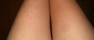
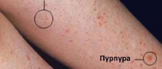
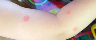
When malfunctions in the functioning of any internal organs occur in the body, it gives alarm signals in the form of clinical manifestations. The appearance of spots on the legs that look like bruises is one of these symptoms. They can have different shapes and different sizes. An experienced doctor can use these characteristics to determine what caused the pathological changes. There are many triggers that can cause pinpoint bruising. Knowledge about them helps to understand how to act if hematomas are detected.
Why do they appear?
They are classified as pigmentation disorders that arise due to a long-term severe form of a chronic disease. Excessive amounts of melanin are deposited in the skin, causing dark spots to appear.
What diseases lead to the formation of dark spots:
- Endocrine melanosis. Begins to develop due to congenital or acquired dysfunction of the endocrine system.
- Cachectic melanosis due to a severe form of tuberculosis.
- Malfunctions of the sex glands.
- With hepatic melanosis, dark areas appear on the skin due to liver dysfunction or cirrhosis.
- Sape-Lawrence syndrome.
- Diabetes.
Vascular
Vascular spots have a pink, purple, purplish-red hue. They are classified into the following types:
- hyperemic:
- hemorrhagic;
- telangiectatic.
Hyperemic, in turn, is divided into:
- inflammatory spots filled with blood, resulting from inflammation of the walls of blood vessels, called roseola (diameter less than or equal to 2 cm) and erythema (diameter greater than 2 cm);
- spots of non-inflammatory etiology, manifested as a result of nervous shocks, worries, experiences, called marks of shame.
Hemorrhagic formations occur due to:
- external influence - bruises that disappear within a few days;
- manifestations of signs of vascular diseases - traces of internal hemorrhages that require serious medical therapy.
Telangiectatic spots are a consequence of dilation of the walls of blood vessels, a violation of their elasticity due to the influence of diets, smoking and alcohol, diseases of the gastrointestinal tract and heart, and excessive exposure to ultraviolet radiation. They represent the so-called “vascular network”.
Can be localized between the legs
Darkening of the skin between the legs and other parts of the body may appear.
Their occurrence is due to a disruption in the functioning of blood vessels or due to pigmentation of the skin.
Find the answer
Are you having any problem? Enter “Symptom” or “Name of the disease” into the form, press Enter and you will find out all the treatment for this problem or disease.
Changes in skin color on the legs are classified as age spots, which are often found in people.
Only the attending physician can identify the cause and prescribe treatment. There is no need to self-medicate, because there are many causes of pigmentation on the lower extremities and each of them requires individual treatment.
Common reasons:
- Increased production of melanin leads to the formation of brown spots on the skin. The shade depends on the amount of pigment produced.
- Due to the use of modern methods of hair removal on the legs, darkening occurs due to malignancy, that is, malignancy of age spots.
- Pathological disturbances in the functioning of the body indicate the development of several diseases, which leads to increased pigmentation. The list of diseases is huge - from lichen to vitiligo. The doctor selects treatment depending on what caused the pigmentation on the legs.
- Malfunctions in the functioning of the circulatory system.
- In case of insufficient intake of ascorbic acid into the body, rutin.
- Diseases of internal organs.
- Insufficient physical activity leads to weakening of the walls of blood vessels and causes pigmentation on the skin in the form of dark spots.
- If the vessels are located close to the surface of the skin.
- Taking medications that cause blood vessel destruction.
- For varicose veins.
Patients often make the mistake of thinking that the change in pigmentation was due to an allergy. If it does not go away after avoiding contact with the allergen, you can be sure that the cause is something more serious than an allergic reaction.
Pigmentation disorders are accompanied by severe itching.
Diagnostics
First, the doctor will conduct a visual examination of the spots on the legs, study the medical history, in particular, he will be interested in problems with blood vessels in the past. To confirm the preliminary diagnosis, instrumental diagnostics will be required. The most popular methods:
- angiography or radiography of blood vessels - it can be used to identify blood clots, plaques, areas of narrowing of the lumen of the veins and disturbances in the speed of blood flow;
- duplex scanning – allows you to evaluate the anatomy of the vascular bed and the dynamics of blood circulation;
- Dopplerography is a non-invasive ultrasound examination technique that allows you to identify problem areas and assess the condition of the walls of blood vessels.
An MRI may also be ordered. This is the most informative, but expensive method for diagnosing spots on the legs.
Pigmentation in the form of darkened spots
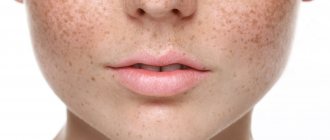
Represents the development of dark areas of various shapes on the skin. They will also range in color from lighter to darker brown.
Causes:
- Skin aging;
- Hormonal changes;
- Use of incorrectly selected cosmetics;
- Skin insolation;
- Taking medications that cause pigmentation changes.
If there are many such spots, then they represent a serious cosmetic defect. Sometimes they are accompanied by roughening of the skin, dryness, the appearance of wrinkles, and the appearance of blood vessels. You should be careful, because this is often how malignant neoplasms are masked.
What can trigger the development of dark spots:
- Metabolic disorders;
- Diseases of internal organs;
- Neuropsychiatric disorders;
- Gynecological diseases.
If they appeared during pregnancy, then there is a high probability that they will quickly go away after childbirth.
Angiokeratomas
Angiokeratomas are malformations of capillaries and blood vessels.
In most cases, they appear as pink, purple or red spots that rise above the skin. There are several forms of such formations. Types of angiokeratomas:
- Limited angiokeratoma. A vascular nodule not exceeding half a centimeter in diameter and having an irregular shape. It usually forms on the feet, thighs or legs and bleeds at the slightest injury. The color of such a spot can vary from dark red to purple.
- Furniture angiokeratoma is characterized by small red spots on the feet, fingers and hands. Over time, vascular formations can increase up to 5 mm, darken and rise above the skin. In some cases, stratum corneum and ulceration may form. This is a hereditary disease that is transmitted primarily through the female line.
- Angiokeratoma papular appears with trauma to the usual form of this disease. In this case, it can increase to a centimeter and take on the appearance of a wart. Usually has a purple color.
- Fordyce's angiokeratoma appears on the scrotum. Characterized by bright red vascular spots reaching 4 mm.
- Angiokeratoma of the vulva forms on the labia majora. The spots are the same as with angiokeratoma of the scrotum.
- Fabry angiokeratoma. It appears in the form of small dark red nodules that rise above the skin. Such formations can also occur on internal organs.
Sun marks
Causes of dark spots:
- After tanning, dark patches may appear on the skin due to excess melanin production.
- The cause will be various diseases of the internal organs.
- Doctors do not advise people with liver disease to stay in direct sunlight for long periods of time.
- If there are problems with the thyroid gland, then there is a high probability that this is the cause of increased pigmentation.
5 useful tips to help avoid pigmentation:
- While on vacation at sea, stop taking hormonal medications.
- Do not use antibiotics.
- Before sunbathing, it is forbidden to use perfume.
- Preference should be given only to reliable tanning products.
- Do not take sedatives.
Compliance with the described rules will significantly reduce the risk of developing increased pigmentation. If it does appear, you need to consult a dermatologist.
How to remove a patchy tan:
- Take hot baths often using a hard washcloth;
- Exfoliating scrubs and gels are effective;
- Post-exposure medications and toning agents will help.
Features of treatment
Any hematoma can be treated at home. If the bruise has just formed, you need to apply cold to it. It will stimulate vasoconstriction, reduce the severity of blood flow, and relieve swelling. Such treatment will be effective only if no more than an hour and a half has passed since the formation of the blue spot.
Important! When using ice, avoid direct contact with the skin and wrap it in a light cloth or handkerchief.
When the swelling of the tissues around the bruise subsides, dry heat should be applied to the stain (a bag of heated salt, a boiled egg, a piece of gauze folded in several layers, heated with a hot iron). Warm compresses made from a decoction of plantain or chamomile help to resolve hematomas. You need to warm up the bruises at least four times a day. The duration of one session is 15 minutes.
Traditional medicine recipes
The goal of local treatment of a hematoma is to speed up the process of resorption of thrombosed blood. This can be done by applying to the bruise:
- Compresses made from onion pulp and salt. Both ingredients are mixed in equal quantities, the resulting mixture is wrapped in gauze and applied to the damaged skin for twenty minutes three times a day.
- Starch compresses. The powder is mixed with water until it reaches the consistency of sour cream. The substance is applied to the sore spot and left for four hours. If a bruise appears on the skin from a blow, it is better to use raw, grated potatoes instead of starch.
- Applications using beets and honey. The vegetable must first be boiled and grated, mixed with a spoon of liquid honey. Apply the resulting mass to the bruise for three hours. In two days it will become noticeably lighter.
It is also recommended to do wiping with a solution prepared from 9% vinegar (two parts), salt (one part) and five drops of iodine.
It is better to remove bruises under the eyes using lotions made from herbal decoctions. To prepare them you need to use plantain, sage, medicinal marigold, St. John's wort.
Pharmacy preparations for stains
There is a wide range of medications that can effectively combat bruises on the body. Among them, the most popular are creams, ointments and gels with leech extracts. Heparin and Troxevasin ointments, bodyaga powder, and “Rescuer” balm help well to resolve hematomas. Their use allows you to eliminate skin defects in two to three days.
Radical treatment
Traditional medicine and local conservative therapy can be successfully used to treat small blue spots. Hematomas with a large affected area are treated using surgical methods. The choice of elimination tactics is made taking into account the characteristics of the clinical picture of skin lesions.
If the doctor sees that the blood inside the hematoma continues to remain in a liquid state (fresh bruise), he can remove it from under the skin using a puncture - a puncture, which is carried out with a special needle with a wide hole. The ejection is suctioned through it.
The doctor removes the clotted blood (more than five days have passed since the formation of the hematoma) by opening the blue area of the skin. For these purposes, he can use a scalpel or laser. The second tool allows you to perform the operation efficiently and avoid the risk of bleeding.
An autopsy with the installation of drainage is chosen if the hematoma begins to fester. During the operation, blood clots and accumulated pus are removed.
If blue spots on the skin are a symptom of the development of an internal disease, therapy is aimed at treating it. You should seek help from a doctor when the blue spots on the skin are large and form swelling, and if a hematoma appears on the head or abdomen.
Blemishes on the skin that look like a bruise
More often, pigmentation on the skin, which looks like bruises, occurs in older people.
Causes:
- Phlebeurysm;
- Impaired blood circulation;
- Fragility of blood vessels;
- Hemorrhagic vasculitis;
- Lack of platelets;
- Low blood clotting.
6 tips to help you get rid of stains quickly:
- Cure the original disease that triggered the development of the symptom.
- If this is an allergic reaction, then eliminate the allergen and undergo a course of therapeutic treatment.
- If pigmentation has developed due to lichen, then take oral and external medications;
- Use special hygiene products for care,
- Increase overall immunity;
- Use external preparations and cosmetic procedures.
Foot injuries
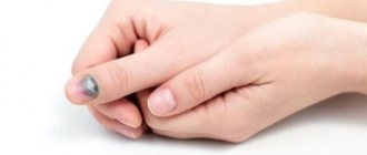
Sprains, sprains, sprains, or dropping something on the foot can cause bruising, causing the skin to appear blue or purple in color. This injury also often causes pain and swelling. People can usually treat minor foot injuries at home with RICE therapy:
- Rest. Avoid unnecessary activities and prolonged stress on the injured leg.
- Ice. Apply an ice pack to the injured leg.
- Compression. Wrap your injured leg in a bandage. The bandages should be snug, but not tight enough to prevent circulation.
- Height. Use pillows or leg cushions to elevate your leg whenever possible.
Over-the-counter nonsteroidal anti-inflammatory drugs, such as ibuprofen or aspirin, may help reduce pain and swelling.
For more severe injuries, your doctor may order an X-ray to check for broken bones in the foot. Treatment for a broken foot depends on the type and severity of the fracture.
Prevention of possible diseases
It is impossible to protect yourself from all diseases, so all that remains is to maintain the body’s functioning at a high level. To avoid failures and the development of pathologies, you must follow a few simple rules:
- Proper nutrition and absence of bad habits
- Moderate physical activity, more walking
- Personal hygiene, cleanliness in the house, regular ventilation
During the winter, you can additionally take minerals and vitamins to maintain the body's protective functions at the highest level. Be healthy!
Watch a video about exercises to prevent varicose veins:
Thus, spots on the legs that look like bruises can appear due to various diseases. In order to determine the true cause of their appearance, it is worth turning to specialists and not self-medicating.
Noticed a mistake? Select it and press Ctrl+Enter to let us know.
For various reasons, dark spots may appear on the skin of the arms, legs, or other parts of the body. This often occurs after sunbathing. To get rid of this problem, you need to visit a specialist (dermatologist) and listen to his recommendations.
The site provides reference information. Adequate diagnosis and treatment of the disease is possible under the supervision of a conscientious doctor. Any medications have contraindications. Consultation with a specialist is required, as well as detailed study of the instructions! Here you can make an appointment with a doctor.
Diagnostic measures
To make a correct diagnosis, the doctor conducts a general examination of the patient and collects anamnestic data.
After this, a microscopic examination of scrapings from the affected area is carried out, this allows us to exclude the presence of a fungal infection. A general blood test is also required.
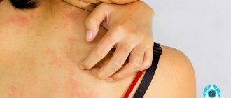
If an allergic reaction is suspected, tests are performed to determine the type of allergen. If necessary, a biopsy of a particle of the skin where the elements of the rash are localized can be performed.
Lichen
Symptoms of many infectious diseases are reduced to the appearance of spots on the skin. Ringworm is very common. It has both fungal, bacterial and viral etiology, and can manifest itself due to weakened immunity and hereditary predispositions.
Experts determine the type of lichen by the characteristic signs of spots on the skin.
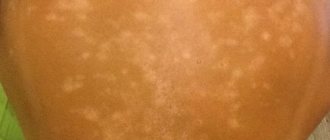
- With pityriasis versicolor, yellow and brown marks with a crust appear on the body.
- With pityriasis rosea - round pink with a red border, flaky.
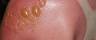
- With shingles, red inflammation resembles an allergic rash with blisters, which is localized along the nerve trunks on the back, under the ribs.
- With lichen planus, the neoplasms look like nodules that connect with each other, forming large-scale foci of inflammation.
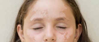
- With lichen alba, white spots appear without clear boundaries.
- Ringworm is characterized by the formation of shapeless bald patches on the head.
The symptoms of lichen are very similar to the rash associated with chickenpox, measles, and rubella. Sometimes lichen is confused with eczema and allergic skin dermatitis.

