Types of trichophytosis, sources of infection
Based on the type of pathogen, a distinction is made between anthropophilic and zoophilic trichophytosis. Trichophyton fungi spread through flakes of skin, hair, fur, and pieces of nails of a sick person or animal.
Anthropophilic types of trichophytosis
Superficial trichophytosis is caused by fungi that parasitize human skin - the anthropophilic fungi Trichophyton violaceum, Trichophyton tonsurans.
They are located mainly inside the hair and do not cause acute inflammation of the skin. The course of trichophytosis is superficial, not affecting the deep layers of the skin.
The disease is contagious and is easily transmitted from person to person through contact or exchange of personal belongings. Shelled skin flakes and broken hair infected with the fungus are transferred through a common comb, hairpin, or hat to the hair of a healthy person.
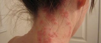
Trichophytosis mainly affects the skin and hair on the head, the smooth skin of the torso and limbs (shown in the photo), and very rarely the nails.
Most cases of superficial trichophytosis of the head occur in children. They get sick more often at the age of 5-12 years, which is explained by age-related characteristics of the acid-base balance of the scalp.
Occasionally, anthropophilic trichophytosis is observed in newborns and adults.
In adults, scalp hair is protected from infection by trichophytons by organic acids that have antimycotic activity. Therefore, ringworm, as trichophytosis is also called, is less common among them.
There have been cases of spontaneous resolution of trichophytosis in sick children as they grow older. This mainly applies to boys.
In girls, the disease lasts for years, taking a sluggish chronic course. The lesions are masked by healthy hair, the disease often remains unrecognized until old age, and is detected accidentally when other family members, usually children, are infected with trichophytosis.
Superficial trichophytosis of the smooth skin of the trunk and limbs occurs equally often in children and adults, and does not differ by gender.
Predisposing factors for chronic trichophytosis in adults are diseases of the peripheral blood vessels - acrocyanosis, erythrocyanosis, dysfunction of the gonads.
Zoophilic trichophytosis
Infiltrative-suppurative trichophytosis develops as a result of infection with zoophilic fungi that parasitize animals, Trichophyton verrucosum, Trichophyton gypseum.
Zoophilic fungi multiply around the hair, cause profound changes in the skin, lead to suppuration of the hair follicles, and the formation of an extensive focus of inflammation.
A person becomes infected with infiltrative-suppurative trichophytosis from wild and domestic animals. The carriers of the disease are mice, rats, sheep, and cows.
The fungus is not only transmitted through direct contact. Much more often, infection occurs through objects contaminated with the fur of a sick animal.
Trichophytosis in humans, caused by zoophilic fungi, is characterized by a severe course.
Risk factors
The risk of trichophytosis increases with fresh cuts, scratches, and skin damage. Endocrine diseases and weak immunity contribute to infection.
The activity of the fungus increases at elevated body temperatures and high ambient humidity.
Epidemiology
- Microsporum canis fungi are infected from sick cats and dogs (usually kittens). Mostly children get sick. They become infected through direct contact with animals. By the time of puberty, self-healing is noted, which is associated with the normalization of cellular-humoral immunity.
- Infection with the fungi Microsporum gypseum and Microsporum nanum occurs in adults cultivating the soil.
- Anthropophilic species of fungi that cause microsporia and trichophytosis are infected from sick people and infected household items (pillowcases, hats, scarves, scissors, combs, etc.).
- The source of Trichophyton mentagraphytes mushrooms is domestic animals: cattle, horses, calves, goats and donkeys, as well as wild animals. The source of Trichophyton verrucosum are small mouse-like rodents, rabbits, gophers and guinea pigs. Transmission of infection from a sick person is possible. Infection from animals occurs through direct contact, as well as through the hair and scales that animals leave on the soil, hay and feed. Rural and urban residents - workers of hippodromes, circuses, zoos and veterinary institutions - are getting sick. In addition to animals, the role of grasshoppers in transmitting infection has been proven. Trichophytons were found in corn stalks and straw.
- Favus spreads mainly among members of the same family. Low levels of sanitation and personal hygiene contribute to the appearance of mycosis.
Anthropophilic trichophytosis
The fungus initially penetrates the hair follicle of coarse hair on the head or vellus hair on the body. The incubation period of anthropophilic fungi is 7 days. After this time, the first signs of the disease become noticeable.
Trichophytosis of the scalp
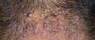
At first, one focus of infection appears on the scalp, and later their number increases. They are located over the entire surface of the head in isolation, without merging with each other.
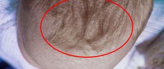
The diameter of the lesion in human trichophytosis is 1-2 cm and has no clear boundaries (see photo). The area of diseased skin loses hair. They thin out, fall out, break off, leaving stumps up to 3 mm high, covered with a gray coating, or black dots.
Characteristic black dots in the lesion have diagnostic value for trichophytosis.
The skin at the site of infection is swollen, flaky, and covered with scales reminiscent of dandruff in seborrhea or psoriasis.
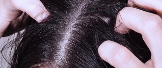
In the chronic course of trichophytosis of the scalp, the only clinical signs of the disease may be the presence of black dots broken off at the base of the hair (see photo).
It is difficult to detect signs of a chronic process, since women suffer from this form of trichophytosis in 80% of cases; it is difficult to detect signs of infection by a parasitic fungus among long, healthy hair.
Black dots are located mainly in the back of the head; it is almost impossible for a woman to detect them on her own. In addition to blackheads, a sign of chronic infection with a trichophyte fungus is slight peeling in the lesion, weak, delicate scars from lost hair.
Superficial trichophytosis of smooth skin
Mostly with superficial trichophytosis, exposed areas of the body are affected. The disease usually begins with the appearance of a clearly defined, round, pinkish spot on the face, shoulders or back, slightly raised above the skin.
There may be several spots with trichophytosis of smooth skin (as in the photo), they do not bother the patient, and there is practically no itching of the skin. Bubbles filled with exudate appear on the surface of the spots. When the bubbles open, they form crusts.
In the center of the spot, inflammation quickly subsides, giving way to slight peeling. A red ridge of inflamed skin remains along the periphery of the spot, due to which the lesion takes on the appearance of a ring.
The hair follicles located at the site of infection are affected. They fester, complicating trichophytosis with folliculitis.
Without treatment, trichophytosis of the skin of the body becomes chronic. The presence of foci of infection with the trichophyton fungus on the scalp and torso is often noted.
Chronic trichophytosis is characterized by predominant damage to the skin of the extremities. Pink-bluish spots of irregular shape without clear boundaries are scattered on the skin of the legs, buttocks, shoulders, and forearms.
Spots are found on the face, back, and chest. The soles, palms, and nail plates are affected. The stratum corneum thickens on the soles and palms, and deep cracks may occur.
When a nail becomes infected, a grayish spot first appears on it. Gradually it grows, the nail acquires a yellowish tint, its surface becomes bumpy. A nail infected with fungus thickens, changes shape, and crumbles easily.
With a weakened immune system, chronic superficial trichophytosis takes on a deep course.
Deep trichophytosis of smooth skin is characterized by the formation of boil-like nodes - trichophytosis granulomas or Majocchi granulomas. With this form of trichophytosis, purulent lesions of the lymph nodes are possible.
Recovery forecasts
If the pathology is diagnosed in the initial stages, and the treatment regimen is chosen correctly, taking into account all the nuances, the infection can be eliminated forever and in a short time. But in the case of an advanced stage, when a person has suppuration and infiltrates have appeared, dealing with the problem will be much more difficult and will take quite a long time, sometimes up to six months or longer. It is important to make sure that the infection is completely destroyed, otherwise trichophytosis threatens to become chronic, which will manifest itself with frequent relapses.
Zoophilic trichophytosis
The incubation period of trichophytosis, caused by the zoophilic fungi Trichophyton gypseum, lasts up to 2 weeks. When infected with Trichophyton verrucosum, it can take up to 2 months from infection to the appearance of the first symptoms of trichophytosis.
Trichophytosis is the most aggressive and capable of causing acute forms of the disease when transmitted from a sick animal to humans.
Subsequently, when people become infected from each other, the strength of the pathogen decreases, and the disease becomes similar to the clinical manifestations of superficial trichophytosis.
The infiltrative-suppurative form is characterized by the appearance of pink spots with a peripheral ridge, flaking in the center. The lesion grows due to inflammation at the periphery of the spot.
The lesions multiply, grow, merging, becoming covered with pustules and crusts. The vellus hair on the affected skin area breaks off, leaving stumps.
The formation of massive purulent infiltrates covered with crusts is characteristic of the scalp, beard, and mustache. The surface of the infiltrates is bumpy due to ulcerations, inflamed follicles, and erosions.
The skin of the lesion acquires a bluish tint, and pus is released at the site of hair growth. When pus appears, the fungi die, remaining only on the peripheral ridge along the border of the spot.
The acute stage of the disease subsides; without appropriate treatment, the disease becomes chronic.
The purulent form of trichophytosis is accompanied by high fever, enlarged lymph nodes near the site of infection, an increase in ESR, and deterioration in health.
Treatment
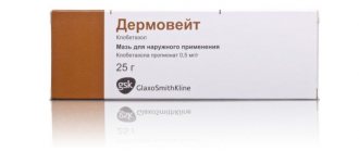
To treat all forms of trichophytosis, use ointments or creams Lamisil, bifonazole, clotrimazole, exoderil, ketoconazole, mycospor, sulfur ointment.
The main antimycotics for the systemic treatment of trichophytosis in humans are lamisil and griseofulvin. During treatment, hair on the affected areas of the skin is removed after 7-10 days.
Griseofulvin is taken orally daily until the first negative test for trichophyton fungi. After which they take the drug for another 2 weeks every other day, then another 2 weeks - every other day.
The full course of treatment takes about 2 months and is considered complete after 3 negative tests for trichophytons done with an interval of 7 days.
In the treatment of trichophytosis, external agents are widely used. The affected area is lubricated daily with a 2% iodine solution, and Wilkinson's ointment and sulfur ointment are applied in the evening.
For treatment of pus and crusts, furatsilin, ichthyol, rivanol, and potassium permanganate are used.
In case of chronic trichophytosis, the stratum corneum layer of the skin is detached. To do this, use milk-salicylic ointment. A compress is made with it for 2 days, after which a compress with 2% salicylic ointment is applied for a day.
The infiltrative-suppurative form of trichophytosis is first treated with antiseptics and anti-inflammatory drugs to reduce inflammation.
Infected skin areas are washed for 3 days in a row 2-3 times a day with solutions of ichthammol, nitrofural, ethacridine.
After this, sulfur ointment with tar is applied to resolve the infiltrate, and then treated with antifungal drugs.
Diagnostics
Laboratory tests will help identify the pathogen.
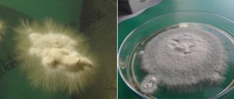
Diagnosis consists of cultural examination of affected tissue samples. To do this, infected tissues are placed in a special environment that promotes active growth of the fungus. The diagnostician examines the samples, monitors their growth, shape, and color. Thanks to these parameters, the type and variety of the pathogen can be determined. Also during the research process, it is important to carry out differential diagnosis and exclude the development of other fungal infectious diseases.
Prevention
Measures to prevent trichophytosis include preventing the spread of fungal infection and timely treatment of animals with ringworm - calves, cows, sheep.
Prevention of trichophytosis includes deratization of residential and industrial premises, as well as mandatory veterinary examination of domestic cats, dogs, hamsters, and rabbits.
To prevent trichophytosis, it is necessary to limit children’s contact with stray animals and use only personal combs and hats.
Prevention of disease relapse
The main measures aimed at preventing the recurrence of the disease are:
- Disinfection of living space and hygiene items used by the patient.
- Thorough examination and treatment of animals that could provoke a re-outbreak of the disease.
- Disinfection of hairdressing instruments.
- Regular sanitization of premises.
To prevent relapse, it is important to carefully examine everyone who has been in contact with the sick person or who may become a carrier of the disease, including pets
If trichophytosis is not treated, the disease can provoke an outbreak of a school or family epidemic. Therefore, at the first signs of the disease, you should consult a dermatologist or infectious disease specialist.