Each of us closely monitors our health, because maintaining it is the main component of success in any endeavor. As popular wisdom says: “If you have health, everything else will follow.”
It may, of course, not work itself out, but if a person is sick, then, as a rule, there is neither the strength nor the mood to achieve any global goals in his personal life, career, and so on.
Unfortunately, it is very difficult to constantly maintain health, so sometimes you have to fight diseases in their various manifestations. One of these symptoms may be the appearance of a ball or lump under the skin. In fact, such lumps appear in many people, so do not panic - first consult a doctor and find out what the nature of the lump is.
Types of seals
A lump under the skin may be harmless and go away on its own or with the use of some cream or gel. However, there are situations in which such compaction means serious health problems. In this case, it is important to identify the disease in time to avoid complications.
Atheroma
It's a cyst. The cause of its occurrence may be an infection in a wound on the hand, or some small foreign body, for example, a wooden or metal splinter.
Very often such compaction is accompanied by suppuration.
Treatment is possible traditionally - compresses with ointments, folk - compresses with coltsfoot leaves, baked onions, garlic, and also through surgery.
There are two types of surgery:
- An incision to remove the contents of the lump;
- Complete removal of the seal - laser cutting.
Treatment depends on personal preferences, capabilities and complexity of the problem.
Hygroma
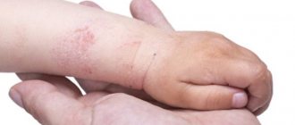
This is a lump on the hand that occurs in people very, very often. It is located on the outside of the wrist. In the normal state it is almost invisible, but if you lower the brush down, this tumor-like formation will be clearly visible. In fact, there is nothing wrong with this compaction - it is joint fluid that has come out of the joint under the skin.
The most common cause is joint injury. However, this often occurs in people who work at a computer or have begun to constantly carry heavy loads. It also occurs in women during the postpartum period, when there is a need to constantly carry the child in their arms.
Hygroma can be almost invisible or reach two centimeters. Depending on the degree of its development, it may not cause any inconvenience, but it can be very painful. As a rule, with this type of lump on the hand it is very uncomfortable and in some cases it is impossible to perform some actions related to the movement of the hand - leaning on the hand, standing on the palm as if doing push-ups - the lump simply prevents you from bending the hand.
Several types of treatment for hygroma are also possible - the use of ointments, compresses, tablet medications, surgery, etc.
As a rule, surgical intervention is not recommended, since the risk of recurrence of hygroma is high. For women during the period of feeding and lactation, the use of a bandage is prescribed whenever possible - it fixes the hand and prevents hygroma from developing, and the massage effect helps to reduce it.
Neurofibroma
This is a ball that, as the name suggests, is associated with nerve bundles. It is a benign tumor that occurs on the skin, under the skin, often found in the elbow area.
In general, neurofibromas are not a cause for concern, and often a person is not even aware of their presence in the body.
However, they can manifest themselves in the form of darkening of the skin, the appearance of a large number of moles in one place, etc. In addition, the ball can compress the nerve and thereby cause the body part to malfunction.
Treatment can be carried out with radiation therapy or through surgery.
Folliculitis
It is a dense ball located in the shallow layers under the skin. The cause of its formation is generally inflammation of the hair follicle. But it can already develop due to severe sweating, uncomfortable tight clothes that rub against the skin and put pressure on it, etc. It is clear that most often folliculitis occurs in the summer during the heat.
Otherwise it is called lipoma. It is a small ball. If you feel it, you will notice that it is a little soft. In this case, such a tumor, unlike others, may move slightly to the side. It is benign, but must be treated as soon as possible.
There are many folk remedies that are very effective. However, in difficult cases, when the tumor has reached a large size, doctors recommend surgery to prevent pinched nerves.
Dermatofibroma
Has a direct connection with the skin, is usually dark in color and increases over time. The skin in this area becomes more sensitive. As a rule, doctors advise removing such a formation to avoid unnecessary injuries and inconvenience.
Electrocoagulation, laser technique, direct surgery, cryodestruction are the main methods of removing dermatofibroma. There are also folk remedies to combat such a lump, but they are less effective.
As you can see, there are a large number of reasons for the appearance of a ball under the skin on the hand. Each disease is treated differently.
If you have a ball under your skin, it is very important not to make a mistake in making a diagnosis. Therefore, do not panic, go to the hospital as early as possible - this will avoid possible complications.
Many people may notice a ball suddenly appearing under the skin. Education may not hurt without causing any inconvenience. Subcutaneous inclusions are localized in various areas of the body. However, people of all ages and genders are not immune from their occurrence.
Benign growths are not dangerous. They need to be removed only if they create discomfort. But it is important to know the signs that should definitely alert a person.
Classification of seals on the foot
If a lump appears on the foot that causes pain, it should be classified in order to prescribe adequate therapy. The characteristic shape of the seal resembles a ball, the dimensions of which range from 1-2 mm to 3-4 cm. When you press on the seal, a sharp pain occurs.
The most common factors when a lump appears on the foot and hurts may be the following conditions:
Corns
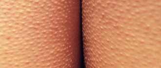
With keratinization of the skin on the feet, which is caused by improperly selected shoes, obesity, excessive sweating, flat feet, arthritis, etc. This reason is the most common. Corns can be flat, dry, raised, with or without rods. They have a yellowish-brown tint, a rough skin surface with reduced sensitivity to external influences.
In some cases, cracking of the foot is possible, accompanied by pain during increased physical activity. In the absence of timely treatment, the foot may become deformed, displaced, and the ball of the big toe may become enlarged.
Corn
A distinctive feature of calluses is their shape, which is a round, limited area of skin on the heel or toes, which is accompanied by pain even with slight pressure. Often, calluses can appear in a child, which is associated with the wrong choice of shoes, flat feet and injuries.
Calluses can be wet or dry. Wet calluses are the most dangerous, since they contain an accumulation of fluid (blood, serous exudate), which can leak out when the callus is traumatically damaged. As a result, there is a possibility of infection of the wound surface. In addition, wet calluses can transform into dry calluses, but with deeper roots.
It will take a lot of time to completely get rid of calluses. If the inflammatory process develops, antibiotic therapy may be prescribed, and if the symptoms are severe, surgical intervention is recommended.
Warts
In appearance, warts are very similar to calluses. The color of the resulting bumps practically does not differ from the color of the skin, and the foot is rough on palpation. When pressing on the plantar wart, acute pain is observed. The difference between a wart and calluses is the presence of a dark dot in the middle, but if the wart grows deeply, it is quite difficult to notice it.
The strains of the virus responsible for the formation of warts on the sole of the foot are not dangerous. The virus is not capable of transmitting from person to person and is activated only in a humid, warm environment. For a wart to appear, a starting point (an abrasion, injury, etc.) is required, as well as a weakening of the body’s immune defense.
Important! After surgical removal of warts, a long rehabilitation period is required, since the roots of the wart grow quite deeply, which requires more radical surgery.
Atheroma
A seal of this type is quite movable and soft to the touch. The cause of atheroma formation is the inflammatory process in the sebaceous gland. During physical activity on the lower extremities, pain symptoms are likely to occur, as well as various types of deformities in the arch of the foot.
The main condition for the success of therapy for atheroma is careful adherence to hygiene measures, prevention of damage to the integrity of the skin, normalization of immune defense and the choice of the right shoes.
Thrombophlebitis
This disease is accompanied by compactions in the lower extremities and is characterized by a chronic course. In the process of disruption of blood flow, a clot (thrombus) is formed, diseased veins become inflamed, and a dark blue compaction appears at the site of development of the pathological process. Even with slight pressure on the tubercle, sharp and severe pain occurs.
Bursitis
This disease in most cases is characterized by a benign tumor-like formation filled with gel-like contents. On palpation, the seal is round, mobile, elastic and soft. Painful symptoms occur as a result of physical stress on the affected leg. Therapeutic measures for bursitis include immobilization of the affected limb, wearing comfortable shoes, and physiotherapy.
In case of chronic development of bursitis, a surgical puncture of the bursa is performed, followed by washing the cavity with antibiotics and prescribing hormonal drugs.
Phlegmon
Even small wounds or cracks in the foot can cause the formation of a seal with phlegmon. Often, phlegmon develops after boils, carbuncles and long-term non-healing ulcerative neoplasms. The site of inflammation is red and hot to the touch, there is acute unbearable pain, weakness and deterioration in the general condition of the patient.
Phlegmon requires mandatory treatment, including surgery, since various complications may lead to amputation of a limb, and in some cases the patient may die. Therefore, at the first symptoms of the disease, you should immediately contact a medical facility.
Heel spurs
With increased calcium deposition, heel spurs may form. In this case, the soft tissues of the foot compress the nerve endings, causing unbearable pain to the person. Typically, the incidence of heel spurs within the joint increases in older age, due to wear and tear of the fatty tissue on the bottom of the foot.
Treatment is carried out with external medications and intra-articular blockades, which effectively relieve the inflammatory process, relieving pain symptoms. In the absence of a positive result, surgical intervention is recommended.
Keratoderma
This disease is extremely rare and the causes of its occurrence are not fully understood. Presumably, genetic mutations can lead to keratoderma. This group includes several types of dermatoses accompanied by impaired keratinization. Externally, the growth resembles a yellow-brown tubercle, slightly rising above the skin with the presence of small depressions on it.
Seals cause discomfort to the patient, and in some cases they may indicate the development of a serious inflammatory process in the body. It is recommended to treat the disease as an internal autoimmune disorder. Characteristic symptoms of keratoderma are lack of pain and constant itching.
Lipomas
A moving ball under the skin on the face or body may be a lipoma. It is a benign tumor of a white or flesh-colored color. The formation is easy to feel, and it is distinguished by its softness and clear boundaries. Some wen (as lipomas are popularly called) have a lumpy structure. The skin in the area of the growth is easy to gather into a fold.
A lipoma can appear:
- on the head in the scalp;
- under the arm;
- on the chest;
- on the back;
- in the hip area.
Wen is often found on the face, especially under the eye, on the eyelids. They form at any age, so they can be found in adults and children.
In a calm state, the growths do not pose a danger. But when neighboring organs and muscles are compressed, pain may occur. Therefore, it is important to show the formation to a doctor in order to resolve the issue of its removal.
Atheroma often resembles a lipoma. In fact, it is a cyst, a stretched sebaceous gland. Her excretory duct is blocked.
Inside the skin formation there is an accumulation of sebum. As it gathers, it seems to stretch the capsule of the gland.
In the central part of the atheroma there is a hole for the contents to exit out
If you feel the bulge, you can feel its roundness and clear boundaries. The skin above the surface does not bunch up. Sometimes there is a blue tint and the presence of a dot in the center. This is what a blocked duct looks like.
Often the atheroma becomes inflamed and festers. It is important to remove it surgically to prevent the development of a serious process.
A ball under the skin that does not roll is called a hygroma. You can find it in the wrist area.
The formation does not pose any danger, as it does not hurt or itch. But when placed on the palm or finger, it can create inconvenience.
If you accidentally hit the affected area, you may notice the disappearance of the tumor. This is caused by the bursting of fluid between the tendon fibers.
Hygromas often form in the wrist area
Reasons for the appearance of a lump
There are many reasons for the appearance of a neoplasm in the form of a lump. It can be immediately noted that most of them, one way or another, are related to skin diseases.
The most common cause of a bump is. This is a benign tumor that consists of adipose tissue. These types of bumps can be of almost any size, from a few millimeters to several centimeters.
As a rule, a lipoma does not develop into a painful tumor and does not degenerate into a malignant neoplasm. When such a lump develops, the patient does not notice a change in color on its surface, inflammation or other subjective symptoms.
A bump on the skin can also appear as a result of the growth of a mole.
Important! It is necessary to consult a doctor to accurately diagnose the benign nature of the mole.
As a rule, such a lump is painless and soft to the touch and always occurs on the skin, and not under the epidermis layer.
Problems with lymph nodes. If this is the reason, then the lump under the skin is always located above the lymph node. It can be defined as a dense neoplasm with a locally high temperature.
Such a lump is always painful to the touch and can be the size of a pea or up to a walnut. However, at the same time, the lump remains free from adhesion to surrounding tissues. A distinctive feature of this formation is always the fact that it appears against the background of the presence of an infection in the body; accordingly, the patient experiences an increase in general body temperature and intoxication with all the ensuing consequences.
The cyst is intradermal. This type of lump may resemble a lipoma in its type, structure and consistency. As a rule, cysts come in completely different sizes and have a fairly dense consistency. To the touch you can feel a clear compaction under the layer of skin. When a cyst forms, the color of the skin also does not change, however, unlike a wen, the cyst can become inflamed from time to time, and its contents can come out.
Skin abscess. The suppuration that turns into an abscess also looks like a lump. However, with such a lump, it is always not only dense, but also extremely painful. The skin over the surface of the lump is always hot, and the formation itself contains a large amount of pus. With a serious and complicated abscess, a lump under the skin can be combined with a general increase in body temperature.
Hemangioma is another lump under the skin, which is a painless formation under the epidermis layer. The cone in this case can be either dense in consistency or quite soft.
A certain type of cone can also be classified as. In fact, this is also a thickening of the skin without changing color and is painless.
Joint nodes
If hard balls appear under the skin on the knee or elbow, the development of arthritis and arthrosis can be assumed. The formations are considered rheumatoid nodes.
In the area of the fingers, Heberden's and Bouchard's nodes are observed. They accompany deforming osteoarthritis.
Large subcutaneous growths are tophi, observed in gout. They contain uric acid salts.
A lump on the foot in the area of the big toe can lead to bowing. The growing bone makes it difficult to walk and makes it difficult to choose shoes. In this case, the help of an orthopedist is required.
Surgical method for removing the seal
The surgical method of removing the seal is used as a radical method. The doctor resorts to this method only if the lump on the eyelid is large and the disease has become acute. The doctor makes a small incision in the swollen area, through which the capsule is carefully scraped out. The incision must be made on the conjunctiva or on the outer part of the skin. Sutures must be removed on the fifth day after surgery.
Surgery for chalazion in children is performed under local anesthesia
Removal of a lump on a child's eyelid must be done under local anesthesia. Swelling will be visible for several days after surgery. As further therapy, you should continue to apply eye ointments to the suture sites. All procedures are performed by an ophthalmologist.
Hernia
Hernial protrusion can be detected under increased loads. It manifests itself:
- on the stomach;
- in the umbilical zone;
- in the groin area;
- on the inner thighs.
In a calm state and lying down, it becomes invisible.
Such formations under the skin are easily palpable and lead to severe pain. In some cases, it is possible to straighten them using your fingers.
A hernia manifests itself by squeezing out internal organs through a weakened peritoneum. The process is especially acute in the case of increased pressure as a result of coughing or physical exertion.
Causes and symptoms of seals
Reasons why a lump may appear:
- Uncomfortable high-heeled or high-heeled shoes.
- Rough seam inside shoes, boots.
- Inappropriate shoe size.
- Prolonged rubbing of fingers.
- Heavy sweating.
- Lack of nutrients.
- Poor hygiene.
- Fungal infection of the foot.
- Inflammatory process.
- Diabetes.
- Heredity to keratinization on the skin.
In addition to the above factors, there are a number of ailments that cause compactions on the sole of the foot:
- arthritis – the foot has poor blood supply due to vascular dysfunction;
- fasciitis – deformation of connective tissues in the foot;
- neuroma - thickening of the epidermis with severe pain;
- gout – abnormal metabolism;
- sole with an abnormal structure;
- tendon damage (stretch or rupture);
- intense pain in the foot (metatarsalgia);
- calcaneal spur-like growth.
Enlarged lymph nodes
When you have a cold, your lymph nodes may become enlarged. They are round formations that can be easily felt under the skin. The balls are pea-shaped or reach the diameter of plums. The growths have no adhesions to the surface of the skin.
The formations are located:
- under the neck;
- under the lower jaw;
- under the lower part of the cheek;
- armpit;
- on the elbows;
- in the knees;
- in the groin area;
- near the ear.
Lymph nodes are part of the immune system to pass and cleanse interstitial fluid.
With an increase in the size of formations on the body, one can suspect the development of sore throat, otitis, and flux. Also, such signs are a consequence of wounds and burns.
In case of redness and severe pain, the development of lymphadenitis can be assumed. In this case, a purulent lesion of the node is observed. You will definitely need the help of a surgeon or a course of antibiotic therapy for mild suppuration.
Lymph nodes become inflamed when the body is affected by various infections
Types and features of localization
The lump on the sole of the foot, which is painful to step on, looks like a ball with dimensions ranging from 1 mm to 4 cm. Pressing on the lump causes pain.
| Name of the disease | Description | Location and distinctive feature | Consequences |
| Corns | The skin becomes horny and cracks; acquires a yellow-brown color, dry, convex growths with a rod. | Sole. | Deformation and displacement of the lower leg, large ball of the toe. |
| Corn | A round bladder containing liquid (water, blood, pus). Can be dry or wet. Causes severe pain when pressed. The risk group is an injured child and flat feet. | Heel, toe. | Infection of nearby tissues. |
Wet calluses harden deep into the tissue.
Lumps in the mammary glands
At different periods of life, a woman may feel the appearance of balls under the skin in the mammary glands. This is often observed before menstruation. After the onset of menstruation, the formations disappear on their own.
If peas persist and after bleeding starts, it is important to visit a gynecologist. Sometimes additional examination by a mammologist is required. Tumors are often benign but may require removal.
You will need to urgently contact a specialist if:
- rapid growth of the node;
- pain in any phase of the cycle;
- absence of boundaries and uneven contours;
- ulceration or deformation of the skin;
- the appearance of discharge;
- palpating enlarged lymph nodes.
Lumps under the skin on the lower back
- Atheroma, it is called a sebaceous gland cyst.
It can occur under the shoulder blade or elsewhere on the back. Atheroma is often diagnosed. Occurs due to blockage of the sebaceous duct. If the sebaceous gland is not functioning properly, the ducts close. As a result, an accumulation of purulent fluid occurs. The ball is located under the skin. In advanced cases, the size exceeds a chicken egg; at the initial stage, the size of the atheroma does not exceed a pea. The sebaceous gland expands in size. This is a dense and mobile formation, its diagnosis does not cause difficulties. The danger is that with increased size, the atheroma can burst and infect surrounding tissues. - A ball under the skin on the back means a lipoma or wen.
This is a harmless growth of adipose tissue. Does not develop into a malignant formation. Causes cosmetic discomfort to patients. Wen appears anywhere on the skin where there is fatty tissue. The lump on the back under the skin does not hurt when pressed. On palpation, a soft and mobile tumor is felt. - Fibroma is a benign tumor formed from connective tissue.
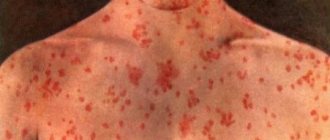
It occurs on the back and any other part of the body. A compaction with clear boundaries that can be felt when pressed. Usually the color of fibroma does not differ from the skin, but sometimes it can be bluish-red. There are two types of fibroids: hard and soft. The first looks like a hard lump, and the soft fibroma hangs over the skin. - A hemangioma is a red bump on the skin.
Can be big or small. It is a collection of small blood vessels. Hemangioma injures surrounding tissues and in some cases can grow rapidly. - A furuncle is a purulent and inflammatory process in the hair follicle.
It develops as a result of bacteria entering the layers of the skin through microtraumas. The boil grows quickly. The lump on the back hurts when pressed and is bright red. Swelling of the surrounding tissues is pronounced. To the touch it is a hard, spherical and hot seal. It is dense and mobile. With severe inflammation, throbbing pain, fever, and deterioration in general health are possible. - Myogelosis is a disease usually found in novice athletes.
For beginners, the back muscles are poorly trained. During high physical activity they experience a lot of pressure. The result is myogelosis or growth on the spine. - Malignant neoplasms have recently become more common.
They can appear in people of any age. A tumor appears on the back due to osteosarcoma. This is a growth on the bone tissue that causes a lump to appear. The main cause of occurrence is chronic osteomyelitis.
Have you been trying to heal your JOINTS for many years?
Head of the Institute for the Treatment of Joints: “You will be amazed at how easy it is to cure your joints by taking the product every day for 147 rubles ...
Read more "
The lump can grow alone or several compactions form at once. They can merge with each other. If the tumor is subcutaneous, then the doctor will not be able to determine with one palpation how many lumps are growing at the same time.
OUR READERS RECOMMEND!
Our readers successfully use Sustalaif to treat joints. Seeing how popular this product is, we decided to bring it to your attention. Read more here...
Types of seals on the back
- Atheroma, it is called a sebaceous gland cyst.
It can occur under the shoulder blade or elsewhere on the back. Atheroma is often diagnosed. Occurs due to blockage of the sebaceous duct. If the sebaceous gland is not functioning properly, the ducts close. As a result, an accumulation of purulent fluid occurs. The ball is located under the skin. In advanced cases, the size exceeds a chicken egg; at the initial stage, the size of the atheroma does not exceed a pea. The sebaceous gland expands in size. This is a dense and mobile formation, its diagnosis does not cause difficulties. The danger is that with increased size, the atheroma can burst and infect surrounding tissues. - A ball under the skin on the back means a lipoma or wen.
This is a harmless growth of adipose tissue. Does not develop into a malignant formation. Causes cosmetic discomfort to patients. Wen appears anywhere on the skin where there is fatty tissue. The lump on the back under the skin does not hurt when pressed. On palpation, a soft and mobile tumor is felt. - Fibroma is a benign tumor formed from connective tissue.
It occurs on the back and any other part of the body. A compaction with clear boundaries that can be felt when pressed. Usually the color of fibroma does not differ from the skin, but sometimes it can be bluish-red. There are two types of fibroids: hard and soft. The first looks like a hard lump, and the soft fibroma hangs over the skin. - A hemangioma is a red bump on the skin.
Can be big or small. It is a collection of small blood vessels. Hemangioma injures surrounding tissues and in some cases can grow rapidly. - A furuncle is a purulent and inflammatory process in the hair follicle.
It develops as a result of bacteria entering the layers of the skin through microtraumas. The boil grows quickly. The lump on the back hurts when pressed and is bright red. Swelling of the surrounding tissues is pronounced. To the touch it is a hard, spherical and hot seal. It is dense and mobile. With severe inflammation, throbbing pain, fever, and deterioration in general health are possible. - Myogelosis is a disease usually found in novice athletes.
For beginners, the back muscles are poorly trained. During high physical activity they experience a lot of pressure. The result is myogelosis or growth on the spine. - Malignant neoplasms have recently become more common.
They can appear in people of any age. A tumor appears on the back due to osteosarcoma. This is a growth on the bone tissue that causes a lump to appear. The main cause of occurrence is chronic osteomyelitis.
The lump can grow alone or several compactions form at once. They can merge with each other. If the tumor is subcutaneous, then the doctor will not be able to determine with one palpation how many lumps are growing at the same time.
Location of lymph nodes
Lymph nodes are small (0.5-5 5 mm) formations consisting of connective tissue and serve to cleanse the lymph of various toxins, metabolic products and pathogens that penetrate the blood. There are more than 600 of them in the human body, and they are located in groups along the lymphatic vessels in the most important areas of the body:
- in the area of the neck and lower jaw;
- in the center of the chest;
- in the axillary region;
- in the groin area;
- near the elbows;
- in the abdominal cavity;
- under the knees.
Depending on the depth of their location, lymph nodes are divided into deep and superficial. The former are found in the deep layers of connective and muscle tissue, the latter in the subcutaneous layer. Superficial nodes in the normal state are not visually noticeable, but can be easily felt by palpation, while deep ones can only be seen with hardware examination, for example, on an X-ray or MRI.
The lymph nodes closest to the spine are located in the chest cavity; they are called mediastinal lymph nodes and are deep. Also quite close to the spinal column (its lower part) are the lymph nodes of the peritoneum and groin.
But they are all located in the front, but on the back there are no such formations, neither superficial nor deep.
Therefore, if a ball-shaped or small lump is felt under the skin of the back, it is most likely a wen or a cyst of the sebaceous glands, and not an inflamed lymph node.
Causes of lumps on the lower back
Subcutaneous formations on the lower back can be divided into several types. Let's list them.
Lipoma
The most common cause of lumps under the skin on the lower back is a lipoma. It develops from adipose tissue, most often found on the scalp and face, but can also form on the lower back. The cause of its occurrence is considered to be a metabolic disorder in the human body.
In general, a lipoma in the generally accepted interpretation is a classic wen that occurs in most people. Lipoma (fat) is a small tumor. This is a round, soft, subcutaneous, rather mobile neoplasm, not fused to the surrounding tissues.
The inner surface of the wen is divided into small lobules filled with fat cells. The growth of the wen occurs slowly; it can reach significant sizes only after many years. The tumor is not inflamed, no pain or redness is observed.
The degeneration of a lipoma into an oncological formation is an extremely rare occurrence.
Atheroma
Also, a fairly common cause of the formation of lumps in the lumbar region is atheroma. This neoplasm is a consequence of blockage of the sebaceous glands. Atheroma is otherwise called a follicular (epidermal) cyst, which is filled with its own pasty secretions.
Upon closer examination, such a subcutaneous capsule consists of a cheesy mass with an unpleasant odor. In some cases, a hole forms in the middle of the surface of the atheroma, from which odorous contents are released. Atheroma is in the subcutaneous layer in a mobile state and grows slowly.
Sometimes multiple rashes occur.
Fibroma
It happens that a fibroma appears on the surface of the lower back. Such a lump is a benign formation consisting of connective and fibrous tissue. It appears due to uncontrolled cell growth after any skin irritation or injury. In general, the true origin of fibroma is still unknown.
Most experts are inclined to believe that this is a hereditary predisposition. Fibroids usually do not require treatment. However, if the fibroma bothers the patient, or there is a suspicion of a sarcoma (malignant formation), it can be removed.
Only a special histological examination (biopsy) should first be carried out to establish an accurate diagnosis.
Trigger points
This is another reason for the appearance of lumps in the lumbar region. These are areas located in muscle fibers with noticeable tension nodes. When pressed or touched, pain or unpleasant sensations appear that cause discomfort. The blood supply to the muscle fibers is interrupted due to the accumulation of waste products from the body.
This has an irritating effect on the nerve endings. To the touch, trigger points are a kind of compaction that resembles muscle lumps, a dense mass, or veins.
They are mainly formed from muscle overstrain due to neurological diseases, various injuries, problems with the musculoskeletal system, and incorrect posture.
Types of subcutaneous bumps
This is a seal that comes in several varieties. Some of them appear almost instantly - within a few hours, others are characterized by slow growth, so their increase in size can be noticed only after a certain amount of time.
In any case, if you notice a thickening under the skin, you need to monitor its behavior and, if necessary, consult a doctor. This symptom should not be ignored, since a subcutaneous lump may be the first sign of an incipient disease.
The most common types of subcutaneous neoplasms are:
- A fatty tumor or lipoma is a benign neoplasm that consists of fat cells. It has clear boundaries and is soft to the touch. It can appear on any part of the body (back, shoulders, elbows), without having any effect on the internal organs. The tumor has its own capsule.
- Hygroma. This is an absolutely safe lump, one of the types of cysts. The lump on the hand under the skin (on the wrist) looks like a ball or lump. Hygroma is the result of the accumulation of fluid between tendons that are constantly under tension, and therefore most often appears on the hands.
- Lumps on the leg under the skin. If such a lump appears, you should immediately consult a doctor and undergo an examination. A neoplasm on the lower leg may indicate the development of quite dangerous pathologies.
- Joint nodes or growths. These are immobile, hard neoplasms that accompany the development of gout or arthritis.
- Cyst or atheroma. It is formed as a result of blockage of a violation of the outflow of secretions secreted by the sebaceous glands. The fluid gradually accumulates and forms a lump, shaped like a cyst. The neoplasm has a capsule filled with a thick mass. Most often, atheroma appears in places where sebaceous glands accumulate and near the hairline. May spread throughout the body. Threatens the development of the inflammatory process and suppuration.
- Painful lumps. Appear within a few hours due to an insect bite, bruise or infection under the skin (through a cut, scratch). In this case, there may be an increase in body temperature. Such bumps are treated quite quickly. If the lumps appear as a result of an impact, long-term therapy may be required.
Ball-shaped lump under the skin
Lump-like densities form on different parts of the body, they are found on the limbs, on the face, and appear in the folds of the abdomen. Their growth is imperceptible; often a person feels a lump that has already reached a large size.
Without symptoms, benign skin tumors grow, which are a compaction under the skin in the form of a ball.
The growth of lumps with pain indicates their cause - infectious tissue damage. The general or local temperature rises, the skin turns red at the site of the bump. Associated complications appear: general health disorder, headaches. Timely treatment allows you to remove growing compactions.
The photo shows the difference between a ball and a cone.
When a lump under the skin is detected on the leg, in the form of a ball, patients go to the surgeon. Its task is to determine the type of tumor. Malignant growths are less common, but what to do, they need to be recognized in a timely manner so as not to trigger the development of the disease.
Source: https://lechenie.asustav.ru/narodnye-sredstva/uplotneniya-pod-kozhej-na-poyasnicze/
Purulent and inflammatory formations
A dense growth may be the result of a skin infection. The cause of inflammation may be staphylococcus, which leads to redness and swelling. The skin in the affected area becomes hot and painful.
When inflammation spreads, erysipelas or phlegmon can be observed. If lesions are observed, then it is worth talking about a carbuncle and a boil. They can even appear on the back and lower back in the spine area, creating discomfort.
In the initial stages, treatment of formations consists of taking antibiotics. Large growths can only be removed surgically.
Purulent formations are treated with antibiotics
Treatment with traditional methods
If the chalazion on the eye does not go away within a few days, then treatment should be started. Conservative treatment includes:
- Anti-inflammatory ointments, eye drops, containing antibiotics;
- Antiviral drugs;
- Preparations containing aloe juice;
Aloe is often used in the treatment of chalazion
- Inflammation can be reduced by injecting steroids into the knee at the site of the seal;
- Physiotherapeutic methods.
The patient needs to know that when the stage of suppuration occurs, it is prohibited to use heat compresses.
Cyst
A cyst can form as a result of skin infection or blockage of the sebaceous gland ducts. Inside the formation there is a sac filled with pus or liquid.
A special type is the epidermoid cyst, which is also called sebaceous. It looks like a round subcutaneous sac formed from a hair follicle. There is a black center inside the formation.
The growth can be found on the genitals, back and chest. It must be removed and then treated with antibiotics. Often the growth does not cause pain and goes away on its own. But in case of inflammation, removal in the surgeon's office is required.
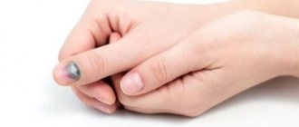
Cysts on the palms may indicate various diseases
What to do if you feel a ball in your lower back?
A lump-shaped lump can occur in the lumbar region at any age. Such neoplasms appear on various parts of the body, not only in the back, but also on the head, chin and even face.
This type of ball in the lower back in most cases does not cause pain, but causes discomfort and is not aesthetically pleasing.
To understand the type of pathology and whether treatment is needed, you will need to consult a doctor for an examination.
Varieties of cones
In order to promptly identify the severity of a developing skin lump, it is worth knowing the types of formations.
Sometimes this kind of discomfort tends to occur in a painful form, causing a sharp increase in temperature, pain in the problem area, pain in the head and general malaise.
Balls in the lumbar region can appear either as a single rash or as multiple rashes. The following types of formations on the skin in the lumbar region are distinguished:
- Lipoma - formed from adipose tissue, is a benign tumor, soft in structure, and easily rolls under the skin. The cause of development is metabolic disorders occurring in the internal organs. Most often women suffer from the pathology.
- Atheroma is a subcutaneous type cyst, small in size, consisting of sebum. It is formed in such a way that it stretches the sebaceous gland, accumulating in the pore. The ball in the hole in the lower back has clear boundaries, but feels dense to the touch. If, when pressing on a lipoma, it can be gathered into a fold, then this action is difficult to perform with a cyst. The skin in the affected area takes on a bluish tint, with a dark dot forming in the center. The neoplasm tends to reach sizes from 5 mm to 5 cm in volume. Painful discomfort develops.
- Hygroma - a small bump is observed on the skin, dense in structure, not causing pain. In some situations it may interfere, but most often, the tumor that appears on the back does not cause any inconvenience. The neoplasm consists of fluid accumulated inside the cavity, which is formed due to an impact or can be triggered by a previous operation. It grows slowly, reaching 2 cm in volume.
- Osteochondrosis - the formation of salt deposits in the form of a lump is observed when a person is inactive for a long period of time. Neoplasms can be triggered by poor nutrition and injury, as well as provoked changes in the metabolic process.
- Nodules on the joints - due to various diseases, there is a possibility of developing subcutaneous bumps, which are most often formed due to mechanical injuries or old age;
- Warts and papillomas - they can affect anyone, regardless of age and gender. They appear in the form of small compactions, painless in nature. Viruses and infectious diseases provoke the formation. Hormonal imbalances. It is difficult to independently identify pathology and distinguish it from a malignant formation, so you should immediately consult a dermatologist.
- Hernias - form in places where scars occur after surgery. It tends to occur due to excessive lifting of weights. For consultation and treatment, you should visit a doctor.
- Tumors of a malignant nature - such compactions occur extremely rarely. These include: melanoma, liposarcoma and lymphoma. The identified subcutaneous ball in the lower back quickly gains size, its boundaries are blurred and it constantly bleeds. If symptoms arise, you need to urgently seek diagnostics and clarify the diagnosis.
Any compaction requires timely diagnosis. Only after identifying the reasons due to which the pathology arose can the doctor prescribe effective treatment.
Reasons for the formation of pathology
Such subcutaneous neoplasms are formed due to various factors. The following reasons are identified that influence the appearance of tumors on the back in the lumbar region:
- Prolonged exposure to direct sunlight during the midday heat;
- Injuries of the deep type of sebaceous glands;
- Hormonal imbalances;
- Lack of simple hygiene rules;
- Abnormal changes in the functioning of the sebaceous glands;
- Cracking of hair follicles;
- Independent squeezing of wen and blackheads;
- A rare genetic predisposition is Gardner's syndrome.
If you experience pain, if the lump does not cause discomfort, when pathology is first detected, you should immediately consult a doctor and accurately identify the cause of the ball.
When should you see a doctor?
In most cases, if a lump occurs in the lower back and does not cause pain, people are not inclined to immediately consult a doctor. The following accompanying symptoms are identified, if detected, you should definitely consult a doctor for advice:
- Pain occurs when you press on the compaction island;
- The size of the ball increases to 5-12 cm;
- The spherical shape has an elastic structure or a dense formation;
- The skin changes its shade.
It is not worth leaving the pathology for further growth; this provokes unpleasant complications.
Therapeutic effect
The main condition for the therapeutic process is a correctly identified diagnosis. Many people, when they detect a swelling, begin to fight the lump with their own hands.
The ball on the lower back must not be pierced, heated, pressed or injured. Mechanical influence can cause more harm and provoke negative consequences.
The following methods of therapeutic impact on the affected area are distinguished:
- Surgery - the surgeon performs an autopsy under general or local anesthesia, using a scalpel, completely removing the affected membrane.
- Liquid nitrogen or laser therapy are the most effective methods and do not cause pain. Thanks to their influence on the surface of the skin, there are no further scars and untidy scars.
- Puncture - a long needle is used, thanks to which the ball is pierced and the contents are sucked out of it. Often such a procedure provokes a relapse.
In any case, after the treatment method, the patient must take anti-inflammatory medications and fortified complexes. These drugs will allow you to restore the body and prevent the formation of a second seal.
Thus, if you find a ball on your lower back, you should first consult a doctor. And then carry out treatment and completely get rid of the unpleasant defect on the back.
Loading…
Source: https://spinanebolit.com/sharik-v-poyasnitse.html
Malignant growths
Malignant tumors appear much less frequently than benign ones. They represent a focal compaction, or a node, constantly increasing in size. The formation does not hurt or itch. On the surface you can see normal skin or peeling, crust, and a dark tint.
Be sure to consult a doctor if:
- uneven and unclear boundaries of the growth;
- enlarged lymph nodes;
- rapid growth of the cone;
- inactivity and cohesion;
- the presence of ulcers and bleeding.
Only a dermatologist can confirm the benign nature of the growth during an examination.
Lumps in the lumbar spine: causes and treatment
A lump on the lower back of the spine is rarely life-threatening.
Most often, the formation is associated with skin conditions and are located in the layers of subcutaneous fatty tissue. In rare cases, bone tumors develop and may not cause significant symptoms. Cones can be located in soft tissues, on the surface, as well as on the sides and directly on the vertebrae. As a result of injury, tubercles, swelling and hematomas can occur in any part of the lower back.
The spasmed muscles surrounding the spinal column sometimes create a sensation of a “knot” deep between the fibers. The inflammatory process in the muscles after physical overload can manifest as swelling or hardening.
Depending on the location of the tumor or lump, you should immediately consult a doctor if:
- the formation increases in size;
- severe back pain appears;
- numbness and loss of sensitivity occurs;
- control of urination and defecation is impaired.
Compaction on the spine in the lumbar region may be a consequence of injury, which manifests itself later when the mobility of the segment is limited.
On right
Lumps or swelling on the right side at the lumbar level may be associated with injury, swelling after a bruise, or muscle spasm. On the right side, spasm can occur in the quadratus lumborum muscle, the internal oblique abdominal muscle, which expands the ribs and is responsible for the formation of the “hump.”
Safe lumps in the lower back include growths with the following characteristics:
- soft to the touch;
- are located in the upper layers of tissues;
- easily move when touched;
- increase with physical activity and decrease after rest.
The lump shown in the photo on the lower back to the right of the spine is an example of a lipoma or wen. On the sides of the spinal column there are three layers of muscles, blood vessels inside them. In the depths there are transverse processes and facet joints, which provide movement in the spine. They are subject to wear and tear.
Left
Lipomas are most often found in soft tissues and are subcutaneous tumors. Sometimes the formations are located in layers of fatty tissue and are therefore difficult to distinguish. When reaching a significant size, they can interfere with rotation, flexion and extension of the lumbar spine. Depending on the location, they provoke inflammation and lymph stagnation.
The photo shows a lump on the lower back to the left of the spine in the soft tissues. A patient with a giant lipoma in the lumbar region is overweight with a body mass index greater than 30.
On the very spine
A rare manifestation of formations on the spine are osteochodromas - tumors usually appear on the posterior parts of the spinal canal and within it, causing a variety of symptoms.
The photo shows an MRI of a patient with a rare form of exophytic formation in the lower lumbar region, namely a solitary osteochondroma. Most often, soft tissue tumors occur in the lumbar and sacral regions. A small lump on the spine in the lumbar region looked like a hard lump in the photo. Typically, swelling over the sacrum is caused by degenerative processes in the joints.
Bone lumps on the spine in the lumbar region between the 4th and 5th vertebrae are the consequences of severe listhesis. The fourth vertebra collapses ventrally (towards the abdomen), and the spinous process of the fifth becomes more visible. This symptom indicates the fourth degree of displacement by 75%, when the department completely loses its function of flexion and extension.
What could the appearance of a lump in the lumbar region mean?
There are various diseases associated with the appearance of lumps in the lumbar region. Oncological manifestations are rare and are distinguished by a number of striking signs.
Malignant and benign tumors
Tumors of the spinal cord and spine can develop in the lumbar region. Extramedullary lesions grow in the membrane surrounding the spinal cord or in the nerve roots that exit the spinal canal.
Tumors can impair the function of the spinal cord by causing it to compress. These include meningiomas, neurofibromas, schwannomas, and nerve sheath tumors. Tumors of bone and neural tissue can manifest as compaction and lumps in the area of the spinous process.
Pain becomes another important sign:
- constant and progressive;
- does not depend on movement;
- worse at night;
- accompanied by nausea, vomiting and dizziness.
Progressive muscle weakness in the legs, changes in bowel and bladder function are characteristic of lesions of the lumbosacral vertebrae.
After CT and MRI, histological examination of tissue samples and confirmation of the diagnosis, a decision is made to remove the tumor or undergo radiation. Some formations are inoperable due to the large amount of nerve tissue. For such cases, radiation and chemotherapy are provided.
Osteochondromas are the most common primary benign tumor of bone tissue, but occur in the spine in 1-4% of cases. According to statistics, 50% of formations appear in the cervical region, and in the lumbar region - only in 3% of cases of total damage to the spinal column.
The pronounced lump on the lower back near the spine in the photo is the result of an MRI diagnosis of osteochondroma. The tumors are the result of a defect in the development of cartilage, which leads to continuous growth of subperiosteal tissue. The lumbar region is less flexible than the cervical region, and therefore suffers less often.
Exostoses develop more often in men and are associated with various symptoms.
Can lead to pain radiating down the legs due to nerve root compression, foot drop, lameness, changes in spinal curvature. Malignant transformation is rare in solitary osteochondromas, and treatment is carried out by resection without the need for reconstruction.
Spinal cysts
Spinal synovial cysts are fluid-filled formations located along the spinal column. They usually appear in areas of joint degeneration and most often develop in the lumbar region. Cysts do not degenerate and rarely manifest themselves. Depending on the location, they can lead to spinal canal stenosis or nerve root compression.
Symptoms increase as pressure increases: pain, cramps in the back and legs. Discomfort increases with prolonged standing.
Ganglion cysts also form due to the destruction of the joint: the body produces more fluid to lubricate it, and it accumulates in the synovial lining. Cysts form in 1-2% of patients diagnosed with degenerative spondylolisthesis.
If the cysts form outside the canal in the muscle tissue, then the only symptom will be a painless lump near the spine in the lumbar region. Only when the formation spreads in the spinal canal do signs of nerve compression appear.
Treatment for cysts depends on symptoms. Epidural injections of steroid drugs are most often used, and physiotherapeutic procedures are recommended. When surgically removed, the cysts recur because their contents adhere to the dura mater.
Soft tissue tumors
Lipomas or lipomas are the most common formations between the skin and muscles. Due to the increased function of the sebaceous glands, they develop more often in men. They are less common in the lumbar region than in the thoracic region.
Lipomas are a mobile and soft formation that is located in a capsule. If it is large, it must be removed because it is a cosmetic defect. During surgical resection, it is necessary to remove the contents along with the capsule, otherwise the wen will spread throughout the body.
Hemangiomas are soft tumors formed by the proliferation of vascular tissue. Due to rapid growth, tumors must be removed. Hemangiomas bleed the surrounding tissues and therefore can cause destruction of the vertebrae, depending on the location.
Conclusion
It is necessary to distinguish between tumor formations, traumatic swelling, spasm and protrusion of the vertebrae. Sometimes a swelling forms at the level of the sacrum, reminiscent of the same “widow’s hump” of the cervical region.
Any pathology associated with the formation of wen, tissue proliferation, indicates a problem with the spinal column.
Lumps that restrict movement and cause pain should be diagnosed promptly.
Source: https://columna-vertebralis.ru/zabolevaniya/shishki/shishka-v-poyasnichnom-otdele-pozvonochnika.html
Which doctor deals with lumps and lumps?
If formations are detected, you should consult a doctor. The patient can be examined by:
- dermatologist;
- surgeon;
- oncologist.
The last doctor rules out the malignant nature of the tumor.
It is important not to delay going to a medical facility. This is especially true for formations that grow quickly or create inconvenience.
In modern society, unlike earlier periods of our history, people seriously think about the state of their health and try to identify possible problems in a timely manner. To do this, you should regularly examine your body in the clinic, as well as conduct self-diagnosis. There are quite a lot of diseases that can be seen during self-examination, but to do this you need to know how they can manifest themselves.
Lipoma is a benign tumor formed from connective tissue and forms a ball filled with fat. This tumor is characterized by the absence of pain when palpated, as well as a soft structure. If you feel such a formation in yourself, you should immediately visit a doctor to identify the exact problem and decide on therapy.
Lipoma is a benign formation
Places of formation of wen
These fat-filled balls can appear in different parts of the body, but treatment options do not depend on the location of the problem. Lipomas often form in places where the fat layer is thinnest and most susceptible to change. Such places are the hips, back, shoulders. Sometimes you can find a formation on the hands or in the armpit area. If you discover something similar to balls that have appeared recently, you need to pay attention to the dynamics of their development. If a tumor grows rapidly, you should immediately contact a dermatologist to immediately begin therapy.
Very often lipomas grow on the back of the head
It is very important that the wen is not subjected to mechanical damage, since in such situations there is a high risk of developing infectious tissue infections. If a lipoma appears on the lower extremities, the likelihood of damaging it is quite high, which will cause complications.
There are some traditional methods of treating lipomas, but it should be remembered that improper self-medication can have extremely negative consequences for health. In the hospital, the patient will receive qualified assistance, and the removal of the wen will take place quickly and without complications.
Self-diagnosis is an important factor in the timely resolution of health problems. Do not neglect your annual examination at medical institutions and regularly examine your body yourself. Be healthy!
Causes of lipomas
Fat globules under the skin can occur for a variety of reasons, including:
- genetic predisposition;
- lipid metabolism disorder;
- hormonal imbalance;
- changes in the condition of fatty tissue (with sudden weight loss or weight gain).
This formation is not dangerous, but it is recommended in any case to visit a doctor to accurately establish the diagnosis and exclude the malignancy of the tumor. In addition, lipoma tends to grow rapidly and there is a risk of it growing into muscle tissue, which will subsequently significantly complicate its removal.
A lump under the skin on the back in the spinal area - what is it?
Sometimes people notice that they have a lump under the skin on their back. Any bump can lead to panic. But don't worry ahead of time. Balls on the back most often mean benign tumors, although there is a possibility of cancerous tumors.
If you notice a lump on your back under the skin near the spine, consult a doctor. He will assess the situation and take the necessary tests for diagnosis. After making a diagnosis, he will explain what to do and how to treat the tumor.
Why do bumps appear on the back?
The causes of compactions depend on the type of disease. The main factors influencing the appearance of balls on the lower back, under the shoulder blade, and near the spine include the following:
- Malfunction of the sebaceous glands. This leads to the appearance of atheromas. The disorder can occur due to hormonal changes in the body or when the hair follicle is damaged (for example, if the patient squeezed a pimple).
- Hereditary predisposition is a leading factor in many diseases. If direct relatives suffer from Gardner syndrome, then their children or grandchildren have tumors in the back.
- The development and progression of osteochondrosis is another significant factor that provokes the appearance of balls on the back. When the disease occurs, increased stress is placed on the spine, and blood supply deteriorates. Lumps and tumors form on the back.
- A lump between the ribs in the solar plexus area.
- The appearance of lipomas is caused by back injuries; this circumstance occurs even in children. The abundance of toxins in the body and disruptions in the metabolic system also have a negative effect.
- Lump on the sternum.
- Leading an unhealthy lifestyle - poor and unbalanced nutrition, a passive lifestyle, bad habits (tobacco and alcohol use), sedentary work negatively affect the immune system and the condition of the spine. This causes vulnerability of the immune system and leads to diseases and tumors on the back.
Treatment of formations
Initially, it is necessary to diagnose a compaction in the spine. Doctors may prescribe an MRI or computed tomography scan, complete a general blood test, or undergo an ultrasound examination of the problem area. Based on these data and examination results, a diagnosis is made and treatment is prescribed.
In the vast majority of cases, this is removal. The cone is opened and its contents are cleared, the capsule is completely cut out. Doctors then stitch up the incision.
After surgery, you must take a course of antibiotics to avoid infection and complications. Penicillin drugs are prescribed.
Vitamins and immunomodulators may be prescribed at the discretion of the doctor. This is necessary if your immunity is low.
If an adult has a boil on his back and the size is small, then conservative therapy is prescribed. Absorbable ointments (Vishnevsky or Ichthyol) are used. Gradually the pus will come out along with the rod.
In case of cancer, the tumor is excised and several courses of chemotherapy are given. In the early stages, the patient is likely to recover completely. In the future, regular examinations and monitoring by a doctor are carried out.
On the Internet you can find many photos with seals on the back. But you should not engage in self-diagnosis and treatment without the knowledge of a doctor. This will cause the tumor to begin to grow, making treatment more difficult and taking longer.
Prevention
Preventive measures do not always help, but they significantly reduce the risk of bumps appearing on any part of the body.
These include:
- regular monitoring by doctors, medical examination and medical examination;
- maintaining an active lifestyle - if work does not allow you to move a lot, then it is advisable to sign up for a gym, swimming pool or sports section;
- balanced diet enriched with vitamins and beneficial microelements;
- rejection of bad habits.
Conclusions
The seal on the body causes confusion and fear. Often it can appear in the back area. If you find a lump, make an appointment with your surgeon. The doctor will conduct a diagnosis, tell you what it is and how to treat the tumor.
Doctors warn! Shocking statistics - it has been established that more than 74% of skin diseases are a sign of parasite infection (Accarida, Giardia, Toxocara).
Worms cause enormous harm to the body, and the first to suffer is our immune system, which should protect the body from various diseases.
The head of the Institute of Parasitology shared the secret of how to quickly get rid of them and cleanse your skin, it turns out that it’s enough... Read more...
Most often, surgeons resort to surgery, opening the tumors, clearing pus and other secretions.
Source: https://kozhamed.com/uplotnenie-pod-kozhey-na-spine-v-oblasti-pozvonochnika-chto-eto-takoe/
Lipoma treatment
There are several possible treatment options that your doctor may suggest, depending on the condition of the tumor. In some cases, if the formation is not dangerous and is located so that it does not cause discomfort to the patient, it is not removed. In this case, you must carefully monitor possible changes in the tumor and, at the slightest suspicion of growth, consult a specialist for advice.
The most reliable method is considered to be a surgical method when the wen is removed directly with its capsule. This guarantees the absence of relapse of the disease. This method is used in any medical institutions, is performed quickly and has reliable results.
Lipomas are most often removed surgically
There are also more gentle and non-invasive ways to remove lipomas, which involve irradiating the formation with radio waves or destroying tumor structures using laser therapy. After this type of procedure, the body’s recovery occurs much faster than after surgical treatment, and no visible scars remain. However, such methods can only be used in the early stages of the disease and may not be performed in every medical clinic.
If the lipoma is small in size, you can get rid of it by introducing a special substance into the tissue, which destroys the structure of the fat and capsule.
After the tumor has already been removed, a histological examination of the tissue is required to exclude its malignant nature. Although lipomas do not tend to develop into cancer, the risk still exists and should be minimized.
Preventing the appearance of seals
Prevention, including:
- compliance with hygiene measures;
- It is recommended to treat your feet with nourishing creams and herbal decoctions;
- a special gymnastics complex for the feet has a positive effect;
- To prevent your feet from hurting, a massage is recommended;
- doctors recommend swimming, light jogging, cycling and walking barefoot on sand and grass;
- any abrasions, wounds and cuts on the foot, and especially under the toe, should be treated in a timely manner;
- shoes should be comfortable and the heel should be of optimal height.
If you follow preventive measures, it is quite possible to prevent the formation of a more serious seal (rod) on the foot. In the event that, despite all the advice, it was not possible to avoid compaction, professional medical assistance is needed to adjust further actions.
You should not try to cope with the tumor on your own (with the exception of dry calluses), since such intervention can lead to infection and further spread of the inflammatory process. With careful attention to the state of your own body, it is quite possible to avoid negative consequences in the future.
A lump on the foot is a neoplasm that causes discomfort. Located on the lower surface of the leg. It is important to find out the cause of the pathology and begin treatment.
Lump in the groin, thighs and buttocks
The appearance of a ball-shaped lump in the groin under the skin always alarms a person.
Seals in these places appear in the form of:
- infectious inflammation of the inguinal lymph nodes;
- the growth of a fluid-filled cyst, a completely harmless neoplasm;
- development of a painful abscess with accumulation of purulent contents;
- infectious genital warts in the form of fleshy growths transmitted through sexual contact;
- hanging moles and warts.
What to do with dense subcutaneous areas depends on the factors of appearance. It may be a result of HPV, so a visit to the doctor is a must, especially when the balls grow quickly. Consultations with a surgeon, dermatologist, and oncologist will help.
Self-medication is not allowed!