Before starting the fight against mycoses, that is, fungal infections on the affected nails, it is necessary to perform an analysis to reliably confirm the presence of this same fungal infection. It is not always possible to identify a fungus by visual signs; it can be either nail dystrophy aggravated by trauma to the nail plate, or another pathology caused by reasons unrelated to a fungal infection on the nails. In order to exclude cases of erroneous treatment of nail fungus due to its absence, and mistaking another pathology for a fungal infection, a microscopic examination of particles of the nail plate is performed.
What is nail fungus?
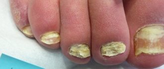
Fungal infection of the nail plate is called onychomycosis. This is an extremely common disease that is diagnosed in people of any age, including children. The pathological process is caused by the active proliferation of infections on the feet and hands, which leads to yellowing, delamination, changes in the structure of the nail, as well as damage to the skin tissue around it.
The disease is classified depending on its pronounced manifestations:
- onycholytic – characterized by damage to the nail plate and its rejection from the nail bed;
- hypertrophic, in which the plate loses its glossy shine, turns pale, turns yellow, darkens, and also changes its structure: it thickens, becomes deformed, and collapses at the edges;
- normotrophic, when a seemingly healthy nail plate becomes covered with small spots;
- total, in which the entire nail is affected;
- distal, when the lesion is located only at the edge.
It is very easy to become infected with onychomycosis. Pathogens are transmitted from a sick person, as well as through the things he uses.
Increase the likelihood of infection:
- weak immunity;
- violation of the integrity of the skin and nails;
- vascular diseases;
- wearing low-quality, tight shoes;
- failure to comply with personal hygiene rules;
- severe stress.
If signs of illness occur, you should consult a dermatologist or mycologist. The sooner therapy is carried out, the faster it will be possible to cope with mycosis.
Types of Fungal Infection
Depending on the location of the fungus and the types of pathological microorganisms, nail fungus is divided into several types and types.
Location:
- Distal. The fungus affects a separate part of the free end of the nail in the center. It all starts with a small spot that gradually increases in size. The surface of the affected area becomes rough, flakes, and crumbles.
- Lateral . Develops on the lateral parts of the nail plate of one or two legs. It is characterized by delamination of platinum, increased fragility, and discoloration.
- Surface white. White spots appear on the surface of the nail, increasing in size and disrupting the integrity of the top layer. The nail becomes soft, vulnerable, constantly peeling and flaking. In the absence of qualified treatment, the deep layers of platinum begin to be affected, and the nail falls off.
- Proximal. Affects the cuticle. The skin becomes inflamed; at the base of the platinum growth, a darkened spot is clearly visible over the entire surface. The nail flakes, peels, crumbles.
- Total. The fungus affects the entire nail. Discoloration and peeling occur.
Thickening amount:
- Normotrophic . The affected nail does not change thickness, the top layer remains smooth. You can suspect the presence of pathology by a change in color.
- Hypertrophic. Following the change in color, the stratum corneum grows. The nail becomes deformed, peels, interferes with putting on shoes, and hurts when walking.
- Atrophic. The affected nail is gray-brown in color. The plate peels off greatly, becomes thinner, and almost completely disappears.
Type of pathological microorganisms:
- Trichophyton red . Affects nails, skin on hands and feet. It all starts with the appearance of small spots and loss of elasticity of the plate. If left untreated, it spreads to other parts of the body.
- Trichophyton mentagrophytes . Infects the skin between the fingers. Causes cracks, redness, severe itching. Smoothly transfers to the nail. The location is the big toe or little finger. It gets worse in the hot season.
- Trichophyton violet . Found in countries with hot climates. Affects the scalp. In the absence of therapy, it spreads to the nails.
- Flake-shaped epidermophyton . The pathology begins with peeling skin on the fingers. There is redness and severe itching, which intensifies in the evening. When moving to the plate, the nails take on the shape of a bird's beak.
- Favus fungus . The disease is common in North America. Pathological microorganisms penetrate through cracks in the skin of the hands. Forms yellow-brown crusts on the nails. There is an unpleasant odor.
- Geophilic microsporum . Rarely seen. Penetrates into the body through microcracks. It affects the head and spreads to the nails.
Yeast-like fungi cause white spots to appear on the nails, while mold fungi cause black spots. If there are any changes in the nail or surrounding skin, you should seek help from a specialist. The disease is quickly treated at the initial stage, but difficult to treat in the advanced stage.
When should you get tested?
A fungal infection manifests itself differently in everyone. In more sensitive people, itching and burning immediately begins in the affected area, and redness of the skin appears. If you continue to ignore the unpleasant symptoms and do not test the nail for fungus before therapy, the plates become deformed, thicken, become covered with longitudinal or transverse grooves and stripes, take on an unpleasant hue, and emit a putrid odor.
Further:
- folds in the interdigital area crack;
- thickenings appear on the skin;
- severe, painful itching and burning appears;
- the nail becomes very fragile and brittle.
When the first signs of pathology appear, you will need to conduct an analysis for the presence of fungus on the nails.
Testing is also necessary when:
- the dermatologist doubts the diagnosis;
- the treatment, which may take a long time, does not give positive results, and the disease progresses;
- the course of treatment has ended, but there are doubts about the complete destruction of the infection;
- clinical manifestations of the fungus are still observed, although in a blurred form;
- the infection became chronic.
Diagnosis is required after therapy is completed.
Important! Clinical testing of biological material is prescribed to identify infections in the body. If fungal flora is present, systemic treatment is prescribed, since the use of external agents alone will be ineffective.
How to protect yourself from fungus
- After a bath or shower, dry the skin of your feet, especially in the spaces between your toes;
- If you wear closed shoes, change your socks/knee socks daily;
- Change your shoes once every two to three days, do not wear the same pair every day;
- Do not walk barefoot in public places (swimming pool, bathhouse, sauna, fitness club);
- If someone in your family has a fungal infection, provide them with a separate set of towels and linen. Wash them separately at maximum temperature;
- If the skin of the feet or nails are infected with fungus on one leg, use two different sets of manicure/pedicure tools to avoid spreading the infection to healthy areas;
- If you have diabetes, monitor your blood sugar. “High sugar” reduces the rate of healing of skin wounds (“diabetic foot”), which facilitates the access of fungal infections.
Be healthy!
Preparing for analysis
Having decided on the problem, where the necessary tests can be taken and knowing the price of services, the patient needs to find out how the procedure goes and how to properly prepare for it.
Often the examination takes place in stages:
- the scraping is examined under a microscope;
- bacterial culture is carried out;
- do a PCR test;
- hemotest.
The biomaterial for study is particles of the affected nail and venous blood.
A scraping for nail fungus will be as informative as possible if you follow these recommendations before going to the laboratory:
- do not wash the affected areas with soap or other cosmetic detergents for three days before the test;
- do not use antifungal drugs during the same time;
- do not drink strong tea or coffee drinks 6 hours before testing;
- 10 days before the examination, do not cut your nails;
- remove decorative and medicinal varnish from the nail surface;
- Do not wet your hands/feet 4 hours before the test.
When taking a blood test for fungus, you need to consider that:
- 8 hours before testing you should not eat;
- do not drink alcohol for 48 hours;
- do not overwork and avoid severe stress;
- do not smoke before taking blood.
You also need to tell the laboratory assistant what medications the person is taking. You may have to stop taking some medications the day before.
Polymerase chain reaction method
Using PCR research, it is possible to determine the presence of a certain type of fungus. The positive aspects of the survey are its accuracy, reliability and speed. Disadvantage: narrow focus. The material to be examined is a scraping from the affected area of the skin or mucous membrane; urine, blood, and prostate secretions can also be examined.
The examination can be qualitative (the results will indicate the presence or absence of pathogen DNA) or quantitative (the number of pathological cells is determined). The study is carried out within 1-3 days. It is impossible to diagnose candidiasis based on PCR analysis.
Types of blood tests
Having discovered changes on the nail plates, you should find out their origin, which helps to analyze the nail for fungus.
Diagnostics includes several types of tests:
- PCR test (polymerase chain reaction);
- clinical and biochemical test of venous blood or urine.
Venous blood is collected in the manipulation room. The laboratory technician puts on sterile gloves and uses a vacuum tube with patient information labeled on it. Tubes with collected blood are sent to the laboratory for careful examination and evaluation of indicators.
Important! Diagnostic procedures should not be postponed for a long time, since fungal infection is extremely contagious and is characterized by rapid progression. It is transmitted through household contact and has a high index of contagiousness.
Bacteriological culture
By taking a bacteriological test to determine nail fungus using the culture method, you can determine the causative agent of onychomycosis. The laboratory assistant collects the necessary biomaterial and places it in a nutrient medium in which microbes begin to multiply intensively. The growth of pathogenic flora is maintained by a thermostat at a comfortable temperature for it. Fungal colonies grow within 3-14 days.
There are negative factors that affect the reliability of the survey:
- type of collected biomaterial. Fungi are rarely detected in the blood. They are mainly localized in scrapings from the affected plate;
- use of fungicidal oral medications on the eve of the examination. In this case, the analysis will give a false result.
Polymerase chain reaction
This is the most reliable nail fungus test, allowing you to identify the pathogens that cause the development of onychomycosis:
- Any biomaterial is suitable for the test;
- using this reaction, you can determine the DNA of the pathogen and identify its type;
- get reliable results within five hours after taking the test;
- the ability to detect even single pathogenic particles.
Despite its many advantages, the PCR method can give false results. This mainly happens when particles of an already destroyed pathogen are detected.
Hemotest
This is an assessment of blood parameters for the compatibility of foods with the body. Allowed foods should be included in a person’s menu, and junk food should be avoided or its consumption reduced to a minimum.
Testing is carried out after taking antifungal medications, when the mycosis has been completely overcome. This diagnostic procedure is considered an excellent prevention of both fungal and other diseases.
Microbiological methods
With the help of microbiological studies, it is possible to determine whether a fungal infection led to the formation of the disease. For examination, biological material is collected:
- particles of the nail plate,
- hair,
- scraping from the surface of the skin.
Microscopic examination
The indication is a suspicion of mycosis of the skin, hair or onychomycosis of the nails. In case of fungal infection of the nails, scrapings from different parts of the nail plate are examined. It is painted and examined under a microscope. Often it contains mycelium threads, fungal spores and yeast cells.
If the skin is affected, then a scraping is carried out from the area between the healthy and the affected area, this feature is explained by the fact that it is in this area that the largest amount of the pathogen is localized.
If the pathological process is localized on the scalp, then hair and skin scales become the material for examination. The study lasts on average 3-5 days. It allows you to find out what exactly triggered the disease; the type of pathogen and its quantity are rarely determined.
A variant of the norm is the complete absence of fungi in the examined material; a small amount of them may indicate carriage.
Culture method
With the help of culture, it is possible to obtain more accurate information about the pathogen. The duration of the study can range from 2 days to a month. The material is collected in a manner similar to the previous method. After delivery to the laboratory, the biological material is placed on a special nutrient medium. In the presence of fungi, growth of colonies is observed. Fungi from each colony are examined microscopically, determining the genus and species, as well as their number. In some cases, even sensitivity to medications can be determined.
Urine examination
The urine of a healthy person should not contain fungal infections. Their identification means that a malfunction has occurred in the body, and the patient requires systemic treatment.
Laboratory methods can detect the following types of fungus in urine:
- moldy;
- yeast;
- radiant;
- candida.
To conduct tests for fungus, you must bring morning urine to the laboratory in a sterile container designed for this purpose.
Treatment
Fungal infections are often more difficult to treat than bacterial ones. Treatment is divided into:
- etiotropic - aimed at destroying the fungus;
- symptomatic.
Antifungal drugs include:
- Ambizome;
- Amphoglucamine;
- Ampholip;
- Amphotericin B;
- Levorin;
- Levorina sodium salt;
- Mycoheptin;
- Nystatin;
- Pimafucin;
- Travogen.
The drugs are used externally and internally, for systemic mycoses (histoplasmosis, aspergillosis, mucorosis) intravenous and inhalation.
Treatment of fungal diseases takes a long time - from 2 weeks to a year - depending on the identified pathogen and the clinical picture.
Symptomatic treatment of the fungus is aimed at:
- to maintain life;
- reducing symptoms of organ dysfunction.
Appointed:
- glucose-saline solutions;
- antihistamines and decongestants;
- antidiarrheal drugs;
- corticosteroids, etc.
Surgical treatment is used for aspergillosis - lesions in the lungs are excised. If the vessels are affected by the fungus, arterial embolization is performed.
Nail analysis
In case of external damage to the nail plate at any stage of the disease, analyzed for fungus:
- microscopy of scrapings (approximate price 600 rubles);
- sowing the fungus for nutritious flora (approximate cost 1550 rubles).
Test for mycosis
When learning how to get tested for nail fungus, you need to find out what material is used. Its careful examination will help determine the infection that affects the plates.
The test for mycosis is carried out as follows:
- Before figuring out how to get tested for nail fungus, the patient should see a dermatologist. The doctor examines the damaged nails and decides whether further diagnostics are needed. Sometimes an initial examination is enough to understand how advanced the pathology is and what drugs are needed to combat it;
- First, the plate is scraped to determine the etymology of the pathogen;
- then bacteriological culture is used to determine the degree of sensitivity of pathogens to antimycotics;
- Based on the results of the research, the specialist determines treatment tactics, which may include taking tablets, using creams, ointments, fungicidal varnishes, and folk remedies.
What is tank seeding?
Nail particles affected by the fungus are placed in a nutrient medium, where colonies of the pathogen begin to actively grow. At the same time, the resistance of the crop to fungicidal substances included in all antimicrobial preparations is determined. The resulting infections are treated with a certain type of drug and see how they react.
It is not advisable to prescribe fungicidal agents without this test, since other dermatological ailments affecting the skin of the hands and feet have similar clinical symptoms: eczema, vitamin deficiency, intoxication, etc. Also, the nail could be severely injured, and an infection could penetrate into the wound, causing severe swelling, redness and suppuration. In this case, antimycotics will not provide a therapeutic effect, but will only worsen the situation.
How is scraping performed?
The sample is taken with a medical spatula, with which the top layer is removed along the furrows or under the nail folds. Biomaterial is also collected under the nail itself. If a small part of the plate is affected, toenail and fingernail fungus is identified by scraping. If the lesion is large, cut off a piece of the nail.
Does scraping hurt?
Patients who are scheduled to have their nails tested for fungus by scraping are interested in whether the procedure is painful. Taking biomaterial this way does not hurt. The test is quick and painless. The resulting material is sent for bacteriological culture, which determines the presence of pathogenic flora.
What does the analysis show?
If a nail fungus test is carried out with blood donation, this is done so that the specialist can understand:
- whether inflammation occurs in the body;
- what stage is the pathological process at?
A biochemical examination shows the condition of the internal organs: liver, pancreas, kidneys. After all, fungi can affect not only the skin and nails, but also important systems of the body, gradually destroying and poisoning them. The pathological process negatively affects the mucous membranes, joints, ligaments, and bone tissue. In addition, this way you can determine which drug should be prescribed: local or oral.
Fungicidal drugs are processed by the liver, excreted by the kidneys and put a strain on the pancreas. To prevent drug therapy from aggravating their effects, it is necessary to conduct a blood test for the fungus. Microbiological examinations of the nail plate itself help to identify the pathogen and determine its sensitivity to drugs.
It is also recommended that the patient donate blood for culture. Normally, it should be sterile, without the presence of fungal flora. If a generalized fungal infection develops, elements of the pathogen are found in the bloodstream, and on the legs and arms the fungus will only be a symptom of the general pathological process. A clinical blood test is required both at the beginning and at the end of the therapeutic course, which helps to monitor treatment measures and determine the degree of their effectiveness.
A dermatologist explains how to get tested for nail fungus. He tells the patient how long the test takes and what specific tests will need to be performed. If a person does not remember what the test for fungal pathogens is called, they will be helped in the laboratory. There you can find out how much the procedure costs and when the results will be ready.
Decoding the results
In healthy people, any test for a fungal infection will result in a negative result. Sometimes it becomes false positive if the scraping rules are violated. If there are no clinical manifestations of pathology, but the examination shows a fungus, most likely the person is a carrier of the infection. If the result is positive, we are talking about dermatophytosis of the nails.
If dermatophytes are found in the scraping, dermatophytosis is diagnosed. The pathological process affects the nails on the toes, and less often on the hands. In this case, the plate thickens, loses its natural transparency, and against the background of deformation, yellow and white inclusions of an unhealthy appearance form on it.
The detection of yeast fungi is not a direct confirmation of onychomycosis. For a final diagnosis, the patient must have external signs of the pathological process. With candidiasis, parasitic fungi penetrate the nails through the skin at the very bed. Most often detected in elderly people.
Mycoses are accompanied by an inflammatory process in the area of the nail fold and tissue degeneration. Mixed fungal infections often occur - in this case, dermatophytes, mold and yeast pathogens are simultaneously detected in the patient’s biological material.
Based on the results of cultural diagnostics, a medicine is selected to which the microorganisms that cause onychomycosis are sensitive. It is this medication that the doctor will prescribe for subsequent treatment.
Can I do the test at home?
If it is not possible to carry out diagnostics in the laboratory, you can do an analysis for the presence of fungus at home:
- prepare a pink solution of potassium permanganate;
- put your feet or hands in it for three minutes;
- wipe the limb thoroughly;
- examine the nail plates: if they are yellowed, then there is no fungal infection. If the color of the nails has not changed, then this is a sign of developing onychomycosis.
With such a test it is impossible to determine the type of pathogen, but it is quite possible to suggest a diagnosis. Therefore, visiting a dermatologist and conducting a laboratory examination should not be postponed.
Some patients don't like going to hospitals. The reason for this is the lack of free time and long waits outside the offices. Also, few people want to voice their problem, because it is still believed that only unscrupulous people can get fungus on their nails. To make the examination as comfortable as possible, some private medical centers offer testing at home. A person will not need to waste time traveling and will not have to wait for his turn. Specialists come to your home, collect material, and send the result by mail.
If changes appear on the nail plate, it has lost its healthy shine, is covered with cracks, spots, or has changed color, then this may be a sign of a fungal infection or onychomycosis. When contacting a specialist, the patient is referred for an examination, which should not be refused. Using the results obtained, you can make an accurate diagnosis and determine what medications you need to use.
Types of tests for fungus
During the patient’s initial appointment, a dermatologist examines him and asks him about complaints about the existing disease. If we are talking about onychomycosis or suspicion of it, the specialist recommends taking the laboratory tests listed in the table at the next visit.
| SCRAPING | Confirms the absence or presence of a fungal infection, the type of parasite, its quantitative composition and sensitivity to drugs. |
| PCR TEST | With high accuracy, it diagnoses the causative agent of the disease based on its DNA and helps determine which medications should be prescribed in a particular case. |
| SOWING | Isolates a specific parasite to study its properties. |
Important! The listed tests will have to be taken more than once - at least twice. In the first case, for a competent diagnosis, in the second - in order to track the effectiveness of the therapeutic procedures.
Painfulness of the procedure
The overwhelming number of people who decide to undergo the study experience some fear of the procedure for collecting biological materials from the skin and nail plates. However, there is absolutely no need to be afraid of such procedures . Such manipulations are absolutely painless and do not cause any discomfort.
Such manipulations mostly resemble the usual cutting of the edges of the nail plates. In addition, a laboratory technician may scrape the area of skin over the infected nail to take a scraping. However, even this impact does not cause pain.
Etiology
The etiological factor is fungi, saprophytes and absolute pathogens. The first group exists in the human body in quantities of up to 3 logarithms and normally does not cause discomfort. The second group includes fungi from the surrounding world, which, if they enter the macroorganism, can cause disease.
Infection occurs in the following ways:
- from environmental objects;
- from animals;
- from a person;
- by activating its own opportunistic microflora.
When encountering a fungus, not everyone can get sick. First of all:
- persons with reduced immunity;
- persons with altered immunity.
If local protection is violated, dysbiosis develops (for example, vagina or oral cavity) and candidiasis develops against a pathological background.
With severe immunodeficiency caused by HIV, the use of cytostatics, and oncological therapy, the fungal flora does not stop at damage to the dermis.
Severe visceral mycoses develop:
- aspergillosis;
- histoplasmosis;
- mucorosis, etc.
Chronic diseases deplete the immune system, and they also have a constant source of inflammation. Add here periodic courses of antibiotic therapy and we get ideal conditions for the development of a fungal infection.
Antibacterial drugs kill not only pathogenic bacteria, but also representatives of normal flora. The mushrooms themselves are sensitive to a specific group of drugs. But against a sterile background they grow beautifully, causing an inflammatory process.
Patients with diabetes are a tasty morsel for mushrooms with a “sweet tooth.” In addition to the body's impaired resistance to infection, people with diabetes have permanently elevated blood sugar and altered acidity.
The need to undergo research
A symptom such as a change in the shape or color of the nail plate is evidence of a fungal infection . However, in some cases, such changes may indicate other pathological processes.
To be able to determine the exact cause, the patient must take a blood test to detect nail fungus. At the same time, the doctor may prescribe an immune status test.
If there is a suspicion of an infectious lesion, laboratory tests are mandatory. If the results confirm the diagnosis, the attending physician will select an individual therapeutic course.
Important!
Self-medication in this situation is absolutely excluded. What you can do yourself is to prevent the disease.
Clinical picture
There are 5 types of fungal infections:
- dermatomycosis;
- keratomycosis;
- candidiasis;
- systemic or visceral mycoses;
- pseudomycoses.
Dermatomycoses
This disease causes skin lesions. The causative agents are a group of dermatophyte fungi:
- favus;
- athlete's foot;
- microscopy;
- trichophytosis.
Infection occurs through contact with soil, animals and a sick person. Rounded areas of hyperemia appear, accompanied by itching. Subsequently, the spots are covered with scab.
With dermatophytosis, the hair follicle is involved in the inflammatory process:
- it is destroyed;
- hair falls out;
- bubbles with cloudy purulent and hemorrhagic contents and crusts appear.
Inguinal dermatomycosis manifests itself:
- pustular rashes;
- redness;
- peeling.
Keratomycosis
With lichen versicolor, pink-coffee spots with scalloped outlines appear, which subsequently become covered with peeling.
Actinomycosis appears
upon contact with cereals, mill workers get sick and the following appear:
- cyanotic nodes;
- gummas;
- fistulas
In addition to skin manifestations, visceral pathologies are characteristic. Piedra affects the hair, but only causes aesthetic discomfort.
Candidiasis
This disease can be different:
- seizures in the corners of the mouth;
- thrush in women;
- severe systemic diseases (esophagitis, pneumonia and sepsis).
They are united by an etiological factor - “sweet tooth” fungi of the genus Candida. Normally, they exist in the body of every person and do not cause harm.
Visceral and systemic mycoses
Visceral develop:
- when encountering a large number of fungi;
- due to decreased immunity;
- in the absence of normal flora - the so-called “good” bacteria.
All fungi are sensitive to the pH environment, and this acidity is controlled by other microorganisms of normal human flora.
Systemic mycoses include:
- histoplasmosis;
- coccidioidosis;
- blastomycosis;
- cryptococcosis;
- geotrichosis;
- chromomycosis;
- rhinosporidiosis;
- aspergillosis;
- penicillinosis;
- mucorosis
Their main symptoms are associated with pathology of the lungs, heart or gastrointestinal tract, and skin lesions are not the main symptom.
At the same time, they develop:
- pneumonia;
- enteritis;
- sepsis.
All of them are difficult and difficult to diagnose and treat.
Pseudomycoses
This disease occurs under the guise of fungal infections, but the etiological factor is bacteria.
How is the scraping taken?
To begin with, the patient will be prescribed a general and biochemical blood test. In addition, the doctor will take a scraping to determine the causative agent of the nail disease. In some cases, a culture of the nail plate affected by the fungus is done.
Along with these tests, the patient is prescribed PCR, a test that will make it possible to identify the pathogen by its DNA.
It has become an established opinion among people that curing fungus is quite difficult. And to alleviate the condition, expensive pharmaceuticals are required. However, in reality this is not always true.
Preparation for scraping
If we talk about preparing for scraping the skin, as well as nails, then before taking it, experts recommend :
- 3 days before the analysis, do not perform hygienic manipulations using soap and other aggressive cosmetics.
- Do not treat areas where the physician intends to take material for analysis. However, it is better not to apply care products, including cream or lotion, to these areas.
- Before collecting the material, you must remove the manicure.
- Experts also advise refraining from taking pharmaceuticals that can distort test results.
It should be noted that the procedure itself will not cause pain to the patient.
Using sterile instruments, the attending physician will carefully collect flakes of skin or nails from the area that is most intensely infected with the fungus. The collected biological material is placed in a cylindrical container and sent to a laboratory for research, where the scales are placed in an alkaline environment for microscopic examination.
General information
It is recommended to take a nail fungus test at the first sign of mycosis:
- with itching;
- with redness of the skin around the affected nail plate;
- with primary deformation of the nail and change in its color.
A scraping for fungus is performed at any skin and venereal dispensary at your place of residence; this analysis does not take much time and is ready in 15-20 minutes. The test shows the presence of mycelium threads and the presence of mycosis in principle.
Carrying out such diagnostics is quite accessible, the only drawback is the possible error in the results, which can be allowed in some cases:
- non-compliance with the rules of the test on the part of the patient;
- inaccuracy of scraping performed by medical personnel.
The purity of the sample taken for the test depends on the compliance with the rules that the clinic client must follow before diagnosis.
As a rule, the inaccuracy of the study is excluded, however, with multiple associations of infectious agents, the results may be distorted.
You can take a PCR test, the results of which are ready after 3-4 days, and which guarantees an accurate result.
Defined by:
- DNA structure of the pathogen;
- resistance.
Therapy after such a diagnosis will be the most accurate.
