It is very rare to meet a person who does not have moles on his body. Small spots ranging from light to intense brown in color may appear on different parts of the body. They usually do not cause discomfort. That's why, If all of a sudden the mole changes color, becomes inflamed, begins to ooze or crust over, this immediately causes concern, and not without reason: the consequences can be very serious.
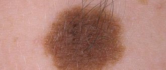
Photo 1. A crust on a mole is not a reason to panic, but a reason for careful observation. Source: Flickr (Donq question).
What is a mole and what is the mechanism of its appearance?
A mole is a benign skin neoplasm that forms in the epidermis due to the uneven distribution and accumulation of melanocytes. These cells react to ultraviolet rays, protecting the body. Excessive exposure to ultraviolet radiation on the skin is the most common cause of the formation of a large number of moles.
The mechanism of mole formation has several stages:
- Initial – accumulation of melanocytes in one place, which is manifested by the appearance of a brown spot.
- Progressive - the mole increases in diameter and can also rise above the surface of the skin.
- Regressive - the mole remains at the same size for quite a long time without causing discomfort to the person.
Moles are an intermediate stage between healthy skin and melanoma. They must be monitored and, if alarming symptoms appear, examined by a dermatologist.
Causes of crust formation on the surface of a mole
The most common reason for the formation of a crust on the surface of a nevus is its mechanical damage. Injured cells succumb to the natural process of regeneration, where the crust acts as a characteristic barrier between the body of the mole and the environment. It acts as a kind of protection for an open wound from pathogenic microorganisms in the environment.
Mechanical damage is a possible cause of a crust on a mole
Also, crust formation occurs when a mole falls off. There are cases when the nevus spontaneously dries out and separates from the body. At the site of the formed wound, a crust appears, under which regeneration processes take place. After spontaneous removal of the scab, the mole area becomes covered with epithelial cells, leaving a small scar.
Excessive exposure to sunlight on the nevus, which provokes the formation of a burn, can also cause the formation of a crust. But the most dangerous option is the malignancy of the neoplasm into a malignant tumor, which is extremely difficult to diagnose based on appearance.
Types of injuries
Many people mistakenly believe that injury can only be dangerous if the nevus is cut off. However, medical removal is much safer since all parts of it are surgically removed. The following are considered household injuries that have bad consequences:
- regular rubbing of the mole (for example, with underwear);
- scratching (in cases where a person scratched himself unsuccessfully);
- constant pressure (belt, tight clothing, shoes);
- cutting off nevus (during shaving, if they are on the head, face, or armpits).
A person does not always notice that he has accidentally caused damage, so when he sees changes on the body, he does not attach any importance to them. It is important to monitor the pigmentation of moles and consult a doctor immediately.
The danger of degeneration of education
Since the rough surface of a nevus may indicate malignancy into melanoma of the skin, it is extremely important to pay attention to symptoms such as:
- The appearance of discharge from the nevus from a transparent color to blood. Discharge can form due to mechanical damage to the crust, or during movement.
- Painful sensations in the area of the mole, as well as enlarged nearby lymph nodes.
- The formation of a white or pink halo around the tumor, which is accompanied by itching.
- Hyperemia and hyperthermia of the skin of the mole and the area around it.
- The hairs on the mole began to fall out on their own.
- The neoplasm rapidly increases in size, has uneven, torn edges and other new growths on its surface.
If you have these symptoms, you need to see a doctor as quickly as possible and undergo a comprehensive diagnosis.
It is strictly forbidden to remove the crust from a mole on your own, as this can provoke the addition of an infectious-inflammatory process (at best), or trigger the processes of degeneration of the mole into melanoma.
Risk factors
According to recent, numerous scientific experiments, it was found that women, unlike men, have a greater tendency to develop melanoma. It is important to highlight that a malignant tumor can be localized separately on an area of the skin or on the nevus itself. Most often, the legs are affected by the pathological process, and a little less often the upper limbs and abdomen. A mole on the face and neck can also become inflamed, swollen, fester and cause many other problems.
People who are characterized by fair skin covered with freckles, as well as patients with a hereditary predisposition, constitute the main risk group. It is in these people that inflammation of the nevus or redness of the mole can result in malignant degeneration.
The above category of people should undergo regular examination by an oncologist and pay maximum attention to any change in the birthmark.
Pale skin and freckles are one of the risk factors for melanoma
Diagnostics
Identifying the causes of the appearance of a crust, as well as making a diagnosis, is carried out in several stages:
- Taking an anamnesis - the doctor asks the patient about the conditions under which a crust appeared on the mole, how long this process has been going on and what additional symptoms are accompanied.
- Dermatoscopy - allows you to examine the skin tumor as closely as possible, which helps the doctor determine the diagnosis and possible causes of the development of the pathology.
- Cytological examination of a smear from the surface of a mole - this analysis is most effective if any liquid is released from under the crust. Its sowing makes it possible to detect the presence of pathogenic microflora, and its quantitative and qualitative composition will indicate a possible pathology.
- Histological examination is prescribed if external signs indicate pathological processes in a mole associated with the degeneration of its cells into a malignant tumor. For histology, a section of the mole may be required, but most often it is completely removed, which helps make a more accurate diagnosis and understand whether additional examinations are needed and what therapy is required.
Dermatoscopy is a method for diagnosing a crust on a mole.
If there is a suspicion of the development of cancer, a blood test for tumor markers may be required. This analysis will reveal antibodies in a person’s blood, the appearance of which is characteristic of cancerous tumors.
How to recognize deadly moles? 6 signs of malignant nevi
Nevi - this is the scientific name for moles - most people of the Caucasian race have. Typically, the average adult will have about 20-30 moles throughout the body, but some people have more than a hundred moles. Sometimes moles and birthmarks are completely harmless, but in some cases it is better not to overlook them.
At the end of the article, we have collected 6 signs of potentially dangerous moles. If you find moles with similar signs, we advise you to consult a doctor.
…………………………………
We are advised by dermatologist Olga Dobronravova, Moscow
…………………………………
………………………………………………………… ………………………………………………………………
Do you know about our website – Amedeya.com ? There you can get a free interpretation of dreams from our expert, read interesting articles on various topics, exciting stories from ordinary people, and find out the latest news from the world and Russia. We will be glad to see you!
………………………………………………………… ……………………………………………………………..
When do moles appear?
Children almost never have moles: they begin to appear on the body only during puberty, when total hormonal changes in the body occur.
By the way, due to surges in hormones, moles may become larger during pregnancy: don’t worry if you find a few new spots
. Take a closer look at them: moles that are symmetrical with flat edges should not cause concern.
However, any suspicious nevus should be closely monitored. Why? The thing is that moles are, in fact, a cluster of skin pigment cells - melanocytes. And under certain conditions they can turn into melanoma cells - one of the most dangerous malignant tumors, which tends to recur and “grow” into other organs
.
When can melanoma develop from a mole?
On average, in 5-20% of cases, large moles larger than 2 centimeters in size turn into malignant tumors.
Doctors say that you need to be careful with such moles, especially if they are in places of contact with shoes or clothing: they say that trauma to the nevus - a cut, bruise, abrasion - or constant rubbing of it can lead to the development of a tumor.
In fact, there is something to argue about here. Some patients whose moles have already degenerated into melanomas and began to bleed often think that the blood appeared precisely because of the damage. They immediately consult a doctor; in fact, it turns out that the patient has cancer. It is important to understand here that it was not the damage that led to the disease, but the disease that caused bleeding
.
On the other hand, some moles can actually grow after an injury. But histological studies indicate that such a mole is not always melanoma. Theoretically, tissue restoration processes take place in the area of damage to the mole, and this is always associated with the presence of a mixture of biologically active substances that stimulate cell growth and division.
Cancer cells can also grow on such a “cocktail,” but so far there is no practical evidence of an increase in the risk of developing melanoma due to trauma to a mole. So if you accidentally cut a mole, don’t panic
. Yes, and run to the oncologist with a request to remove the unfortunate tumor too.
But dynamic observation will not hurt
– You need to visit a doctor from time to time. You need to go to an oncologist if a mole peels off, ulcers appear on it, it bleeds, or there are unpleasant sensations - tingling, itching, burning.
Do moles need to be removed?
By the way, there are quite mixed opinions in the medical community regarding the removal of moles with laser in beauty salons. For many, the aesthetic issue is important.
If you have made this decision, discuss it with your oncologist.
. It is very important not just to remove the mole, but to conduct a subsequent histological examination: doctors must make sure that the nevus does not contain dangerous cells.
It is not so easy to determine “by eye” what kind of mole is in front of them: there are also clinically atypical melanomas, hiding under the mask of completely harmless spots, which are very difficult to recognize at the stage of a simple initial examination. So detailed research never hurts.
Laser mole removal
Avoid direct sunlight
There is such scientific evidence: if a person has received three or more serious skin burns during his life, the likelihood of developing melanoma increases by 2-6 times. Who may be at risk?
In total, there are six groups of people depending on the color of their skin and its ability to tan.
Two of them are the most susceptible to skin cancer: people of the first group will never get a golden skin tone, no matter how much they sunbathe, and representatives of the second phototype may darken a little, but will have to lie in the sun for hours. So, before going out into the scorching rays, these people should stock up on sunscreen.
Those who have many moles – more than 50-100 – should also be on alert
. In a good way, it is better for them not to sunbathe at all, because even creams with UV factors do not always protect against ultraviolet radiation. If your body is strewn with moles, oncologists recommend seeing a specialist after your vacation. Moreover, it is advisable to do this twice a year.
Owners of moles need to monitor whether their size, color, or height have changed. If the mole has paled, become darker, or even multi-colored, make an appointment. You need to be wary if the mole has completely disappeared: this happens both with absolutely harmless halonevus and with spontaneously disappeared melanoma
.
6 MAIN SIGNS OF DANGEROUS MOLES
· asymmetrical shape;
· uneven edges;
blur;
· appearance of gray, white, brown or red stripes or dots on the mole;
· inclusion of cracks (including bleeding ones) or crusts;
· increase in size.
Source: //zen.yandex.ru/media/id/5c93cb937d4e3f0740ce0dda/5ce3a9c23f91de00b3742805
Algorithm of actions when a crust appears on a mole
If a crust appears on a mole, you should immediately consult a doctor. If you ignore this alarming symptom, there is a risk of developing oncology, the diagnosis of which in the later stages is practically untreatable and has an unfavorable prognosis.
Absolutely forbidden:
- Remove the crust mechanically.
- Apply compresses and treat with various ointments and creams.
- Heat the damaged area of the mole or expose it to chemical liquids, which can cause a burn.
If for some reason the crust falls off the surface of the mole, you must:
- Apply a piece of bandage soaked in hydrogen peroxide to the damaged area of skin. This will not only stop the bleeding, but also disinfect the surface.
- Cover the wound with a bactericidal plaster and immediately consult a doctor.
- Avoid direct sunlight entering the wound.
It is not recommended to use iodine and brilliant green for treatment, as this can provoke a pronounced painful effect.
Prevention measures
The following preventive measures will help prevent the degeneration of a mole into melanoma and the formation of a crust on the surface:
- Avoid excessive sun exposure, which is typical for frequent visits to solariums and sunbathing.
- Wear clothes made from natural fabrics, which minimizes the risk of injury to the mole. If neoplasms are located in areas of increased risk of injury, doctors recommend removing them.
- Carry out hygiene procedures carefully, using only those cosmetics that do not cause irritation or damage to the surface of the mole.
- Do not try to remove skin tumors yourself, as this may cause complications.
Measures to prevent a crust on a mole include avoiding tanning beds.
The crust can come off on its own, which is a completely natural process. However, if it occurs repeatedly, and the mole itself changes, you should visit a dermatologist and make sure there is no malignancy. Self-medication can be fraught with the development of a number of dangerous complications, and can also become a trigger for the degeneration of a benign neoplasm into a malignant one.
Methods for removing nevus
Malignant nevus is dangerous by metastases to the lymphatic system.
Removing a nevus for no reason is dangerous. If, after consultation with a dermatologist and oncologist, there is no other option, you need to think about a method for removing the skin lesion. You can get rid of skin lesions using:
- laser skin correction;
- cryodestruction (freezing with liquid nitrogen);
- radio knife;
- electrocoagulation (cauterization);
- complete removal by the surgeon during surgery.
Regardless of the method of mole removal, it is recommended to follow certain rules after the procedure. You cannot wet the area where the nevus was for a week. It is recommended to limit sun exposure, even when using sunscreens with a filter of 60-100. It is important to stop using cosmetics for a while. Under no circumstances should you tear off the crusts that have appeared at the site of the removed mole. Over time, she comes off on her own. Even multiple symptoms do not indicate 100% that skin lesions are malignant. Therefore, consultation with a specialist is important.