A birthmark is a benign growth formed on the skin. It is subject to the mechanical influence of external factors that provoke the transformation of healthy cells into cancer cells. This process occurs at lightning speed, so if there is the slightest change in the structure, color, or shape of the formations, you need to consult a doctor. He will prescribe a set of examinations to take treatment measures. One of them is the histology of the mole.
The importance of histology in the early diagnosis of cancer
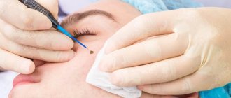
Histological examination of a growth on the skin is carried out on the basis of a referral issued by a physician, or on the basis of the personal will of the patient. The procedure is only permissible within the walls of the clinic. For analysis, preliminary collection of biomaterial from the growth area is required.
There are 3 examination methods:
- using a needle (in this case, part of the tissue structure is excised using a specialized instrument, anesthesia measures are first carried out);
- incious method (a part of the material being studied is taken and several fragments of it are sent to the laboratory);
- excision method (used during surgery, however, to the full depth of the skin with excision of healthy elements that are located near the growth, the subject of study is the complete body of the nevus, which was surgically removed).
Small in size and regular in shape, moles do not pose any threat. At least this lasts until changes occur in them. If the slightest abnormalities are noticed, you should not delay a visit to a doctor, as they may indicate the initial stage of a dangerous pathology - melanoma. It is a skin cancer. If detected early, it responds well to treatment (traditionally removal is used).
What to do if the histology is bad
Poor histology of moles after removal is not a final diagnosis or verdict. The correct diagnosis will be made by an oncologist based on the following factors:
- age, gender;
- visual inspection results;
- tissue histopathology;
- individual, family history of the patient;
- history of skin cancer;
- data from other examination methods.
During a histological examination, the presence of a malignant process, the type of carcinoma, and its degree are determined. Based on these data, the doctor chooses patient management tactics and develops a treatment plan.
Indications for histology of a mole
During the visual examination, the doctor has the right to refer the patient for additional tests.
These include:
- study at the molecular level,
- skin examination,
- radioisotope type scanning,
- CT,
- histology.
It is the latter option that is considered the most acceptable and preferable. This is due to the fact that the technique helps to identify malignant cells and prescribe timely treatment measures.
Here are the indications, when they occur, the doctor will refer you to a histological procedure:
- intensive growth of the tumor;
- changes in structure;
- the appearance of tubercles;
- the appearance of a shiny crust on the mole;
- excessive bleeding;
- dark plaque or darkening of the mole;
- spreading of the stain over the surrounding skin areas;
- the occurrence of pain due to mechanical impact.
If glomus angioma is suspected, tactics are rarely performed; it is appropriate only if a cancerous tumor is suspected.
When is histological analysis required?
A referral for histological analysis is given to a person after undergoing an examination by a dermatologist or oncologist.
This is an additional diagnosis after dermatoscopy, radioisotope scanning, computed tomography. Among these methods, histology has many positive reviews. This is explained by obtaining an accurate indicator regarding the risk of nevus transformation.
You can independently request a histological examination. The reasons are:
- the spot has acquired a blurred pattern, clearly defined boundaries have disappeared;
- the formation increases sharply in size and becomes inflamed;
- upon palpation or contact with items of clothing it begins to hurt;
- tuberosity appeared;
- the nevus is covered with a shiny film;
- the hairs that were located on the mole fell out;
- the nevus has acquired a dark shade;
- the birthmark is subject to constant mechanical stress;
- a white halo appeared near the formation;
- blood or pus comes out of the mole;
- itching and burning appeared;
- the flat spot has turned into a raised mole;
- peeling appears on the surface of the mole or near it;
- the mole became hard to the touch.
Histology of a mole can be carried out if a person decides to remove it for aesthetic reasons. Nevus can be located on the face, head, neck, which spoils a person’s appearance. In the absence of dangerous symptoms, a histology analysis is still performed. This helps determine the presence of melanoma, which can be treated at an early stage without consequences to health.
Methods of histology of moles
The lion's share of medical institutions conduct histological examinations free of charge. As a result, it becomes possible to make a final verdict and confirm or refute the fact of cancer or glomus angioma. The procedures are carried out within the walls of the laboratory according to classical schematics.
It looks like this.
- The doctor removes the problematic tumor.
- After removal, he takes the biomaterial for himself.
- The growth is put into a special solution and taken to the laboratory.
- The material is applied to the glass and must be painted.
- After a certain period of time, the doctor examines the cells under a microscope.
The research process can be carried out in several ways.
- Freezing the medication. This method is appropriate in a situation where an urgent study of a mole is required. For example, during surgical treatment. If the results of the analysis do not correspond to the doctors’ assumptions, it remains possible to change the course of the operation.
- Paraffinization of biomaterial. This technique makes it possible to preserve the removed growth for a long time. This is required for it to be studied by several specialists at once.
Using modern equipment, it becomes possible to remove and examine tumors of any complexity, and the result is guaranteed.
The classic scheme of an event consists of a number of stages. Let's look at them in more detail.
- Sampling (direct excision of biomaterial).
- Fixation (placing the sample in a container with water, which is subsequently replaced by ethanol, formalin). This measure helps prevent cellular decay and preserve the intravital structure of the mole.
- Wiring. In order to give the taken “raw material” the optimal shape and structure for examination, it must be dried and filled with paraffin.
- Cutting. When the material hardens, it is cut using special techniques until thin plates are obtained. Excess paraffin must be removed.
- Coloring. At this stage, the plates are lowered into a special solution with dyes that were prepared in advance. This helps determine the cellular structure.
- Microscopic examination. The most important stage of the entire procedure. Modern optical instruments are taken, and the laboratory assistant uses them to obtain a series of data that subsequently needs to be transferred to the attending physician.
Carrying out the technique requires about 7 days.
If it is necessary to deliver the material to another locality, the period is extended for the entire transportation time.
If a physician suspects aggressive types of pathology, urgent surgical treatment is necessary in order to save human life. For this purpose, an accelerated method is relevant.
How long does it take to perform histology?
The duration of processing of tissue samples should be checked with your doctor. There are 2 types of analysis:
- Emergency - the cut is examined during the operation. This allows, if necessary, to expand the scope of surgical measures and remove more tissue. And immediately after removal of the tumor, begin drug treatment. Used in controversial cases, when new circumstances are identified during surgery.
- Planned - used when the diagnosis is beyond doubt, and an emergency study confirmed the doctor’s assumption. The processing time for results is up to 7 days.
The timing of the study is influenced by the following factors:
- legal status of the laboratory or clinic - private or public;
- analysis methods and equipment used in a particular medical center;
- laboratory workload;
- force majeure circumstances.
A long sample processing time is not synonymous with bad news!
What does poor histology of moles mean?
If glomus angioma is not confirmed, but cancer is detected, we are talking about melanoma. When it occurs, doctors recommend surgical treatment. Conservative tactics are not used. The removal procedure is performed in a clinical setting using the following strategies:
- laser excision;
- electrocoagulation;
- radio wave method;
- using an electric knife.
If surgery is not performed in a timely manner, there is a risk that infected cellular elements can form metastases to other organs.
It is important to know! Some time after surgery, you need to see a doctor so that he can eliminate the possibility of relapse.
A scar appeared after mole removal
When excision with a scalpel, modern, atraumatic absorbable suture materials are used to eliminate a wound defect and, often, the patient cares for the wound independently.
In this case, the scars remain as aesthetically pleasing and delicate as possible, but nevertheless they are still scars. After removing a mole with a laser or electrocoagulator, the marks are usually invisible on the body (when nevi up to 5 mm are removed), or are visible, but are perceived not as scars and scars, but as minor changes in the skin (there are no associations with surgical procedures).
The appearance of a scar after mole removal depends on the properties of the patient’s skin, the size of the lesion being removed, its location, the duration of healing, and the characteristics of caring for the wound during healing.
The most unpleasant moment is the appearance of a keloid scar at the site of the removed nevus - this is a frequent reason for repeated visits and many questions. A keloid scar after removal of a mole occurs due to the predisposition of the patient’s skin, in certain areas of the body (sternum area, for example), when complications arise in the form of suppuration of the wound, or during prolonged healing due to improper care of the wound.
A keloid scar can be disturbing not only due to its sloppy appearance, but also due to pain or severe itching.
Modern technologies make it possible to correct a keloid scar. For example, we use injection correction in combination with surface laser resurfacing. If treated in a timely manner, one procedure is sufficient.
How to prepare for histology
No special preparation measures are required before the removal procedure. If the doctor did not have time to conduct this analysis, the material can be taken during the removal procedure. This is done before or after the operation – it doesn’t matter. Sometimes the condition is assessed after surgery, and the patient does not even know about it.
Thus, in order to avoid glomus angioma, melanoma and other complications, a specialized examination is required, which will help in making a diagnosis.
How long does it take for wounds to heal after mole removal?
It all depends on the area of the base of the removed formation, that is, on the size of the wound itself, as well as on the characteristics of the skin where the mole grew (on the face, neck, and scalp, wounds heal the fastest, and in injured areas or on natural folds of the body - longer) .
On average, the skin after mole removal takes 10-14 days to heal. During this time, the wound should be covered with a dense scab-like crust. It is formed by an oncodermatologist after removal, and then gives the patient detailed instructions for care.
A strict restriction is not to wet the scab during the first 24 hours after mole removal. Then you just need to repeat the treatment after each water procedure.
The wound is considered healed when the crust has fallen off (on its own!) from the surface and the wound site is no longer wet. Healing after mole removal will proceed faster if you follow all the doctor’s care recommendations.
If after removal the mole takes a long time to heal (more than 3 weeks), you must contact the doctor who performed the removal. The sooner a problem is addressed, the less effort it will take to fix it.
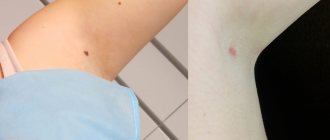
Changes at the site of the removed nevus
If, after removing a mole, strange changes occur at the healing site, immediately contact the doctor who removed it.
Reasons for concern may be compaction, vascular growth, itching, burning, pain, or colored areas at the removal site. All these changes do not necessarily indicate recurrence of the nevus. These may be the same keloid changes, rough scarring or scar pigmentation (darkening of the young skin covering the scar - this often happens when the scar is exposed to sunlight immediately after it has healed.)
Are there errors in the histological examination of nevi?
Yes, unfortunately, they are possible. I wrote about this. Histological examination is carried out by people, and they can make mistakes. If you have the slightest doubt about the results of the conclusion, take your histological preparations and send them for review to another laboratory. The preparations include both paraffin blocks with mole tissue and glasses with sections.
You ask: “How can I go and pick something up? They’ll send me…” If they don’t give you histological preparations, then you need to go to the head doctor and demand it. In my experience, after that everyone gives away.
When should a mole be removed?
If a mole or other formation on the skin increases in size, changes color, or is constantly exposed to trauma, then you need to consult a specialist. For example, a mole or nevus may be located in places of frequent injury - head, face, neck, axillary area, palms, feet, lower back. Recently, a patient came to us with a small growth on her scalp that began to bleed when scratched. Another case is that a man’s nevus was often injured while wearing a belt. Such cases are not rare and have direct indications for removal, since they can degenerate into a malignant neoplasm.
Who makes mistakes more often?
What I say below is my personal opinion. This is based solely on my experience and is not supported by research.
It seems to me that the probability of a doctor’s error when removing moles is higher, the further he is from skin cancer. A cosmetologist, surgeon or dermatologist is more likely to mistake a melanoma or skin cancer for a regular mole. This probability is much lower for an oncologist. The error for an oncologist specializing in skin tumors (dermato-oncologist) is close, but also not zero.
Dear colleagues, dermatologists, surgeons, cosmetologists! If you are reading these lines, please do not be offended. I know for sure that among you there are those who will give many oncologists (including me) a hundred points ahead in diagnosing melanoma. Unfortunately, my experience in conducting advanced training courses for doctors of these specialties suggests that this is rather the exception than the rule.
One mole – one container with full name
If histological examination of the mole reveals melanoma, the removal site should be re-excised more widely. The capture of healthy tissue is determined depending on the Breslow thickness.
I would like to draw your attention to the fact that if all removed moles are placed in one container, they will be able to undergo histological examination. But in this situation it will no longer be possible to establish where exactly the melanoma was and it will be necessary to do a wide excision at the sites of all removed moles.
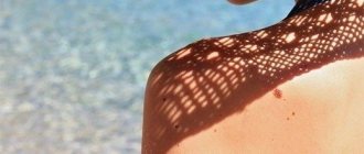
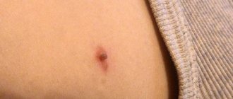
To have a correct idea of the volume of tissue removed, you can look at the photo and see what an average scar looks like.
If you have several moles removed, when sending them for histology, the location of each nevus must be clearly indicated.
But it’s not worth removing so many moles at once, as in the photo. Why? I'll tell you in the following publications.
Summary, or Briefly about the main thing
Histological examination is the key to safe removal of moles and other skin formations. Always insist on histology for all but small soft papillomas. Preference should be given to laboratories of large oncology institutions. The histological report should always be collected in person and shown to the doctor who performed the removal.
Other articles:
- Radio wave removal of papillomas
- Removal of any skin formations by a dermato-oncologist
- Removal of warts by a dermato-oncologist
- Removal of moles by a dermato-oncologist
Useful article? Repost on your social network!
Why are papillomas not sent for histology?
The main property of a malignant tumor is uncontrolled cell division. Papilloma is a small (1–3 mm) formation on a thin stalk of soft consistency.
If the papilloma cells were malignant, they would grow a “leg”, and the papilloma would lose it. In addition, the consistency is softer than the surrounding skin, in my opinion, always indicates good quality.
This case is the only one when, in my opinion, the material need not be sent for histology.
Why examine a mole if it has already been removed? Will it be too late?
A logical question arises - what to do if the histology shows melanoma? Will we do any harm by removing it? Will this lead to tumor progression?
No.
A large number of clinical studies have found that performing an excisional biopsy
does not worsen the course of melanoma.
In other words, the chances of recovery in patients who first underwent such a biopsy, and then wide excision, are the same as those whose melanoma was immediately excised widely.