Skin lesions can come in different sizes, colors, and shapes. We are talking about moles (nevi), which can be either congenital or acquired in a person. These formations are benign, but sometimes they can become malignant (melanoma), so they require constant monitoring.
Redness around the mole may be alarming. It may indicate either the degeneration of a nevus into melanoma, or that it was simply subject to mechanical damage. In any case, this fact should not go unnoticed.
What causes the growth to become red?
The red tint is given to the growth by disturbances in the integrity of the vessels supplying the formation with blood. The reasons for the modification are internal and external.
External factors influencing the condition of the birthmark:
- mechanical impact: friction, bruise, scratching;
- exposure to sunlight or its artificial substitute;
- the effect of low or high temperatures on the skin;
- electric shock;
- the action of caustic chemical or plant elements.
The reddened area, subjected to a single influence of external irritants, will eventually return to its original state. Regularly repeated exposure can lead to the degeneration of a mole into a malignant formation.
Internal factors leading to redness of the mole:
- hormonal imbalance in the body, taking hormones as medicine;
- metabolic disorders, diabetes mellitus;
- lack or excess of vitamins;
- allergic response of the body;
- decreased immunity, severe infectious skin diseases, fungal infections;
- problems of the vascular system;
- heart failure.
Why does a mole turn red, enlarge and become painful?
The fact is that inflammation can begin in the area of hair follicles and sebaceous glands. As a rule, the sebaceous gland becomes inflamed, the excretory duct of which opens into the hair follicle.
In medicine, inflammation of the sebaceous gland and hair follicle is called a furuncle, popularly called a “ pimple.”
«.
This happens for a variety of reasons, from pollution and microtrauma of the skin to metabolic disorders. As you can see in the picture below, all inflammation phenomena appear in the area of the mole:
What should you be wary of?
If the redness of the skin around the growth is associated with internal problems of the body, it is recommended to immediately consult a dermatologist and other specialists. If a red spot appears, you should not try to eliminate it yourself, especially if there are accompanying signs:
- The inflammatory process of the skin around the mole is clearly visible: swelling, pain when pressed, dry flaky surface, small bleeding cracks.
- The formation changed its color, a spotty color appeared, alternating light and dark areas.
- The growth visually increased and exceeded 5-6 mm in diameter.
- The boundaries of education have become blurred without a clear outline.
- The surface of the mole is covered with bumps and pits.
- A lighter circle has formed around the nevus.
The degeneration of a growth into malignant melanoma occurs with every thousandth mole.
Considering that a person sometimes develops up to hundreds of moles of different sizes, skin cancer affects every 10-15 patients who notice a modification of the nevus.
When do moles first appear in children?
Moles in children can appear at different ages, even if there are many of them on the body.
But it happens when a child develops a large mole with a white spot around it, which can lead to serious consequences.
Despite the fact that science has long found an explanation for this phenomenon, nevi for many people remain some kind of mysterious mark, in connection with which many signs and beliefs have been invented.
This is especially true for children when a child has moles.
But medical research can certainly explain why and when moles appear in children, and what you need to pay attention to.
When they appear
While still in the womb, the fetus already develops new growths.
Every hundredth baby is born with nevi on various parts of the body, and the spot can be invisible, and with age, as pigment cells with melanin concentrate in it, it acquires color. Both are normal.
Congenital nevus is very rarely dangerous for a child.
By the age of 25, up to 80% of suspected nevi form in a person, but the timing of their appearance is not limited - from the moment the baby is born until old age.
The exact reasons for the age at which spots become visible on the skin have not been found.
It is believed that they appear as a result of genetic predisposition and certain external factors.
- The first ones begin to appear from six months to two years.
- The second period of active appearance is from 5 years to 6 years.
- Their main quantity is formed in adolescence - from 12 to 16 years.
Photo
What are there
Typically, neoplasms in children are safe (except for isolated pathological cases), and more often they are flat or slightly convex pigmented formations up to 1 cm in diameter.
A common birthmark in a small child is light brown in color and does not affect the baby’s health.
Benign neoplasms can be of the following sizes:
- up to 1.5 cm – small;
- up to 10 cm – medium;
- 10 cm or more – large.
The latter are usually located on the buttocks or thighs.
Unlike small moles, their medium and large size should cause concern and require constant monitoring.
They degenerate much more often, and up to 50% of such processes lead to cancer, therefore, regardless of the age at which they appear, they require consultation with a dermatologist and oncologist.
Dark spots
Photo: pigmented congenital nevus
All neoplasms containing melanin are called pigmented.
Some skin cells are disrupted in the production of melanin, which is why they acquire a characteristic brown or black tint.
Most nevi occur in children when signs of puberty appear.
Pigment spots on the body are divided according to their location in the skin layer:
- borderline (with accumulation of melanin in the epidermis) - the most common first moles in babies;
- intradermal (when melanocytes are concentrated in the dermis);
- mixed (in the outer and inner layers of the skin).
In young children, borderline moles that are harmless to health normally appear in the form of brown or yellowish spots of regular shape.
As you grow older, these spots may change..
As for convex neoplasms like in adults, if you notice such spots appearing in a child, you need to urgently consult a doctor.
These are pathological changes and must be constantly monitored.
Normally, ordinary flat age spots do not cause harm, no matter how old they appear, but you need to monitor new growths in any case.
Reds
Photo: strawberry hemangioma
Infants often develop so-called red nevi.
Very often such vascular neoplasms are located on the head.
- They occur as a result of childbirth, when the baby's delicate skin passes through the protruding pelvic bones of the mother.
- This vascular red mole, as a rule, disappears by the age of one, but in the case of vascular tension it can temporarily remind itself again.
Parents have a question whether such a neoplasm is dangerous.
No, it usually does not require intervention and will not cause concern to the baby.
The local pediatrician will tell you what to do and will recommend who to turn to for advice as needed.
Vascular moles include hemangioma:
- “strawberry” - soft, convex, red in color with a strawberry tint. Its appearance is typical in 1-2 months, but can also occur at birth. There are not many of them on the body; usually there is only one vascular cluster. The unpleasant thing is that it appears on the face, and as the child grows, it can increase and fade to a grayish tint. Only a doctor can judge whether a mole can be removed, since this treatment can lead to dangerous consequences;
- “Cavernous” is an accumulation of mature large vascular elements, penetrating deeply into the skin, with unclear boundaries. Such a spot may be grayish or bluish, but there is usually no danger or inconvenience from it. It may grow at first, but removal is not required, since it disappears by adolescence.
This applies to congenital hemangiomas.
If it arose during life, it will not disappear on its own, and you need to find out what to do from a doctor.
Wine stains
Photo: wine stain
A type of flat mole is wine mole, usually large in size.
- This is a part of the skin with dilated blood vessels that is red with a purple or pink tint, usually covering the baby's head and face.
- As a rule, spots grow as children grow, and their color does not change.
This tumor is not dangerous, but doctors recommend removing it as soon as possible since it causes a cosmetic defect.
Treatment is usually carried out using a laser.
Anemic nevus
Characteristic features are irregular outline and pale color, similar to vitiligo. It is caused by underdevelopment of blood vessels.
It is located on the face and back and is treated with surgery.
Are moles dangerous for children?
Hemangiomas require treatment very rarely, since most often they disappear without intervention, the same can be said about “stork bites” - red-yellow postpartum spots that disappear by two years.
The degree of danger of pigment spots depends largely on the size.
- Small spots on the skin are not dangerous, unlike medium and large ones.
- The latter in 40% of cases degenerate into melanoma.
Therefore, if you have such moles, you should urgently contact an oncodermatologist and find out whether treatment is required.
Price for laser removal of a mole on the face in Moscow clinics.
What to do if black dots appear on a mole? Find out here.
What to pay attention to
Parents should constantly monitor their child’s nevi.
Red hemangioma usually disappears on its own, but there are exceptions, so it requires observation.
- A child with neoplasms should spend as little time as possible under ultraviolet radiation, since it is the main factor in the degeneration of a nevus into melanoma.
- Make sure that the child does not accidentally or deliberately pick off a mole or injure it with clothing.
- If you suddenly start bleeding, you need to treat the wound with an antiseptic like chlorhexidine, stop the bleeding and get an appointment with a dermatologist or oncologist.
It is strictly forbidden to cover it with an adhesive plaster - suppuration may begin, and this is very dangerous.
What to do if the stain grows
Moles can increase in size both in the case of pathological transformations and during normal development of the body.
- As the skin renews and grows, a benign formation may also grow slightly, but if the growth is intense, then urgent consultation with a doctor is needed.
- The size of a benign nevus in children is normally no more than 1 cm, with rare exceptions.
If it is larger, begins to increase, or a white halo appears around it, consult a doctor immediately.
Video: “Wine Stains”
Dangerous nevi
Both the color and size of the tumors can vary, and they can be both benign and dangerous.
And the number of nevi is not as scary as their size. A lot of them on a child’s body is not a pathology.
- But if the mole is one, large (from 1.5 cm in diameter) and black, this should alert you.
- Large nevi that protrude above the main covering of the skin are also dangerous.
They require close attention so as not to miss the symptoms of possible degeneration into melanoma.
Melanoma
Diagnosis of melanoma, what does it mean?
The mother of a child with skin cancer urgently needs an answer to this question.
A mole consists of melanocyte cells, which, with a benign status, are present in a certain quantity; the nevus does not change externally and does not cause concern. Under the influence of a number of factors, these cells begin to mutate - grow and multiply without stopping.
This is how one of the most insidious types of cancer begins - melanoma skin cancer.
Photo: squamous cell carcinoma
At this stage, the neoplasm usually changes in both color and size, and a white spot may also form around it.
If you perform surgery in time and remove all pathological melanoma cells before it forms metastases, then there is a high chance of survival and absence of relapse.
If the mole has enlarged and no measures have been taken, the melanoma metastasizes to vital organs and death is almost inevitable.
With seemingly unimportant symptoms such as slight discoloration, gradual, barely noticeable growth, or mild itching, the disease progresses extremely quickly.
How to recognize a malignant mole yourself?
What to do if a mole turns red and hurts? Find out here.
What kind of doctor deals with moles on the body? Read here.
Therefore, it is important not only to periodically examine spots in children by parents, but also to consult a doctor at the slightest suspicion of changes.
Removal
Suspicious neoplasms require a thorough examination by a specialist and appropriate treatment or removal.
Just like adults, children are examined for nevus degeneration.
- If it has begun, then there can be only one treatment - urgent surgical excision with removal of the maximum amount of pathological tissue.
- When a potentially dangerous nevus does not show suspicious signs, and dermatoscopy does not show cancer, the doctor may prescribe laser removal for certain indications.
For example, if a child scratched a potentially dangerous tumor.
Methods for getting rid of nevi
Photo: nevus removal with laser
Neoplasms are removed in exactly the same way - for both adults and children.
- If an oncological cell mutation is confirmed, the operation is performed by surgical excision.
- In all other cases, laser removal, cryotherapy, and radio wave radiation are possible, but any effect on a mole on a child’s body is an extreme measure and should only be prescribed by an oncodermatologist.
Where to delete
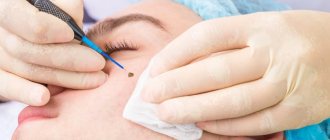
According to the doctor's indications, a mole is removed in a special clinic only after a series of tests and a comprehensive examination to rule out melanoma.
No cosmetologist has the right to violate the integrity of a nevus in any way, just like home removal methods - this is deadly!
Care after removal
Children usually tolerate a well-performed operation to remove a nevus easily, but postoperative rehabilitation and treatment are very important.
- The baby needs a gentle regime, the prescription of a number of immunostimulating drugs, as well as compliance with measures to prevent relapse.
- The wound left after removal is cared for as after any other operation - dressing, examination by a surgeon and antiseptic treatment may be required.
Find out what to do if a mole becomes inflamed.
The meaning of a mole above the lip on the right in women. Read on.
What happens if you rip off a mole on your neck? Read here.
Prevention of degeneration
The main preventative measure is to monitor moles, prevent them from changing and consult a doctor in a timely manner.
- Large elements must be under constant control.
- If a white coating appears on the nevus, it quickly increases in size, changes color, bleeds, becomes covered with a dry crust or festers, you need to urgently go to a dermatologist.
- The main factor in the degeneration of nevi into melanoma, in addition to genetic predisposition, is UV radiation, which is why children need to be protected from direct sun exposure - cover their skin with light clothing in the summer, wear a hat and do not sunbathe on the beach. When exposed to the sun, use sunscreen.
- It is very important to notice immediately if a child has torn off a mole and go to the doctor, since any violation of the integrity (not necessarily) can lead to serious consequences.
- You cannot cover the wound with a band-aid and fill it with brilliant green and iodine - only treat it with an antiseptic like hydrogen peroxide or chlorhexidine and a sterile bandage!
To prevent degeneration, the doctor may also recommend removal, but such cases are quite rare.
Video: “Moles in children. Summer, sun, beach"
kozha.hvatit-bolet.ru>
Types of formations
Moles on a person’s skin can take various forms:
- flat skin formations;
- convex round growths of various sizes;
- hanging, connected to the skin through a thin or thick stalk.
Hanging structures are most susceptible to injury. If the integrity of the growth is compromised, bleeding may begin, the skin around the mole may become red, swollen, and painful. Inflammatory manifestations that last more than three days are a reason to consult an oncologist or dermatologist.
Why does the skin around the flat type of formation turn red? This is due to the following reasons:
- skin diseases associated with uncleanliness, fungal infections, inappropriate cosmetics and hygiene products;
- irritation from the bites of blood-sucking insects;
- allergic reaction to food, animals, dust, etc.;
- exposure to radiation;
- frequent exposure to open sunlight.
Redness around a mole can go away on its own without a trace or give rise to cancer.
To protect yourself from malignant degeneration of the growth, it is recommended to consult a doctor as early as possible, because melanoma skin cancer can be successfully treated only at the first stage.
How to prevent inflammation
Moles that are most susceptible to mechanical damage are those located on the neck, shoulders, chest, in the area of the shoulder blades and in the intergluteal region. In women, the risk zone is considered to be the area of the body where the bra straps are constantly located. You should be careful about nevi, which can be injured when shaving, this is especially true for men.
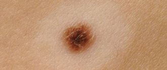
A reddened and enlarged mole is noticeably different from a malignant neoplasm with clear, non-blurred boundaries, which are colored with uniform intensity as the central part of the nevus.
There are simple preventive measures that must be followed:
- regularly examine moles for changes in shape, color and size;
- do not wear tight clothes and avoid accessories that cause friction or put pressure on the nevus;
- limit your exposure to the sun and use UV protection products that match your phototype;
- do not self-medicate, this may cause more serious problems.
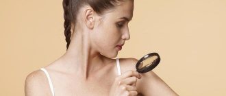
Diagnosis of growth
A dermatologist performs a visual examination of a reddened lesion on the patient's skin. If the malignant nature of the redness is suspected, a histological analysis is performed to confirm or exclude oncology. Computer diagnostics are widely used, allowing one to examine in detail the condition of all layers of the skin and find the cause of redness of the growth and the skin around it.
In cases hazardous to health, it is proposed to remove the nevus using one of the methods used by modern medicine:
- freezing an object with liquid nitrogen;
- burning out the formation with a laser beam;
- removal of a mole by electric current or radio waves;
- surgery to confirm malignant transformation.
While carrying a child, a woman's hormonal levels change, so there are frequent cases of redness of moles. This usually goes away after the baby is born, but you should definitely see a doctor to completely eliminate unwanted consequences.
Treatment
Therapy in this case has two directions. Symptomatic treatment simply relieves redness, and complete treatment involves removing the nevus from a given area of skin. Traditional medicine methods can be used as symptomatic treatment. Removal is carried out only on the basis of a medical institution after examination by an oncodermatologist.
Drug treatment
The following pharmaceutical products are used to relieve redness:
- Zinc ointment;
- Salicylic-zinc ointment;
- Tincture of calendula;
- medical alcohol;
- Syntomycin ointment;
- Streptocide ointment;
- Triple antibiotic ointment.
To remove a nevus, a doctor can use various techniques:
- laser removal;
- cryodestruction;
- electrocoagulation;
- radio wave removal;
- surgical removal.
Photo 2. A conclusion on the removal of a mole is issued by an oncodermatologist. Source: Flickr (Eugene Evehealth).
Important! The treatment method is prescribed by the attending physician. Self-treatment of a nevus and redness around it can cause malignancy of the tumor.
ethnoscience
The use of traditional medicine for redness is aimed at reducing the intensity of blood flow at the site of the lesion and preventing the development of infection. Therefore, anti-inflammatory and antiseptic elements are used:
- A weak solution of potassium permanganate. You need to wipe the affected area 2 times a day until the redness completely disappears.
- Cucumber. Grind the vegetable and apply as a compress for 15-20 minutes.
- Cabbage. Apply as a compress to the area of redness for 20-25 minutes.
- Potato. Make a compress of grated or simply peeled potatoes on the area of redness. The exposure time is on average 25 minutes.
Note! Before using these methods, be sure to consult your doctor! Self-medication can be dangerous.
Traditional methods for inflammation
If it is impossible to visit a doctor right away, and the inflamed spot creates discomfort, you can apply temporary measures to relieve the inconvenience:
- lubricate the reddened area with tincture of calendula or celandine;
- apply a clay cake to the inflamed area;
- make lotions from a warm decoction of chamomile, St. John's wort, plantain, black tea without impurities;
- attach an aloe leaf, cut lengthwise;
- moisten a fresh plantain leaf with clean water and apply the smooth side to the affected area;
- spread the formation and the skin around it with toothpaste with an anti-inflammatory effect;
- mash the leaves of grapes or sorrel and moisten the redness near the mole with the secreted juice.
If the measures taken turned out to be effective in removing the redness of the mole, a visit to the doctor should not be postponed. The dermatologist needs to explain in detail what exactly led to the improvement, so that “home” treatment does not affect the final diagnosis.
Briefly about the main thing:
If your mole becomes inflamed, red, or painful without any external influence, do not panic. Apply compresses according to the above scheme and inflammation should subside within 3-5 days. If this does not happen, see an oncologist immediately.
If you don’t have the mental strength to wait 5 days, you can get my consultation online right now. There is also the opportunity to make an in-person appointment with me at the clinic in St. Petersburg (Asafieva 7/1).
If you find that a mole is swollen and painful, you should immediately contact a medical facility. The reasons may be different and not all of them are dangerous, but checking will help avoid serious consequences.
Preventive actions
The following precautions can help prevent the appearance of redness around the birthmark:
- avoid mechanical damage to the mole: make sure that clothing parts in contact with the skin do not rub the formation;
- It is not recommended to scratch the nevus and the area around it with extended nails, as their sensitivity decreases;
- use razor and depilator carefully near skin lesions;
- do not use a hard washcloth, try not to rub the mole;
- do not wet the growth with salt water or caustic liquids;
- balance your diet so that your body receives the necessary vitamins and microelements;
- to refuse from bad habits;
- drink high-quality, well-purified water.
If a mole on the skin suddenly turns red, it is necessary to entrust its treatment or removal to a specialist doctor.
Self-treatment can lead to irreparable consequences and death.
Where can angioma be found?
They can be found almost everywhere: on the face, body, muscles, bones. Anywhere there are blood or lymphatic vessels.
Bone hemangiomas often form in the bones of the pelvis, skull, and spine (can cause musculoskeletal disorders). The most dangerous are those that grow in the brain and compress the vessels that feed it. Such a formation is difficult to remove, as vital parts can be affected.
A large number of red moles on the body
Types of nevi
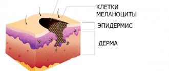
A nevus is a collection of melanocytes - dark pigment cells. Depending on the appearance and location of the nevus, there are:
- Borderline nevi, which look like a spot and may stand out slightly on the surface of the skin, but have a clear outline.
- Intradermal ones, which are located in the deep layers of the skin and can rise above it; when passed over them, a small tubercle is felt, and after a while a hanging wart can form.
- Epidermal, which is formed in the epidermis layer and rises above the skin level, and has different sizes and colors.
Intradermal ones are most often found on the scalp; other types can be found on any part of the skin.
Depending on the structure, a mole can be:
- Not vascular - it is based only on melanocyte cells, therefore such a mole will have a brown or black color.
- Vascular - which covers small clusters of vessels and capillaries, and therefore has a red, burgundy color.
Vascular nevi are most often found on the face and neck, where capillaries are located close to the skin layer.
Swelling after injury
It happens that a mole swells after injury, although its carrier may not remember a single injury. The following effects are considered trauma for moles:
- Scratching, especially after a hot bath or sauna, when the pores are enlarged and the skin is steamed. If you feel itching, you should absolutely not touch it.
- Friction or compression, for example, when wearing tight underwear, constant contact with objects, if it is on the hand.
A mole located on the face, arms, legs, or armpits will swell most quickly. It is injured during hair removal, contact with surrounding objects, and can react sensitively to cosmetics. If seriously injured, it may bleed.
In such situations, you need to disinfect the skin with chlorhexidine and go to the doctor so that he can assess the consequences of the injury.