An easy gait, beautiful posture, good health - all this can be ruined by a seemingly ordinary lump under the skin on the leg. Its appearance often indicates the beginning of the development of a disease in the body. Checking this tumor and eliminating its cause gives a chance to prevent the disease and maintain health.
Bumps on the bottom of the foot near the arch
Plantar fibroma
In human anatomy, the arch of the foot is formed by the metatarsal and tarsal bones. They are strengthened by ligaments and tendons, which allows the leg to support the body weight in a straight position with the least load. The arch is divided into longitudinal and transverse.
A lump on the bottom of the foot near the arch is most likely to be plantar fibromatosis (fibroma). This is a condition in which small nodules (bumps) grow on the arches of the foot on the plantar fascia, the thick connective tissue that supports the arch of the foot. It begins where the heel bone protrudes and extends to the head of the metatarsal bone.
This type of lump is benign, which means it is non-cancerous and will grow slowly. Most of these bumps will grow less than an inch. They are usually painless, but depending on their location and size, they can cause leg pain. If plantar fibroids do not show any symptoms, they sometimes go undetected for years. Common symptoms may include the following:
- A small lump under the skin is the most common visible symptom. It will often be found on the sole of the foot at the highest point of the arch.
- While the lump is still small, it will be painless, but as it grows, it may begin to hurt and make walking difficult.
- If this formation is left unchecked for a long time, it can cause thickening of the plantar fascia, which, in turn, will lead to contracture - insufficient mobility of the toes.
The actual cause of plantar fibroma is unknown, but research suggests that there are a number of factors that may contribute to its development:
- Genetic predisposition
- Long-term foot injury
- Alcoholism
- Medical conditions such as diabetes, epilepsy, liver disease and others
- The condition has been found to be approximately 2 times more common in men than women.
The bump caused by plantar fibroma is benign and can only be removed with proper treatment and medical interventions.
Causes of impaired wound healing
Finally, it’s worth saying a few words about why tissues sometimes recover so slowly. The first reason is the person himself. But violations appear even without his participation. You should consult a doctor if the skin color changes, pus forms, or the severity of the wound increases. This is not normal and an infection is possible. By the way, to prevent it from appearing, it is important to constantly wash the wound.
You also need to know that the skin of an adult heals more slowly than that of teenagers, for example. Also, in order for the wound to heal faster, it is necessary to maintain a normal level of moisture in the tissues. Dry skin does not heal well.
But if the wound is serious and any abnormalities are observed, you need to consult a doctor and not self-medicate.
Lump near the heel
The heel is the back of the foot behind the arch and below the ankle. A lump appearing near this part of the foot can be caused by any of the following:
Blisters
Dropsy
They are a small cavity filled with fluid, often found in the upper layers of the skin, and are usually caused by friction between the foot and shoe or between the foot and the ground. Blisters can also occur due to burns, frostbite, chemical exposure, or infection.
Dropsy often occurs as a result of wearing ill-fitting shoes or as a result of a secondary infection. If the blister bursts, it can be painful and increase the chances of infection. Infected dropsy takes a long time to heal and can be very painful.
To prevent the appearance of such tubercles on the leg, you need to avoid the irritation that causes them. Choose shoes that fit properly, wear cotton socks, and maintain good foot hygiene to prevent infection.
Warts
Warts (spikes)
This is the next common reason for the appearance of a bump near the heel. These are growths on the sole of the foot, found on the heels and balls of the feet. They are also popularly called thorns. They are caused by the human papillomavirus (HPV) and range in size from 1 to 10 mm, according to NHS England.
These growths are considered contagious, but the risk of spreading them from one person to another is low. Your feet are more vulnerable to this infection when they are injured and in humid environments such as swimming pools and locker rooms.
Symptoms of warts:
- They often appear on the soles of the feet
- May be painful
- White, often with a black dot in the center
- Most are flat rather than raised
Plantar fasciitis
Plantar Fasciitis
Plantar fasciitis is the most common cause of heel pain, according to WebMD. The plantar fascia connects the heel to the toes and supports the arch of the foot. When deformed, it becomes weak, swollen and irritated. As a result, the bottom of the foot can become damaged when walking.
Plantar fasciitis is caused by strain on the ligament that supports the arch of the foot. The strain causes the ligaments to tear, causing pain and swelling. The condition is more common in people over 40 years of age. Other risk factors for this condition include:
- Any activity that puts a lot of stress on your heels
- Foot mechanisms such as flat feet, high arches or pathological gait patterns
- Being overweight or obese as this puts increased pressure on the plantar fascia
- Forced standing for long periods of time
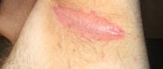
Scars
The medical classification knows more than one type of scar. When a wound heals by primary intention, any scar can actually form. It doesn't all depend on how the fabrics are tightened. The type of scar is determined by the prerequisites for the appearance of the wound itself. Let's say a surgical operation. The man transferred it, and the cut made with a scalpel was stitched up. This is primary healing, since the tissues are in close contact and there are no infections. But it will still be called a surgical scar.
Another situation. A man was cutting tomatoes with a sharp knife and accidentally hit his finger with the blade. A domestic accident, one might say. But the type of healing is still the same, primary. However, it will be called an accident scar.
There are also keloid, normotrophic, atrophic and hypertrophic scars. However, they are not relevant to the topic. It is enough just to know about these types of scars.
Lump on the bottom of the leg near the toes
A foot lump can appear anywhere - near the toes, on the heel, near the arch or on the plantar fascia. It is also common for bunions to appear on the top or bottom of the leg. Perhaps the bump near the toes is just a corn (a rough, dry callus). In other cases, regardless of where the formation appears, you need to show it to the doctor as soon as possible.
Ask a dermatologist or trauma specialist to take a look at the lump to determine what the problem may be. Your doctor will then help you get rid of it and prevent it from recurring. Having a bump on your feet near your toes is likely to be caused by the following conditions:
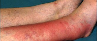
Diabetic foot syndrome
At the same time, trophic ulcers are diagnosed in almost 15 percent of patients with diabetes.
These formations are caused by neuropathic and vascular complications of the disease. Nerve damage that causes changes or complete loss of sensation in the leg is called peripheral neuropathy. With peripheral neuropathy, the effects from cuts, bruises or foot injuries go unnoticed. Thus, diabetic patients do not immediately become aware of any foot injury. Vascular disease is a serious problem in people with diabetes, which results in a lack of healthy blood flow and, as a result, ulcerations. Patients with diabetes also have problems with wound healing.
Bunion of the big toe
Bunion
This is a bony and very painful bump on the first joint of the big toe that bends when you walk. The problem is also popularly called “underwire” and usually affects women as they age. It is considered to be a family disease, as well as one that is caused by weakness of the foot.
The bunions will have the following signs and symptoms:
- The thumb protrudes towards the other fingers
- The appearance of swollen bone on the first joint of the thumb
- Severe pain and swelling of the entire thumb
- Inflamed skin on the bump
- Change and deformation of the normal shape of the foot
- Tough, calloused, and red skin that results from the big toe overlapping the second toe
Taylor deformity
Taylor's deformity
This pathology is also called varus deformity of the fifth toe. It resembles the previous problem with the thumb, but in this case it is the bone of the little finger that is suffering. In this case, a bump forms on the outside of the foot.
Causes also include genetic predisposition, foot problems and wearing unsuitable shoes. In addition to a number of methods, surgery is used to relieve pain and condition, which in 85% of cases is successful and Taylor’s deformity does not return.
Hammer fingers
Hammer fingers
This is a painful deformity of the second and third toes in which they bend at the ends, acquiring a hammer forum. The main causes of these deformities are injuries, wearing poor shoes, hallux valgus and rheumatoid arthritis. If left untreated, this condition can lead to calluses as a result of constant irritation between the feet and shoes.
A crooked toe can also cause tension in the ball of the foot, leading to pain known as metatarsalgia. This is pain in the metatarsal part of the foot.
Cystic inclusions
Epidermal cysts
Also known as epidermoid or epidermal cysts, they can appear anywhere under the skin and often form as a result of mild injuries such as cuts and scrapes. Cystic inclusions between the toes are usually small, ranging from a few millimeters, although sometimes they grow up to 5 cm.
Mycosis of the foot
Fungus
This is a fungal infection of the skin, also called athlete's foot. The condition can lead to severe itching, cracking of the skin, and painful blistering or peeling of the skin on the fingers. If the fungus is not treated, it can spread to other parts of the finger. The condition can also cause thickening and yellowing of the nail if not treated promptly.
Athlete's foot causes the skin to become itchy, red, scaly and dry. It may also cause pain and a small, tender blister on the legs. The main reasons for the development of the disease:
- Failure to maintain proper hygiene
- Shoes that lead to severe overheating and sweating of the feet
- Sharing personal hygiene items with sick people
- Staying barefoot in wet public areas (showers, swimming pools, saunas)
- Other medical conditions such as diabetes
Primary healing
We need to talk about it first. After all, types of wound healing begin with the primary. Next comes the secondary. The last type is healing under a scab.
Initially, the wound heals when its edges are smooth, touch as closely as possible and are viable. Healing will occur successfully if there are no hemorrhages or cavities inside, and there are no foreign bodies. Therefore, it is important to wash the wound. This also helps neutralize infections.
This type of healing is observed after aseptic operations and full surgical treatment of the injury. This stage passes quickly - in about 5-8 days.
Little bumps
Lumps on the feet are very common. They are harmless and most can go away on their own without any treatment. In addition to the reasons listed above, small bumps on the bottom of the foot can be a simple callus caused by friction and irritation of the feet from shoes. The lump can also sometimes be caused by frostbite.
These bumps are painful, itchy swelling on the skin caused by poor blood circulation, especially when exposed to cold. When you have frostbite, you will feel a burning, itching, and swelling around the affected area.
Diagnosis
The following specialists deal with dense neoplasms on the legs:
- orthopedist;
- surgeon;
- oncologist;
- traumatologist.
At the initial stage of diagnosis, the patient is sent for an x-ray. This will allow you to determine the degree of deformation of the lower limb using an image.
To identify the full picture, the patient is sent for an ultrasound examination of the joint, as well as blood tests. If a malignant neoplasm is suspected, a biopsy is used (removal of part of the lump tissue using a special medical needle). In a complex case, the patient is sent for magnetic resonance imaging (MRI). MRI allows you to identify the exact location of the compaction, the condition of nearby tissues, concomitant pathologies (the presence of a cyst, necrosis, arthritis, osteoarthritis, inflammation of the periosteum).
Red cones
A lump on the bottom of your legs may be red, white, or yellow depending on what is causing it. Red bumps on the leg can be caused by the following factors:
Calluses and corns
The Mayo Clinic defines corns and calluses as a thick, rough layer of skin that develops as the feet's defense mechanism against friction and pressure. They often appear on the feet, toes and hands.
The main causes of calluses:
- Wearing tight-fitting shoes for long periods of time
- High heels
- Wearing tight stockings and socks
- Deformed fingers
Plantar warts
These are hard, granular growths that often appear on the heels and balls of the feet. Plantar warts are caused by the human papillomavirus, and pressure placed on the feet can cause the warts to grow inward under the hard, thick layer of skin.
Dropsy
Dropsy appears as a small red bubble on the skin filled with blood serum. These blisters are caused by friction, burns, and other damage to the outer layer of skin. Dropsy can be easily prevented. They can also go away on their own without any medication, but it is important to consult your doctor about a blister that keeps recurring.
Consolidation after fibroadenoma
Good afternoon! In February, a fibroadenoma was removed from the breast, everything went well, according to histology it was a normal fibroadenoma. In May I had a check-up ultrasound with my mammologist, in the place where there was a fibroadenoma - it remained like a lump. he said that these were internal scars and prescribed Wobenzym and Aevit. It’s now November, the compaction remains, it’s become a little smaller, it feels like a single canvas or something, not like a ball, not like a formation. I felt uneasy, I went for an ultrasound a week ago - the ultrasound specialist did not see anything wrong, there were small ductal cysts and performed an FCM. This also wasn’t enough for me and I went to my mammologist-oncologist, who operated. I was already thinking about taking a puncture from the “seal” that remained from the fibroadenoma. He looked at the ultrasound again and said that yes, these are still scars and on the ultrasound they do not show up as something bad, etc., and that there is nothing even to take a puncture from. He also confirmed the FCM and prescribed Mamoclam and come back in 3 months and see how the drug worked. I trust my doctor, but I am very worried that it will soon be a year, and the scars remain. my question is, can an ultrasound and mammologist-oncologist make a mistake and not see something bad? I wanted to take a puncture, but since the doctor himself says that it cannot be taken from scars. Now I touch this lump every day and it seems to me that it has even become red and even a little painful, probably due to the constant pulping? Also a question - as long as I can remember, my ultrasound always shows that the lymph nodes are enlarged, but the ultrasound specialist says that everything is within normal limits and the structure, etc., is all as it should be. sometimes they kind of hurt, I feel them or something. How critical is this, etc.? Thank you!
On the Ask a Doctor service, you can consult a mammologist on any problem that concerns you. Expert doctors provide consultations around the clock and free of charge. Ask your question and get an answer immediately!
If you have a similar or similar question, but you have not found the answer to it, get your 03 online consultation from an expert doctor.
If you want to get a more detailed consultation with a doctor and solve the problem quickly and individually, ask a paid question in a private personal message. Be healthy!
Cancer
Skin Cancer
Skin is the most common form of this disease. It can occur on any part of the body. Just like in other areas, it will have the same features on its legs. These can be slow-growing and often painless bumps on the surface of the skin that, if left untreated, will likely crack, bleed, or ulcerate.
It is believed that leg cancer can be caused by overexposure to UV rays, harmful chemicals, chronic inflammation or irritation, and can also occur in people who are genetically predisposed to it. During most medical examinations, people tend to ignore their feet, but this is not true. It is important to have a healthcare professional check your feet regularly for any abnormalities.
When the underlying cause of a lump on the lower leg is cancer, you are likely to experience the following symptoms:
- White bumps or patches on the skin that may ooze fluid or crust over
- The development of the lump begins as a severe callus-like lesion
- Hard and painless lump
- In people with malignant melanoma, the lump begins as a small, brown or black spot
- Bumps that look like nevi (birthmarks)
- The borders of the bump may appear jagged, jagged, or ragged
- Size greater than 6mm in diameter
Bone cancer
In addition to skin lesions, subcutaneous lumps in more rare cases can cause cancer of connective bone or soft tissue (sarcoma). This disease is less common but can affect people of any age. The most common symptom is soreness, but other signs can vary greatly, including local manifestations such as lumps, swelling, as well as general symptoms such as malaise, weight loss, etc.
Once it is confirmed that you do have cancer, treatment will depend on its stage and type. The most commonly used treatments include radiation therapy, chemotherapy and surgery.
Secondary healing
It can be observed when one of the conditions for the primary one is missing. For example, if the edges of the fabric are not viable. Or they don’t fit closely together. Cachexia (depletion of the body) and a lack of necessary substances in the body can contribute to secondary healing. And this type of tissue restoration is accompanied by suppuration and the appearance of granulations. What it is? Such newly formed glomeruli of blood vessels are called granulation. In fact, this is familiar to every person since childhood, because each of us fell and tore our knees. Everyone remembers that the wounds were then covered with a crust. This is granulation tissue.
In general, types of wound healing and their characteristics are a very interesting topic. Not everyone knows that the tissue repair process occurs in three stages. First, the inflammatory phase of healing takes place (about 7 days), then the granulation phase (7-28 days). The last stage is epithelization. That is, the wound is covered with new, living skin.
Drug treatment
After extensive surgical interventions, the woman remains in the medical hospital for 5-7 days. To prevent complications, she is prescribed the following therapeutic measures and medications:
- Antibacterial agents – broad-spectrum drugs are used that are active against most pathogenic or conditionally pathogenic bacteria. An antibiotic may be contraindicated if you have allergies.
- Non-steroidal anti-inflammatory drugs - drugs are necessary to reduce the severity of the inflammatory reaction in damaged tissues. This helps reduce pain and discomfort. The use of drugs for a period of time exceeding 5 days is prohibited.
- Diuretics - medications are prescribed to reduce the severity of soft tissue edema caused by the release of blood plasma into the intercellular substance against the background of increased permeability of the walls of microcirculatory vessels.
- Multivitamin preparations are prescribed to improve the course of metabolic processes, as well as accelerate tissue healing.
What medications and how much to take are decided by the attending physician individually. In a hospital setting, dressings are required after large incisions are made. They involve treatment with antiseptics (can be washed with a solution or smeared with ointment), which are agents that destroy any microorganisms. Then an aseptic dressing is applied.
The duration of the early postoperative period after removal of breast fibroadenoma coincides with the woman’s stay in the hospital department.
Preventive measures
To prevent an annoying and painful nodule from appearing on your body, you must adhere to a number of recommendations:
- selection of comfortable, proportionate shoes made from high-quality and natural materials;
- select shoes with the most comfortable instep; too high or low a heel negatively affects the health of the foot;
- for increased comfort, you can use orthopedic insoles that support the foot in the correct position;
- will cope with rubbing, and silicone inserts will help reduce impacts on the foot;
- daily hygiene procedures, which include washing feet, baths, removing rough skin with a stone;
- At night, lubricate your feet with a rich cream and put on cotton socks;
- cold and hot shower.
Experts say that moderate physical activity has a beneficial effect on the condition of the legs. It is useful to take walks, ride a bike, jog, ski and skate. Performing special gymnastic exercises. Self-massage has a beneficial effect on the condition of the feet and helps improve blood circulation.
It is very useful to walk barefoot on the grass, especially in the morning, when the grass is still covered in dew. Any injuries, microcracks and cuts must be carefully treated with antiseptics. After all, this is a direct path for infections to enter the body. If, however, a lump appears on the leg, it is necessary not to delay and begin treatment. Constant monitoring of your health will help you cope with many problems.
Neglecting bandages
If you think it's a good idea to let your skin breathe after initially treating any microtrauma, think again. By not covering the wound with a bandage, you expose the skin to infection. New cells must migrate to the appropriate areas for the wound to heal faster. By covering it with a bandage, you make this process easier and faster. The best way to help prevent infection from developing is to apply an ointment to the wound, which you should always have in your home. If you haven't visited a doctor, then at least keep Vaseline in your medicine cabinet. As you know, it prevents the wound from drying out and scabs forming, and accordingly, it heals faster.
Dietary recommendations
Nutrition for breast fibroadenoma affects the rate of tissue healing. Dietary recommendations should be followed for at least 2 weeks after surgery.
- The predominance of plant foods, dietary meats (rabbit, chicken, beef), fish, and dairy products in the menu. Thanks to vegetables and fruits, it is possible to ensure the supply of a sufficient amount of vitamins, fiber, and mineral salts.
- Excluding fatty fried foods, spices, marinades, smoked meats, and pickles from the diet, which increase the load on the digestive system and contribute to the development of putrefactive processes, which ultimately negatively affects the process of tissue regeneration and rehabilitation in general.
- For 10 days after surgery, avoid drinking alcohol, which is a vascular toxin - due to the effect of the compound, the wound will take longer to heal.
- Dishes should not be too cold or hot; it is recommended to eat chopped food.
- For better absorption of nutritional compounds, it is better to eat small portions, but often, at least 5 times a day.
- The last meal is allowed no later than 2 hours before expected sleep.
Corns
A very common disease that is accompanied by painful sensations. Corns occur as a result of keratinization of dead skin and its hardening. Reasons: incorrect selection of shoes, which causes the foot to feel discomfort, excessive sweating of the feet, flat feet, excess weight, and so on.
In appearance, they can be flat or concave, with a yellow-brown stem. This area of skin is almost completely devoid of sensitivity. If the skin is excessively dry, cracks may occur. The lack of drug therapy can trigger the process of foot deformation.
Postoperative period
Removal of an adenoma is an invasive intervention, which is carried out using several techniques:
- sectoral resection;
- enucleation;
- laser removal, for which puncture is performed.
The need to remove the formation, including mastopathy, is due to the fact that the disease may very rarely recur in the future. Each technique has its own level of trauma, which requires a certain amount of pain relief. Before the operation, the woman undergoes preparation, the doctor makes sure that there are no contraindications. Regardless of the chosen operation, further tissue restoration depends on the implementation of rehabilitation measures, which include:
- drug treatment;
- general recommendations with limitations;
- diet;
- wearing special underwear.
Before performing surgery, the doctor must inform the woman about the peculiarities regarding recovery after removal of breast fibroadenoma. The collected tumor is sent for histology. In the laboratory, a histological examination is carried out under a microscope to evaluate the tissue structure of the formation.