White rashes on the skin of the eyes - description
Black spots that appear before the eye are otherwise called floaters and are considered a harmless phenomenon. However, sometimes they can become a manifestation of some kind of malfunction in the body.
Most often, such dots move and fly when the eyes move, and when they stop, they smoothly return back.
Optical interference can occur in the form of filamentous formations. “Worms” and “threads” flashing before the eyes can overlap each other and form a “web”. This phenomenon indicates destruction in the vitreous body.
When, during examination, granular formations are visible in the form of dots, rings, circles or spots with blurred outlines, penetration of hyalocytes into the vitreous body can be suspected. Most often, these opacities are formed as a result of surges in blood pressure or damage to the skull.
At first, flying dots cause only discomfort, but over time they can cause concern for the functioning of the visual apparatus.
Thus, the appearance of black spots in the field of vision is a consequence of vitreous dissection or some other eye pathology. If there is a large accumulation of dots, you cannot postpone a visit to the ophthalmologist: they can become a manifestation of retinal detachment and indicate an imminent loss of vision.
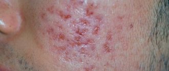
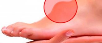
As already mentioned, usually white spots that appear on the eyelids are milia. This is not an infectious pathology and it is not contagious to others. Most often, millet lesions occur in the fair sex.
They do not cause inconvenience or discomfort, and their location is the eyelids. Compared to papules, milia are a little more difficult to remove and effective therapy requires understanding the specifics of this formation.
Black spots in the eyes can appear in the form of both single and multiple (filamentous) formations. In some cases, flies are not noticeable at all and do not cause discomfort, while others seriously interfere with a person’s life, turning into full-fledged lines. Dots in the eyes can be a symptom of more serious diseases.
Black dots before the eyes are divided into two types:
- Granular destruction. Hyalocytes (dead eye cells) enter the vitreous body, which die over time, join together and form black dots.
- Filamentous destruction. The development of pathologies and metabolic disorders lead to the death of some collagen fibers. A person begins to see strings or entire webs in front of him, which significantly interfere with object recognition.
The main feature of the disease is that when the head is sharply moved to the side, black lines or dots move in the same direction, creating a kind of trail. Pathology occurs after suffering from serious diseases affecting the eyes, when using medications with aggressive substances, or after overcoming 50 years of age. Those who are diagnosed with myopia are also at risk; over the years, changes in vision only contribute to the occurrence of defects.
Localization of white pimples on the eyes
Millet is most often located on the upper and lower eyelids, but can appear anywhere there is hair. It looks like a small cyst filled with epithelial cells and sebum. The source of formation is the excretory ducts of the sweat glands or hair follicles.
It appears as single or multiple nodular rashes on the eyelids of a white-yellow color. The elements of the rash are clearly demarcated from healthy skin, without an inflammatory halo. They feel dense to the touch; in the center there may be a black comedon - an accumulation of horny masses. They resemble a grain of millet in appearance, which influenced the name.
The nodules reach a size of 1-2 mm, often located symmetrically. Millet on the skin of the upper or lower eyelid does not cause pain, only aesthetic discomfort. Pronounced rashes are a cosmetic defect.
In the area of the inner edge along the lash line it can cause discomfort. Convex nodules come into contact with the mucous membrane when blinking or moving the eyeballs, causing the sensation of a foreign object in the eye, lacrimation, and burning.
On the lower eyelid, millet manifests itself as chaotic multiple rashes, tending to be located at the outer corner of the eye, spreading to the cheeks and cheekbones.
Milia do not appear on the mucous surface. If a white nodule or inflammation occurs on the inner surface of the eyelids, you should immediately contact an ophthalmologist for diagnosis and treatment.
Possible reasons
The main reason for the appearance of black dots on the eye is the destruction of the vitreous body. It occurs due to problems with the lens, insufficient blood circulation, which contributes to the destruction of collagen fibers with age and in serious diseases affecting the eyeballs. Fiber fragments become concentrated at one or more points in the lens, obstructing the passage of light, creating spots. Bad habits stimulate the manifestation of this pathology. Reasons why black spots appear in the eyes:
- crystalline formations;
- the appearance of malignant tumors in the eye;
- diabetic retinopathy;
- exposure to aggressive substances (acids, alkalis, etc.);
- entry of foreign substances and dirt particles into the eye;
- occurrence of injuries;
- migraine;
- long exposure to bright light;
- depletion of visual organs;
- other damage to the eye shell.
If black lines fly before your eyes, this may be a consequence of more serious pathologies. Such problems are not always immediately noticeable, but due to the fragility of the blood vessels they progress quickly. Peripheral vision deteriorates, areas of clouding in the visual organs grow, sparks and flashes may occur. It is important to see a doctor immediately and surgery may be necessary. Such symptoms may be the result of diseases:
- fungal or viral infections;
- hemophthalmos (bleeding into the vitreous body);
- vascular pathologies;
- tumors;
- retinal detachment or other damage;
- vitreous detachment.
To independently identify vision problems, look at a white or light-colored surface so that it occupies 100% of the visual area. If, in addition to the white color, there are defects, spots, or the appearance of some particles flying before your eyes, consult a doctor. You may not have a pathology, but it’s worth being safe. The doctor will make a diagnosis and prescribe treatment.
This symptom most often occurs as a result of aging of the body and worries people who have crossed the 50-year mark. The cause may be atrophy of the eyeball. If black spots in the eyes cause significant discomfort, you should visit an ophthalmologist: they can become a sign of retinal detachment.
Recently, ophthalmologists have noticed that the symptom has become “younger.” Young people are increasingly complaining about spots flashing before their eyes. This is due, first of all, to long-term exposure to electronic devices and gadgets on the visual system.
Other causes of blackheads include:
- Impaired blood circulation in the vessels. Intense physical activity, drinking alcoholic beverages, smoking, and frequent surges in blood pressure can lead to poor blood flow and damage to blood vessels. Bursted blood vessels often cause blood to thicken, which leads to dots flashing before the eyes.
- Lack of nutrients (especially vitamin A), metabolic disorders. Vitamin deficiency negatively affects all internal organs and is often the cause of black flies flying in the field of vision.
- Sometimes spots before the eyes can become a manifestation of cervical osteochondrosis and arise due to insufficient blood supply to the brain.
- Burns and traumatic damage to the organs of vision can lead not only to the death of vitreous cells, but also to retinal detachment, as well as loss of vision. Visual interference is also often observed when foreign objects enter the eyes.
- Black flies can be a result of exposure to pathogens that cause eye pathologies. This explains the presence of cloudiness before the eyes in some ophthalmological diseases.
- The appearance of black spots may be associated with overstrain of the visual system, while dots, circles and lines are seen by those people who constantly spend a lot of time at the computer or work with small details.
- So-called white spots before the eyes, as well as flashes of light, can be a sign of dangerous diseases of the visual system. These manifestations most often indicate pathologies of the lens, cornea or retina.
- Another reason is intoxication. When toxic substances enter the body, the patient has dots, spots or curved lines floating before his vision.
We invite you to familiarize yourself with Traumeel C tablets: description of composition, instructions for use, dosage, indications and possible side effects, price, analogues and reviews
Also, provocateurs of an optical defect can be:
- migraine;
- diabetic retinopathy;
- tumors of the eye tissue;
- stressful situations;
- retinal disinsertion;
- vitreous hemorrhages;
- penetration of viruses and fungi into the retina;
- vitreous detachment;
- anemia;
- hemophthalmos;
- helminthiasis;
- diabetes;
- uveitis
Pigment spots on the iris of the eye
Vision is an indispensable ability to communicate with the outside world.
Therefore, we must protect the organ responsible for it, reacting sensitively to changes in its well-being or appearance. The eye is an extension of the nervous system and contains many nerve endings, blood vessels and tissues connected to other organs of the body.
Sometimes spots and dots appear on the iris of the eye.
Dot on the iris of the eye
Photo 1: The science of iridology believes that the eye is a miniature screen on which you can see the state of all organs of the body. Small dark spots are indicators of toxins found in the tissues of various organs.
Detoxification measures and a moderate regime of work and rest can remove toxic substances from the body, then the spots will become less noticeable or disappear completely.
To avoid mistakes in diagnosis, you need to consult a specialist, because spots on the iris can be caused by other reasons.
Reasons for the appearance of a dark dot on the iris of the eye
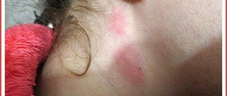
Dark red spots - small hematomas on the iris - are a sign of sudden changes in pressure.
They cannot be treated and may soon go away on their own, but their appearance is a reason to monitor your blood pressure.
Microtraumas that occur when pressure increases during childbirth have similar manifestations. This situation is usually temporary. After some time, the spots disappear on their own.
Increased eye pressure also leads to the appearance of dark spots on the iris. If spots appear, you should consult a doctor. The ophthalmologist will make a diagnosis and prescribe the right treatment.
Floating dots on the membrane of the eyeball are a sign of incipient retinal detachment. Sometimes they feel like discomfort from a speck of debris in the eye. The diagnosis, in this case, will be made by an ophthalmologist. If retinal detachment is confirmed, you will have to resort to laser correction.
A nevus is a benign pigmented neoplasm in the eye, a relative of a freckle or mole. A nevus can appear at any age. Most often observed in fair-skinned people. The point can be flat or convex and measure 1-2 mm in diameter. The danger of a nevus is that it can deform the pupil.
Why do dark spots appear?
Factors contributing to their formation: accumulation of melanin, changes in hormonal levels (for example, in adolescents and pregnant women, women during menopause and taking hormonal contraceptives).
Stress and infections can cause excess pigmentation.
Nevi may remain unchanged or progress.
Those that do not change their size are benign in nature. Progressive ones can impair vision and develop into a malignant form.
Attention! The following situations require immediate consultation with an ophthalmologist: a sudden and rapidly growing nevus, a sharp change in its color, blurred vision, discomfort in the eye
Reasons for the appearance of a white dot on the iris of the eye
White spots on the eye are associated with changes occurring in the lens and may indicate leukoma and cataracts.
If the problem is in the cornea, it is leukoma; if the lens becomes cloudy, it is a cataract.
Leukoma appears as a result of inflammatory processes, injuries, and unsuccessful surgical interventions. Leukoma is also called a thorn. It can be congenital or acquired.
Today, laser technologies and surgery are used to treat cataracts.
It is important! Chemical burns also lead to the appearance of white spots, often leading to significant impairment of vision. Cataract – clouding of the lens
Cataract is clouding of the lens.
It may be complete or appear as white dots. The disease can be congenital or acquired. Most often it occurs due to age-related lens degeneration.
Treatment is aimed at normalizing metabolic processes and improving tissue nutrition.
What measures need to be taken
Prevention is aimed at strengthening eye tissue. Vitamin A, blueberry extract, eye exercises, and elimination of excessive stress are useful for the retina.
To understand whether a dark spot is dangerous, you need to determine how long ago it appeared. Old close-up photographs are a good help for determining the timing of the appearance of dots.
If a spot appeared on the eye in childhood, it may be an ordinary mole; if recently, it may be a sign of a dangerous disease.
Homeopathic treatment of points on the iris of the eye
Important! With the help of homeopathic medicines you can get rid of cosmetic defects and preserve your vision. Please note! Homeopathic treatment clears the skin and mucous membranes of cysts, growths and spots. To achieve a long-term positive effect, it is necessary to take medications prescribed by a doctor for one to one and a half months.
A white pimple appears on the eyelid - what is it?
The shape of the millet resembles small inclusions, which are located on delicate areas of the skin - near the eyes, eyelids, on the lips. The structure of the formations is compacted, and their color is white.
Despite the fact that they are outwardly quite harmless, in fact, millet grasses are not so safe for human health and life. Milia are formed as a result of impaired excretory function of the sebaceous glands of the skin, as well as due to clogging of pores with keratinized particles of the epidermis.
Interesting fact:
When the sebaceous glands function normally, excess fat comes out; when the process is disrupted, the follicles are enveloped in sebaceous fat and whiteheads form.
In fact, there are many different reasons why grasses form, but most often it is:
- hormonal disorders;
- disorders and pathologies of the nervous system;
- reaction of the skin and small glands to hormonal changes;
- diseases of the gastrointestinal tract;
- poor nutrition and consumption of unhealthy foods - fast food, chips, sugary drinks, etc.;
- lack of vitamins and other useful elements;
- hereditary factor;
- poorly selected care products or insufficient hygiene.
With the correct determination of the main root cause, we can talk about effective therapy and results. To accurately find the provoking factor, it is recommended to seek help from a specialist - a cosmetologist or dermatologist.
After an accurate diagnosis and a series of studies, treatment is prescribed, which is completely individual, depending on the current situation.
The main reason for the appearance of white dots under the eyes is a violation of the mechanism of exfoliation of dead skin particles. When this process fails, they accumulate on the surface of the skin, clogging the ducts of the sebaceous glands, which become clogged, and milia appear. Also, the cause may be excessive activity of the sebaceous glands - owners of excessively oily skin are well aware of this, when excess sebum does not have time to be eliminated.
Other factors that cause millet grains are disturbances in the gastrointestinal tract. Often milia are a consequence of hormonal changes in the body - for example, during puberty, pregnancy or menopause. Another version of the appearance of white spots on the skin around the eyes is exposure to ultraviolet radiation.
Milia can occur after a chemical peeling procedure, when using certain steroid drugs, as well as with frequent use of “heavy” cosmetics, in particular professional ones, which clog the pores of the skin on the face, and the process of peeling off the keratinized scales is much more difficult. In addition, doctors name allergic reactions and a lack of vitamin A in the body among the causes.
Millet is often observed in newborns in approximately 40-50% of cases. In this case, white dots can appear not only on the face, but throughout the body. In infants they disappear in the first 1-3 months of life. The cause is usually a blockage of the sebaceous glands; the child’s body adapts to a new, unusual environment for him.
Causes
Millet can be primary or secondary, depending on the etiological factors.
Primary occurs on the skin of the eyelids in people with a hereditary predisposition. Secondary develops against the background of a disease or injury - accidental household or during cosmetic procedures.
Possible reasons for the appearance of millet:
- autoimmune diseases;
- metabolic disorders;
- incorrectly selected eyelid cosmetics;
- disorders of the gastrointestinal tract;
- endocrine system disorders;
- damage to the eyelids;
- seasonal hypovitaminosis;
- oily skin type;
- burns or excessive sun exposure.
The appearance of millet, whiteheads on the skin of the lower or upper eyelid depends on the functioning of the sebaceous glands. They are most affected by the endocrine system and the effects of cosmetics.
When to see a doctor
The temporary occurrence of an optical defect is not a warning sign and does not require a visit to a medical facility. However, in some situations a visit to the doctor should become mandatory:
- if the number of black dots or transparent lines has increased over time;
- if the phenomenon occurs together with flashes and glare in the field of view;
- if this symptom occurs after injury to the skull or visual apparatus;
- if flashing flies cause severe discomfort and interfere with normal vision;
- if the appearance of spots is accompanied by a decrease in vision.
Diagnostics
If the opacities are persistent, you should visit a doctor and talk in detail about the symptoms that appear and the duration of their course. The specialist will conduct a visual examination of the visual apparatus and, if necessary, prescribe additional diagnostic methods:
- to establish the cause of black spots, the ophthalmologist will need to examine the fundus, the functioning of blood vessels and the retina using an ophthalmoscope or a slit lamp;
- Examination with side lighting will help identify small changes in the anterior part of the eye. Transmitted light will detect clouding of the lens;
- if a stroke is suspected, as well as in case of eye damage, a CT or MRI of the head is used;
- using optical coherence tomography, structural disorders of the retina can be detected;
- Ultrasound of the visual organs is used to study the structure of the eye, the condition of the retina and lens.
The specialist can also measure the patient’s intraocular and blood pressure and refer for a general and biochemical blood test. If you suspect a fungus or eye infection, you will need to take a smear for bacterial microflora, demodicosis, and also undergo allergy tests.
Treatment methods
Usually, acne on the eyelid disappears immediately after correcting a person’s diet, as well as after changing care and decorative cosmetics. In addition, it is necessary to enrich the body with missing vitamins and minerals, which can be done through a vitamin complex. With this simple treatment, the grasses disappear within a maximum of two weeks.
But there are also more severe situations in which hardware therapy is necessary. During this treatment, small white pimples on the eyelid are removed mechanically. However, such treatment is prescribed exclusively by a doctor and only after a thorough diagnosis of the disease. There are several options for hardware therapy, which are selected depending on the situation.
Electrocoagulation
Thanks to the influence of high-frequency current, the tumor is excised. In general, this procedure is absolutely painless and is carried out within a few minutes. After the procedure, a small scar may remain, which disappears within two days. Electrocoagulation is a modern and effective method that will allow you to permanently get rid of a pimple on the eyelid.
We invite you to familiarize yourself with the amazing effect of black cumin oil for the face
Laser correction
In fact, this procedure is similar to electrocoagulation, but the cleansing of the dermis is more intense. At the same time, laser correction is less painful and traumatic. After the procedure, there are no traces or scars left on the epidermis.
Curettage
It is the most radical method of removing milia. During the procedure, the millet is carefully opened and the white contents are extracted using a special instrument - a curette.
How to use boric acid for acne? Read more
This option is the most traumatic, after which scars and red spots appear on the skin. Curettage is rarely used to remove white spots on the eyelids. More often it is used to eliminate acne, pimples and comedones on the face.
In addition to drug therapy, you can use folk recipes, which are quite popular in the treatment of millet. Due to its accessibility and good effectiveness, this method stands alongside medication.
The most effective and time-tested folk recipe is an oatmeal mask with viburnum juice.
Directions for preparation and use:
- Squeeze out the viburnum juice and mix it with pre-chopped oatmeal. Oatmeal must be ground to flour. The required proportion is 1:3.
- The resulting homogeneous mass is applied to the most affected areas and left for 30-40 minutes.
- Wash off carefully with warm water.
What pharmaceutical remedies for expression wrinkles are effective?
Finally, it is also recommended to apply viburnum solution to the skin. The procedure can be performed no more than 2-3 times a week. Using this recipe, you can reduce inflammation, if any, and avoid relapses.
Also, if a white dot appears on the eyelid, you can use freshly squeezed lemon juice. This will allow you to achieve excellent disinfection, dry the formations, and also lighten them, making them more invisible. To do this, you need to dilute lemon juice with water in a ratio of 1:3 and apply it pointwise to the affected areas. It is better to do the procedure before bed.
You can also use boric or tar soap as an additional therapy. Tar soap for acne, rashes and blemishes is an effective and inexpensive remedy. But you should be careful not to dry out the already delicate skin of the eyelids.
Traditional recipes will not help to completely get rid of the problem. But the white dots under the lower or upper eyelid will noticeably decrease, and the condition of the skin will improve significantly, especially with complex treatment.
By following certain rules of facial skin care, as well as adhering to all tips and recommendations, you can avoid the occurrence of milia.
Millet rashes are rashes on the epidermis that are of a rather complex nature. In this regard, therapy should only be carried out under the supervision of an experienced dermatologist. Self-medication in this case can be quite dangerous and can lead to aggravation of the clinical picture. As a result, quite dangerous pathogenic processes may even arise. Therefore, take care of yourself, your health and consult with specialists on time.
Treatment
You should also remember to adhere to a special diet. The diet should not contain refined cereals, white bread, coffee, over-salted dishes, or foods containing starch. The diet should include seafood, any leafy vegetables, fish, citrus fruits, nuts, etc.
To treat melanosis, which was caused by an inflammatory process, you can use folk remedies.
You need to put 2 tablespoons of cornflower inflorescences in a mug, then pour boiling water over them and leave to steep for 3 hours. The cooled and infused decoction is filtered through cheesecloth and used as an eye lotion. This procedure should be performed daily for 5-7 days.
Therapeutic measures depend on the underlying cause.
Eye injury
Primary treatment, instillation of antibacterial, analgesic, hemostatic and absorbable drugs.
In the case of a penetrating wound, emergency surgery is performed; in the future, reconstructive ophthalmological operations are possible.
During the rehabilitation period, vitamin complexes, absorbent agents, and physical therapy are prescribed.
Treatment is predominantly surgical and includes cryopexy (freezing) at the site of injury, laser photocoagulation, removal of the vitreous (vitrectomy), sclerotherapy and pneumatic retinopexy in combination with cryopexy, photocoagulation or laser treatment.
Additionally prescribed medications:
- Emoxipin;
- Taurine;
- Taufon;
- Ophthalm-Katachrome;
- Quinax;
- Emoxy-Optic;
- Papaverine;
- B vitamins;
- Acetylsalicylic acid;
- Pentoxifylline, etc.
The basis of therapy is normalization of blood glucose levels and lifestyle correction.
Drugs for diabetic retinopathy:
- angioprotectors;
- vitamins;
- corticosteroids;
- biological peptides.
Corticosteroids for diabetic retinopathy slow down processes associated with inflammation:
- edema;
- fibrin deposition;
- collagen deposition;
- expansion of capillaries;
- migration of leukocytes and fibroblasts.
Representative – Triamcinolone (synthetic glucocorticosteroid). In combination with laser therapy, the treatment effect is higher.
Surgical interventions:
- Photocagulation;
- Vitrectomy;
- Cryotherapy.
Proper nutrition and physical activity help maintain optimal weight, which further helps control diabetes and its complications.
At the initial stage, specific therapy is not required, but it is important to eliminate/minimize risk factors that contribute to the progression of the pathology. Some experts consider the use of physiotherapy justified: ultrasound, phono- and electrophoresis, hyperbaric oxygenation
Some experts consider the use of physiotherapy justified: ultrasound, phono- and electrophoresis, hyperbaric oxygenation.
Along with drug therapy, laser coagulation of the retina is performed, which is the standard in the treatment of age-related macular degeneration.
Elimination of the main provoking factor – high blood pressure.
Behavior correction: proper nutrition, physical activity, taking antihypertensive drugs.
Medicines:
- vasodilators;
- anticoagulants;
- angioprotectors;
- neoangiogenesis inhibitors;
- anti-sclerotic drugs;
- multivitamins, etc.
In the advanced stage, when hypoxia impairs vision, laser coagulation is performed.
Additionally, multivitamins, immunomodulators, etc. are prescribed.
Mishina Victoria, doctor, medical columnist
The course of the therapeutic course is determined after diagnosis and varies significantly.
Pinguecula is a benign formation that does not need to be treated. To eliminate unpleasant symptoms (such as dry eyes, redness and general discomfort), the ophthalmologist prescribes moisturizing drops or mild steroid medications.
Red spots on the eyeball caused by infectious processes are treated with medication. In such cases, the doctor prescribes antibiotics.
DETAILS: Feces for stomach ulcers - Treatment of gastritis
For leukoma, laser removal or keratoplasty will come to the rescue. Implantation of donor cornea is also common today.
A conjunctival cyst can be treated with conservative methods (moisturizing, antibacterial, anti-inflammatory drops) or surgically. In the latter case, they resort to traditional surgery or use a laser.
A yellowish spot on the eyeball is not always a reason to remove it. Surgery is required when:
- There is discomfort when blinking or moving the eyeball.
- Vision decreases.
- The outflow of tears decreases.
- Pronounced cosmetic defect.
- There is a threat of malignant growth.
Preventive measures
To avoid unpleasant symptoms, all people without exception are recommended to:
- lead a healthy lifestyle;
- eat foods rich in vitamins and minerals;
- systematically engage in sports;
- get enough sleep;
- stop smoking and drinking alcohol;
- use sunglasses;
- protect the visual apparatus from injury;
- for women, wash off makeup before going to bed;
- systematically contact an ophthalmologist;
- Don’t forget about eye exercises.
The appearance of blurred vision in the form of black dots, white lines or circles is a common visual defect that may disappear on its own. However, if the symptom bothers you regularly and is accompanied by severe discomfort, consultation with a specialist is required.
Treating milia at home
Millet grass can be removed at home, but to do this you need to be extremely careful and have the appropriate skills.
To remove milia yourself, you must follow certain recommendations from cosmetologists.
- Clean the dermis using a wash gel (soap) and an alcohol solution.
- Steam the skin to open the pores.
- Use your fingers to squeeze out the contents of the millet. Cosmetologists can use a special tool - a puncture needle, but doing this yourself is strictly not recommended.
- The resulting wound is disinfected.
If milia have reached the size of a wen, you need to seek help from a doctor; self-medication is ineffective and even dangerous.
Secondary milia do not require removal; they most often appear in groups. In this case, you only need to treat the affected area with salicylic ointment or salicylic-zinc paste. You can also use badyagu to reduce the outbreak.
A mask made from badyagi and peroxide is quite effective. As a result of such procedures, redness and peeling of the facial skin may appear. This is the norm. After a few days, the pimples will peel off and disappear.
How to get rid of whiteheads at home
You can get rid of corn on the eyelid at home using different methods:
- chemical peeling;
- ointments;
- masks;
- extrusion
Chemical peeling is done using acids: AHA, malic, trichloroacetic, salicylic. It is recommended to consult a doctor before treating a rash that has appeared on the eyelid.
A more gentle method is keratolytic agents that accelerate the exfoliation of epidermal cells: ointments with salicylic, lactic acid, urea-based preparations. Apply to cleansed skin and leave for several hours. Before this, treat the healthy skin of the eyelids around the grass with a protective ointment, zinc is suitable. Keratolytics thin the covering of the lesion, making it easier to remove.
Squeezing is done after using keratolytics and steaming the eyelid. First, the skin is treated with an antiseptic, then the millet is carefully pierced with a sterile needle. Squeeze out the contents with smooth movements. If salicylic ointment has been applied several times in a row, it can be removed without a needle - thinned skin breaks through easily.
To soften and self-cleanse the skin of the eyelids from milia, you can use masks based on bodyaga and peroxide. To prepare 50 g of dry bodyaga, mix with peroxide to obtain a thick paste. Apply directly to the grass, rinse off after 10 minutes.
The product may cause an allergic reaction. If symptoms appear, you should immediately wash off the mask.
Prevention of millet
White pimples on the upper eyelid can be prevented. There are certain prevention recommendations that will help avoid their occurrence:
- constantly take care of your facial skin;
- choose the most suitable cosmetics;
- prevent clogging of pores - do peelings, masks, use scrubs, etc.;
- Healthy food;
- to refuse from bad habits.
It is recommended to choose skincare products that contain salicylic acid. This will help prevent the formation of milia or reduce their number if they have already appeared.
Causes and symptoms
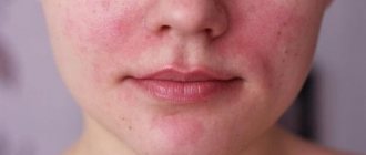
Vitiligo
The most common cause of white spots around the eyes is vitiligo. This is a skin disease, the mechanism of development of which is poorly understood. Why the spots under the eyes are white is explained by the peculiarities of skin coloring.
The color of skin, hair and eyes is determined by the amount of melanin pigment. The more of this substance, the brighter the color. White markings are areas lacking melanin. Vitiligo is characterized by the appearance of such areas on the face, arms, and torso.
The disease can appear at any age. It is observed quite rarely in young children; it usually begins after puberty. The following factors can provoke the development of vitiligo, and therefore white marks:
- hormonal fluctuations, especially during pregnancy and childbirth;
- prolonged emotional stress;
- liver diseases;
- helminth infection;
- metabolic problems;
- frequent skin injuries;
- excessive exposure to sunlight.
Vitiligo is of hereditary origin, and these factors contribute to its manifestation. White spots appear on any part of the body, including under the eyes on the skin. They have an irregular shape, are clearly defined, and occasionally merge with each other. White markings vary significantly in size - from a few millimeters to tens of centimeters.
Vitiligo spots are absolutely painless and are not accompanied by any other manifestations. It is extremely rare for a person to be bothered by mild itching. A characteristic feature is the absence of tanning in these areas. Exposure to sunlight quickly causes the bleached areas to burn.
Additionally, watch a video about vitiligo disease, its causes, symptoms and treatment:
Xanthelasmas
White or yellow spots on the upper eyelids are xanthelasmas. Their appearance is explained by the accumulation of cholesterol under the skin. Xanthelasmas are observed in people with atherosclerosis.
White spots most often appear in the area of the upper eyelids, closer to the eyelash line. Sizes from small grain to pea. There are usually no subjective symptoms. But if there are several xanthelasmas, discomfort appears when blinking.
How to get rid of blackheads in eyes
To get rid of unpleasant rashes, several methods are used, but their essence is the same: opening the milia and removing its contents. The choice depends on the number and location of whiteheads, as well as their size and skin type. The effectiveness of all methods is the same, the difference lies in the cost and duration of the procedures.
- Mechanical removal. A dermatologist or cosmetologist, using a thin long needle or curette (a surgical spoon for scraping out soft tissue, removing suppuration, etc.), opens the milia and removes its contents. Before the procedure and after it, antiseptic skin treatment is carried out. At the site of each removed formation, small wounds remain, which soon heal. This method has its pros and cons. Of course, the advantage is the low cost of the procedure, but at the same time, mechanical removal is not suitable for people with thin or overly sensitive skin. Another disadvantage of this method is that in one procedure it is possible to remove only 10-15 milia, after which a break of several days is taken. Treatment can take quite a long time. Also, the mechanical method is not suitable if the white dots are located on the eyelids very close to the roots of the eyelashes. The skin in this area is extremely delicate, and exposure to it with a needle is undesirable, and noticeable redness will remain. Currently, this method is used only for medical reasons when other types of procedures are not suitable.
- Electrical coagulation is the destruction of milia by cauterizing them with high-frequency electric current. This procedure has much greater advantages compared to the first method. Using electrocoagulation, you can get rid of fairly deep formations, which is difficult to do with a needle or curette. But this method has its own characteristics: after cauterization, a dense crust forms at the site of the milium, which must be treated for 10 days until it falls off. Also, shallow scars may remain at the spots - small, but still noticeable.
- Laser coagulation is the most modern and safe method of getting rid of milia. It is suitable for large clusters of points, as well as when they are located too close to the organs of vision. To carry out the procedure, a CO-2 (carbon dioxide) laser is used, which has been successfully used in aesthetic cosmetology for almost three decades. It eliminates pathological formations layer by layer through thermal action. After its use, there is a lack of redness and suppuration, which can be observed in the case of a mechanical procedure, since the laser beam also has a bactericidal effect. In one or two procedures you can completely get rid of milia. Laser coagulation has the highest cost among the described types of treatment.
We invite you to familiarize yourself with Language in small red dots
Vision is a key resource of the body, and you should not treat it with disdain. Treatment of black spots and dots with folk remedies is impossible; the problem itself also does not go away. You are allowed to drink herbs approved by your doctor; they will help you recover faster and prevent the recurrence of vision problems. Do not self-medicate - it is dangerous.
If you suspect the presence of black spots and dots, contact an ophthalmologist who will conduct an examination and confirm or deny the symptom. Contact a public clinic or a trusted private clinic. It happens that black spots arise due to another disease (tumor, infection, etc.), and in a private clinic you can be treated only for symptoms, without paying attention to the pathology itself that caused this condition.
During treatment in the hospital, existing defects are eliminated and the source of the pathology is sought. In most cases, it is possible to get rid of the disease that led to the appearance of black spots and dots, but it is not possible to clear the eye of defects. Dead cells are not removed from the vitreous body on their own.
In rare cases of floaters appearing before the eyes, for minor problems, vitamin drops are prescribed: Taufon, Quinax, ethylmorphine hydrochloride solution. Effective removal of dark spots is possible with the help of potassium iodide drops. If it is necessary to accelerate the regenerative component of the vitreous body, Wobenzym and Emoxipin are used.
If the disease is detected in the initial stages, it is recommended to take multivitamin complexes. The doctor prescribes examinations and procedures that eliminate the causes of vision problems. After getting rid of them, the black spots on the eyes completely or partially disappear on their own. If the disease is advanced, serious measures may be needed.
- Vitrectomy is a surgical procedure in which the vitreous humor is partially or completely removed. It is being replaced by an artificial environment. This extremely dangerous operation can lead to cataracts, retinal detachment, and hypotension. It is used only in cases where other methods have not produced any results.
- Vitreolysis - using a laser, the threads are broken, that is, clusters of dots are destroyed. The eye is completely reanimated. The operation is complex and is performed only by experienced doctors.
Treatment of milia
Professional removal of eyelid marks is performed by a dermatologist or cosmetologist. The procedure is available in aesthetic medicine centers.
Treatment methods:
- electrocoagulation;
- laser removal;
- cryodestruction;
- mechanical extraction of millet grains.
Electrocoagulation is based on exposure to high temperatures. The nodules in the eyelid area are burned with an electrode. A crust forms and will fall off within a few weeks. Before the procedure, the surgical site is injected with an anesthetic. The formations are small, there are no scars left. The procedure is bloodless.
The laser can accurately control the depth of burning. The method is more gentle; you can remove millet in hard-to-reach places - corners, folds of the eyelids, where the skin is thin and delicate. The beam simultaneously cauterizes the vessels, eliminating bleeding. Before the procedure, local anesthesia is given. The success of treatment and the risk of scar formation depend on the qualifications and experience of the doctor.
Need advice from an experienced doctor?
Get a doctor's consultation online. Ask your question right now.
Ask a free question
Cryodestruction. Previously, they used the reed method: they wrapped cotton wool around a wooden stick, soaked it in liquid nitrogen, and applied it to the formation without the ability to control the depth of impact. This method caused a high risk of scar formation on the eyelid and was not used when localizing nodules near the lacrimal canal or the outer corner of the eyes. Nowadays they use applicators: the device delivers the refrigerant precisely to the pathological area. Several repetitions of freezing and thawing ensure the death of the millet cells. In place of the milia, a vesicle with serous contents appears. It soon subsides and a crust appears. Recovery lasts about two weeks, and the method can be used without anesthesia.
You can remove millet on the lower eyelid manually using a scalpel: the cyst is pierced and the contents are squeezed out with a special spoon. The method is accessible, but traumatic. Pressure on the skin around the milia leads to damage to healthy tissue and increased swelling. This method does not guarantee complete removal of content. If not everything is removed, an inflammatory process may begin, suppuration, complications are especially dangerous in the eye area. It is difficult to carry out manual manipulations near the upper fold of the skin due to the mobility of the tissues. Hardware methods are more suitable for removing a nodule.
In case of a massive rash, you will have to cleanse the skin again: you can remove no more than 10-15 rashes in one session.
If milia on the eyelid appears in a child in the first year of life, do not treat at home. Cysts may disappear with age, consult your doctor and monitor the condition of the nodules.
Congenital spots
In humans, spots on the cornea of the eye can be detected immediately after birth. Their appearance is associated with a congenital disorder in the production of melanin pigment. The spots are called nevi; they are black or brown in color and are located on the sclera.
If a birthmark has formed in the eye, you should consult an ophthalmologist. The spots tend to grow, causing vision impairment. Pigment formations can degenerate into malignant ones. This happens quite rarely, but pathology must be excluded.
A child sometimes develops darkening of the iris. This is how iris tumors manifest themselves - benign or malignant. Urgent consultation with an ophthalmologist is recommended.
Necessary treatment
For minor dysfunctions, drops are prescribed, for example, “Taufon”.
Appearances on the iris of the eye must be treated promptly if they cause cosmetic discomfort to the patient and negatively affect visual function. For minor disturbances associated with fatigue and other non-pathological factors, you can use medications. For spots on the iris, the patient is often prescribed the following eye drops, which quickly eliminate the problem:
- "Taufon";
- "Emokipin";
- "Quinax";
- Wobenzym.
If the deviation cannot be removed with the help of medications, then a radical therapeutic measure is required. Spots formed on the iris can be removed through vitriolysis. The procedure involves exposing the pathological neoplasm to a laser beam, which eliminates only damaged tissue without affecting healthy tissue. In particularly advanced cases, when the spot has grown to a large size and impairs vision, surgery is required.
Acquired spots
If dots appear in the eyes throughout life, the reasons for this may be different conditions. These are not necessarily diseases - spots are the result of overwork or the use of medications.
Reds
The appearance of red spots is usually associated with the reaction of blood vessels:
- eye injury with capillary damage;
- a sharp increase in pressure leading to rupture of the vessel;
- an inflammatory process accompanied by capillary swelling.
They appear suddenly and disappear within a few days. They rarely stay for a long time.
Dark or black spots
A dark spot on the cornea of the eyeball appears due to macular degeneration. It is associated with impaired blood supply to the vitreous body. The causes are usually bad habits and age-related changes in the body.
The disease is accompanied by decreased vision and pain. Dark areas gradually increase in size, indicating the progression of the disease. Lack of treatment leads to irreversible blindness.
Brown and black spots on the whites of the eyes occur against the background of impaired pigment metabolism. Melanocytes begin to intensively secrete melanin, which forms black spots on the eyes. It is observed in hereditary diseases and oncological pathologies.
Quite rarely, black dots on the whites of the eyes are formed due to a fungal infection. Aspergillus spores, a microscopic fungus, can enter the eyeball. There they grow, causing pain and blurred vision. It is extremely rare that the eye is affected by helminths - Toxocara or tropical parasites.
A dark spot on a child's eye is usually not a disease. This is a physiological process associated with a change in the color of the iris. The condition goes away with age.
Therapeutic approach
Black spots in the eyes require mandatory treatment, which directly depends on the cause of their appearance. Symptomatic therapy alone will only help get rid of the “flies” themselves, but will not eradicate the existing problem, so you should not select medications on your own.
Treatment also depends on the structure and size of the threads that fly before the eyes. If they are small and are not the result of serious pathologies, then you can get rid of them with the help of eye drops:
- Taufona;
- Emokipina;
- Quinaxa;
- Wobenzima et al.
However, treatment of black spots in the eyes with the above medications is possible only in case of impaired functioning of the vitreous body
They need to be buried carefully, in a lying position. After the procedure, it is recommended to lie down for 1–2 minutes so that the drops have time to penetrate into the underlying tissues of the organ.
These products help normalize local metabolism and resolve floaters, but it should be remembered that they help some patients, while others still continue to complain about floating black dots flashing before their eyes.
Other therapeutic techniques
If the drops do not help get rid of the black spot in the eye on the cornea, the patient may need surgery. But this method is not as popular as instrumental procedures, and it can also lead to blindness.
Alternative methods are very popular in treating a blackhead that appears in the eye:
- Vitrectomy, during which partial or complete removal of the vitreous occurs. Manipulation is carried out only according to indications, mainly if black spots appear in the eyes due to ophthalmological diseases.
- Vitreolysis is a procedure performed using a special YAG laser. Using a laser beam that is aimed specifically at a dark spot located on the white of the eye, the “spot” is carefully removed without harming the integrity of healthy tissue.
The second manipulation is, of course, safer and more effective, but it is not cheap, which is its main disadvantage.