Urticaria pigmentosa is one of the most common forms of such a rare systemic disease as mastocytosis, and its name is only consonant with the common urticaria. In 75% of cases, this pathology is observed in children (mainly young children), but it can also develop in adult men and women.
For the first time, this disease, accompanied by pigmentation disorders and leaving brown spots on the patient’s body, was described as “chronic urticaria” in 1869, and in 1953 the disease was called “mastocytosis.” This disease in 90% of cases is accompanied by urticaria pigmentosa and can occur in the form of a cutaneous, systemic or malignant (mast cell leukemia) form. Mastocytosis is caused by the accumulation in tissues of an excess amount of mast cells (special forms of white blood cells), which, when destroyed, release histamine and other inflammatory mediators. As a result of their entry into tissues (for example, into the skin), an inflammatory reaction develops, manifested by characteristic rashes.
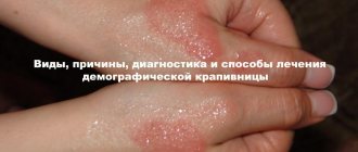
In this article we will introduce you to the causes, varieties, symptoms, diagnostic methods, treatment and prognosis of the cutaneous form of mastocytosis - urticaria pigmentosa. This knowledge will be useful to you, and you will be able to suspect its development in time and seek qualified help.
Causes
The reasons for the development of this disease have not yet been sufficiently studied. There are several assumptions. Some experts are inclined to believe that hereditary factors may be a possible cause of urticaria pigmentosa. This is because the disease often develops in people who are closely related.
Other scientists suggest that the disease is provoked by inflammatory processes caused by infectious diseases or toxic effects. However, many cases of urticaria pigmentosa develop against a background of unclear causes, and that is why there is no consensus yet on the prerequisites for the occurrence of this disease.
Mastocytosis can result from both immune and non-immune reactions. Its development is provoked by excessive accumulation of mast cells in various tissues, including the skin. When they are destroyed, which can be caused by various factors, histamine, heparin and other bioactive substances enter the tissues. They actively affect tissues, increase vascular permeability, cause swelling of tissues and dilation of capillaries. As a result, the release and action of these inflammatory mediators leads to visible skin changes and a possible decrease in blood pressure, the development of paroxysmal tachycardia, an increase in temperature, increased secretion of gastric juice and the appearance of attacks of skin itching.
In non-immune forms of mastocytosis, the release of histamine and other inflammatory mediators can be caused by the following factors:
- temperature effects (cold or heat);
- friction;
- squeezing;
- direct sunlight;
- climate change;
- stressful situations;
- toxic compounds;
- medications;
- some food products.
The peculiar coloring of the skin in urticaria pigmentosa is due to an increase in the number of melanocytes in the skin and the deposition of excess melanin in the lower layers of the epidermis. Scientists suggest that such disorders are caused by complex interactions between mast cells and melanocytes.
Why does the disease appear?
The most common cause of pathology is gene changes. Against this background, the number of mast cells in the body exceeds.
Predisposing factors for the development of the disease during the first year of life include the following reasons:
- heredity;
- unbalanced temperature regime. The pathology is caused by both overheating and hypothermia in the room;
- untimely start of complementary feeding. Including food that is not indicated for the child according to his age;
- wearing tight clothing that rubs the skin;
- infection by pathogenic microorganisms.
In children older than one year, pathology can develop as a result of exposure to the following factors:
- keeping the child under the sun;
- frequent stress;
- treatment with potent medications;
- deterioration of the immune system;
- food allergies;
- adolescent use of alcohol and drugs;
- frequent contact with water.
Mastocytosis can be caused by the habit of taking hot baths.
Forms of urticaria pigmentosa
The cutaneous form of mastocytosis (urticaria pigmentosa) can develop in patients of different age groups. Dermatologists distinguish two forms of this disease:
- cutaneous mastocytosis of infancy: the pathology begins its development in the first two years of the child’s life and is never accompanied by damage to the internal organs; in most cases, all symptoms completely and forever disappear before or during puberty;
- cutaneous mastocytosis of adolescents and adults: pathology develops in adults or older children and is accompanied not only by skin symptoms, but also by damage to internal organs (unlike systemic mastocytosis, such lesions do not progress), in some cases this form of urticaria pigmentosa becomes a systemic form mastocytosis.
Urticaria pigmentosa is classified into the following forms:
- form of mastocytoma - a single tumor appears on the patient’s skin;
- generalized form - multiple rashes appear on the skin.
The most common form of urticaria pigmentosa is the generalized form.
Symptoms
Urticaria pigmentosa develops suddenly, and the severity of its symptoms depends on the age of the patient, the form of the disease and the types of skin rashes. Approximately half of patients complain only of skin changes.
Cutaneous mastocytosis in children
Itchy skin usually accompanies urticaria pigmentosa.
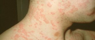
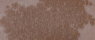
A sick child develops severe itching, and red-pink spots gradually form in the places where it occurs, which over time transform into red-pink blisters with transparent or bloody contents. They are edematous and accompanied by a pronounced exudative component. When rubbed with a finger, a spatula or a needle prick, the Darrieus-Unna phenomenon (or inflammation) appears at the site of the skin element. The bubble becomes more swollen, red and begins to itch more. The same signs of exacerbation of the skin rash can be observed after compression or thermal procedures (hot bath, exposure to direct sunlight, etc.). Sometimes such skin changes disappear without a trace, but more often after they are opened, brown-brown pigmentation remains on the skin.
More often, elements of the rash appear on the torso. They can spread to the face and hands, but in most cases their spread stops there. Sometimes the rash continues to progress and covers the entire surface of the skin.
In some cases, not blisters, but red nodules appear on the skin of a sick child. With this course of urticaria pigmentosa, the symptom of the Darrieus-Unna phenomenon is negative.
With urticaria pigmentosa, the skin gradually thickens, becomes yellow, and the skin folds deepen. With large accumulations of blisters in the groin, which after opening can be covered with cracks or ulcers, the child feels pain during movements.
With solitary mastocytoma, a tumor-like nodule with a diameter of up to 50 mm appears on the child’s torso, neck or forearms. More often, the formation of only one nodule is observed, less often - up to four. Mastocytoma feels like rubber to the touch. It may have a wrinkled or smooth surface. If the nodule is mechanically damaged, pustules or blisters appear on it, and the child feels a tingling sensation.
In most cases, urticaria pigmentosa in children is benign and is not complicated by the development of a systemic form of mastocytosis. During puberty, all symptoms of the disease disappear permanently. Mastocytes are characterized by sudden disappearance: the nodule seems to sink into the skin and smooth out with its surface.
Cutaneous mastocytosis in adults
In adults, urticaria pigmentosa begins to appear in the form of round spots or papules with a diameter of about 0.5 cm. They have clear boundaries and a smooth surface without signs of peeling. The color of the skin changes may be light gray or pink-brown. More often such spots appear on the body.
Over time, skin lesions spread to the face, arms and legs. They become spherical and acquire a dark brown or dark brown color (sometimes with a red-pink tint). The process may pause and persist for many years. Over time, it progresses, and urticaria pigmentosa is complicated by systemic mastocytosis (damage to internal organs). This form of the disease ends in the death of the patient, since damage to organs leads to a significant disruption of their functioning.
Solitary mastocytoma in a child
This form of the disease is rare in children. It is a single tumor-like formation on the baby’s body. In most cases, solitary mastocytoma can be cured without medical intervention. Usually passes by the beginning of puberty of the child.
You might be interested! What diseases does the appearance of red spots on the hand indicate?
With mastocytoma, there is no severe itching, and there are no changes in the organs. If a mastocytoma is scratched or injured, blisters may appear in its place.
Types of skin changes in urticaria pigmentosa
Depending on the type of rash, dermatologists distinguish the following types of skin changes in urticaria pigmentosa:
- Maculopapular (maculopapular). Clearly defined spots and papules appear on the skin. This type can be observed in children and adults.
- Nodal. Flat or convex seals (nodules) with a smooth or bumpy surface appear on the skin, their color can be yellow, pink or red, the diameter of the formations can be from 1 to 1.5 cm. They can be grouped, isolated or multiple, sometimes merge into plaques. This type is more common in newborns or children 1-2 years old.
- Erythrodermic. Dense patches of yellowish-brown color appear on the skin, with clear and jagged edges. Such formations are accompanied by severe itching, which leads to scratching and the formation of cracks and ulcers. It can be observed in children or adults, but in childhood it is not accompanied by the development of cystic reactions.
- Bullous. Multiple blisters appear on maculopapular rashes or areas of their fusion into plaques. This type is more often observed in infants or young children.
- Spotted telangiectatic. Red-brown lesions appear on the skin in the form of telangiectasia, located against a background of hyperpigmentation resembling freckles. Skin changes are most often located on the chest, legs or arms. This type is more often observed in adults (usually women).
- Diffuse. The skin thickens, becomes yellowish, and skin folds deepen. More often, such changes are observed in the inguinal folds or armpits, and when they are damaged, painful cracks and ulcers form. Can be observed in children and adults.
Most often, maculopapular or nodular rashes are observed with urticaria pigmentosa. Telangiectatic and diffuse types of skin changes occur against the background of damage to internal organs (systemic mastocytosis). Sometimes progressive erythrodermal urticaria pigmentosa can lead to the development of a systemic form of mastocytosis. Also, this type of skin changes can cause the development of erythroderma.
Diagnostics
Differential diagnosis of urticaria pigmentosa with other diseases is extremely important.
If the above-described skin symptoms appear, the patient should consult a dermatologist. The external manifestations of urticaria pigmentosa are similar to other skin pathologies, and the doctor will definitely carry out a differential diagnosis of cutaneous mastocytosis with the following diseases:
- pigmented nevus;
- skin reticulosis;
- dermatofibroma;
- congenital Rothmund-Thomson poikiloderma;
- pemphigus of newborns;
- lymphogranulomatosis;
- hemangioma;
- xanthoma;
- histiocytosis X;
- drug rash.
In most cases, the symptom of the Daria-Unna phenomenon helps to suspect cutaneous mastocytosis. To make an accurate diagnosis, the patient is prescribed the following types of laboratory and instrumental diagnostics:
- histological analysis of skin biopsy;
- general blood analysis;
- urine test for histamine.
If systemic mastocytosis is suspected, the patient is prescribed a more comprehensive examination, which includes consultations with doctors of other specialties and ultrasound of internal organs, radiography, CT, MRI, etc.
After a diagnosis of urticaria pigmentosa, the patient must contact a hematologist, therapist or oncologist for an effective course of treatment for this disease.
Treatment
Treatment of urticaria pigmentosa is prescribed individually for each patient and depends on his age, severity of symptoms and suspected causes of the disease. If cutaneous mastocytosis in children is not accompanied by pronounced changes in the skin and systemic manifestations, then therapy is not carried out, since this form of urticaria pigmentosa often heals itself before adolescence.
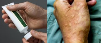
In more severe cases, symptomatic remedies are used for treatment:
- antihistamines: Diazolin, Cetirizine, Suprastin, Tavegil, Pipolfen, etc.;
- antiserotonin drugs: Ketotifen, Bicarfen, Periactin, etc.;
- anti-bradykinin drugs: Anginin, Prodectin, etc.;
- corticosteroids: preparations for internal and external use;
- preparations based on cromoglyceric acid: Nalkrom, Intal, KromHexal, etc.;
- cytostatics: Prospidin, etc.;
- Histaglobulin (for nodular urticaria pigmentosa).
PUVA therapy can be used to treat urticaria pigmentosa. This technique involves the combined effect of photoactive medicinal substances (psoralens) and long-wave UV rays on the skin.
In some cases, with pronounced cosmetic defects, mastocytes are removed surgically.
All patients with urticaria pigmentosa need to protect the skin from mechanical and thermal influences that provoke an exacerbation of the disease.
ethnoscience
For mastocytosis, any traditional methods must be agreed with your doctor. If there are no contraindications, then they can be used as an addition to the main treatment. Typically, the following folk recipes are used to relieve symptoms:
- drink 1 tsp three times a day. freshly squeezed celery juice;
- to improve sleep and relieve itching, take valerian infusion at night;
- To calm the inflammatory process, prepare compresses from oak infusion.
To reduce itching and accelerate the healing of blisters, use nettle infusion, 1 tbsp. boiling water take 1 s. l. dry nettle. It is recommended to treat the affected area with the resulting infusion up to 3 times a day.