Exudative erythema multiforme is considered an independent disease, which is characterized by an acute course, frequent relapses and seasonal exacerbations. The disease got its name due to its characteristic symptoms - redness of the skin, the appearance of blisters, peeling and crusts. No one can name a specific reason for the appearance of this disease, but many experts associate MEE with a viral origin. As with all forms of erythema, MEE is characterized by the appearance of many spots and redness on the skin.
The first signs of the appearance of exudative erythema multiforme
Important! Erythema multiforme is not contagious and cannot be transmitted by airborne droplets or sexual contact. Most often, the disease can be triggered by some chronic foci of infection (caries or skin tuberculosis).
To begin with, I would like to draw your attention to the fact that the disease most often appears in the spring and autumn periods. The first signs are the appearance of redness on the skin, fever, weakness, malaise and chills. In some cases, all of the above symptoms may be accompanied by persistent joint pain.
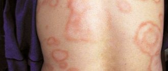
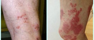
Regarding the rashes, they can be localized on almost every part of the body, but most often they can be found on the backs of the hands, feet, forearms, thighs and legs. In its acute course, the disease can affect the palms, external genital areas and mucous membranes of the oral cavity. Reddish rashes may be accompanied by bluish blisters that rise above the skin.
At the initial stage, the patient may experience single lesions the size of a small grain, but as it progresses, the rash can reach a size of 2 cm, and in especially severe cases, multiple blisters merge into one large lesion. We must not forget about blisters filled with clear watery liquid, which are usually diagnosed in the facial area. As MEE progresses, the color of the fluid may become cloudy and even contain blood. Almost always the bubbles burst, leaving behind gray-yellow or bloody crusts. This is what explains the cracks and painful wounds in the corners of the lips.
Erythema multiforme: characteristics of the rash, diagnosis, treatment
Minor ME has only cutaneous manifestations, while major ME affects one or more mucosal areas.
- Minor ME: Typical raised, edematous papules, with a peripheral distribution, without mucosal involvement, affecting <10% of the body surface area.
- Typical or raised edematous papules, with a peripheral distribution, involving 1 or more mucous membranes, affecting < 10% of the body surface area.
Diagnostics
Although ME is often mild and self-limiting, a thorough history and clinical examination are important to determine if there are triggers that could be avoided in the future. More serious mucocutaneous diseases such as Stevens-Johnson syndrome and toxic epidermal necrolysis (TEN) must also be excluded.
- Minor ME has only cutaneous manifestations, while major ME has cutaneous manifestations and affects one or more mucosal areas.
- Lesions in small and large ME involve <10% of the total body surface area.
Anamnesis
Although the cause is often unknown, a careful history should be reviewed for infections, relapses, and new medications. Possible causes must be carefully examined before a patient is identified as an idiopathic case.
- Infections: The most common associated infections are herpes simplex virus (HSV) and Mycoplasma pneumoniae. Other associated infections include hepatitis B, Epstein-Barr virus, cytomegalovirus, histoplasmosis (with associated erythema nodosum), coccidioidomycosis, gardnerella, and contagious pustular dermatitis (Parapox virus, which can be transmitted from sheep or goats to humans). ME is also associated with herpes zoster.
- Medicines: Associated medications include aminopenicillins, docetaxel, TNF-alpha inhibitors, antimalarials, anticonvulsants, lidocaine injections (which can cause both ME and erythema nodosum), sulfur-based drugs, and nonsteroidal anti-inflammatory drugs (NSAIDs). Statins can cause light-sensitive ME.
- Vaccines and allergens: Hepatitis B vaccines and allergic reactions to contact allergens, including tattoos, are also known to cause ME.
Lesions typically develop several days after exposure to the trigger. Some lesions are initially completely similar to the target lesions; others develop from small erythematous plaques. The lesions develop quickly and usually increase in number within 4 to 7 days. They may cause general discomfort but will not cause itching until healing begins. Involvement of the oral mucosa can be particularly painful for the patient, and in more severe cases causes restrictions on the ability to eat and drink.
Clinical study
ME presents with characteristic target-type lesions (circular erythematous rings with an outer erythematous zone and a central blister, between which there is normal-colored skin) and atypical target-shaped papules (without a central blister). Clinical features of the lesions provide an important diagnostic tool, with characteristic target lesions often characterized by a symmetrical distribution on the extremities. Target-like lesions tend to unite more often. The mucous membranes of the mouth, eyes, nose and genitals also need to be inspected for erosions, which are observed with large ME. Some characteristic target lesions and minimal mucosal lesions indicate ME, particularly in the presence of HSV or Mycoplasma pneumoniae infection.
A general physical examination should also be performed to identify possible infectious causes. HSV infection is characterized by clustered vesicles on an erythematous base. A red eardrum is highly likely to indicate Mycoplasma pneumoniae, as is wheezing and/or wheezing. Hepatomegaly is often observed with hepatitis B infection.
Target lesions on the face and erosions on the mucous membranes with crusts due to relapse of HSV-1
Laboratory research
In most cases, ME disease can be diagnosed based on history and clinical examination alone, and no further testing is required. However, if there is diagnostic uncertainty after clinical evaluation, a hematoxylin and eosin biopsy can be performed. If the biopsy result is inconclusive, immunofluorescence biopsy can also be performed.
If the cause of ME is not established based on clinical examination, laboratory tests are performed to determine the cause. Because the most common infections that cause ME are HSV and mycoplasma infection, primary tests include complete blood count, electrolytes, HSV serology, cold agglutinins, M. pneumoniae titers, and/or chest x-ray (depending on clinical condition of the patient). If the results of these tests are negative, tests are performed for less common infections that may be causing the disease (for example, liver function tests and serological testing for the hepatitis B virus). ME caused by herpes zoster can be differentiated from the generalized form of herpes using a rapid polymerase reaction.
Serological testing for HSV may also be appropriate in the presence of recurrent ME episodes and in the absence of identified HSV-specific lesions. In patients with recurrent ME, the presence of antibodies to desmoplakin is noted.
Differences between Stevens-Johnson syndrome and toxic epidermal necrolysis from ME
Other more severe reactions must be excluded, including Stevens-Johnson syndrome and TEN. Severe restriction of oral food intake and pain when urinating are common in these diseases, but they can occur with large ME. Stevens-Johnson syndrome affects <10% of the total body surface area and is characterized by extensive involvement of the oral and genital mucosa. Exposure to the causative drug is often identified. TEN demonstrates active skin damage, generally more than 30%. If differentiating ME from Stevens-Johnson syndrome or TEN is difficult, 2 tests can be performed:
- The Asbo-Hansen sign for TEN is characterized by enlargement of the blisters with lateral pressure, demonstrating necrosis of keratinocytes. Stevens-Johnson syndrome and TEN are also characterized by Nikolsky's sign with detachment of the epidermis upon touch, which is not present in ME.
- Biopsy and evaluation of fresh frozen tissue may demonstrate necrotic keratinocytes in Stevens-Johnson syndrome and TEN. Monocyte infiltrates and red blood cells are more characteristic of the histopathology of ME; epidermal necrosis and inflammatory infiltrate on the dermis are absent.
Key diagnostic factors
- Presence of risk factors Significant risk factors include previous development of ME and herpes or mycoplasma infection.
- Lesions with 3 zones (red rim, lumen zone, and central blister or erosion) on the distal extremities are highly suggestive of ME.
- Relapse of ME is often observed in patients with disease associated with the herpes simplex virus.
- Observed with large ME.
- Erythematous papules without a lumen zone (2 zones) are often centripetal.
Differential diagnosis
| Disease | Differential signs/symptoms | Differential examinations |
| Hives |
|
|
|
|
|
|
|
|
|
|
|
|
|
|
Treatment
A step-by-step approach to the treatment of ME is a disease that ends in self-healing; its therapy is carried out using the following strategies:
- Maintenance therapy to maintain hydration and prevent secondary bacterial infection of erosions
- Treatment of suspected infections
- Suppressive antiviral therapy if recurrent disease is caused by herpes simplex virus (HSV)
- Topical or systemic corticosteroids to reduce inflammation.
Symptomatic therapy
Minor ME (typical target-shaped or raised edematous papules with a peripheral distribution, without mucosal involvement, affecting <10% of the body surface area)
- The lesions should be cleansed twice daily with soap and water. Topical emollients (softening agents) should be applied to all damage - they moisturize the skin and act as a protective barrier. If open lesions are encountered, they should be carefully cleaned and covered with sterile dressings to prevent secondary bacterial infection.
- Pain relievers (eg, paracetamol, nonsteroidal anti-inflammatory drugs [NSAIDs]) may be taken for general pain relief and to reduce discomfort caused by the lesions.
Large ME (typical target-shaped or raised edematous papules, with a peripheral distribution, involving 1 or more mucous membranes, affecting <10% of the body surface area)
- Topical emollients, gentle cleansing, and sterile dressings of open lesions help prevent bacterial superinfection of blistered and eroded lesions.
- For painful lesions in the mouth, lidocaine solution can be applied directly to the lesions, or a mouth rinse can be used to coat and soften the mouth before drinking liquids or eating.
- Although some patients are severe enough to require hospitalization, and although the disease usually resolves without specific therapy, some patients with large ME may require fluid resuscitation if they become dehydrated due to reduced oral fluid intake.
- If the urethra is obstructed by expelled mucous membrane particles, catheterization can help restore urination.
- Oral analgesics may reduce discomfort caused by lesions
Treatment of suspected infections
Herpes simplex virus
- In cases of oral herpes simplex (herpetic fever) and genital herpes, oral antiviral drugs are used.
Mycoplasma pneumonia
- Clinical guidelines for the treatment of atypical pneumonia recommend the empiric use of macrolides or doxycycline for uncomplicated community-acquired pneumonia to cover atypical organisms
Treatment of inflammation
Topical or systemic corticosteroids may be used to reduce inflammation. Topical corticosteroids for small ME have been helpful in resolving the lesions. Prednisolone may also be used. Large EM may require oral or intravenous corticosteroids.
Relapse Prevention
If recurrent HSV infection is the cause of repeated episodes of ME, suppressive antiviral therapy may be prescribed. At the first sign of an outbreak of oral herpes simplex, you should immediately start taking valacyclovir (course duration is 1 day, taken 1-2 times a day). If this does not control symptoms, a daily dose of valacyclovir may be prescribed for at least six months. Doubling the dose may be necessary to keep symptoms under control. For patients who require the drug in liquid dosage form, acyclovir is the drug of choice.
If suppressive therapy combined with antiviral drugs does not work, immunosuppressive therapy and phototherapy can be tried, although these methods have only been used in a few isolated cases to date.
Patients may be advised to avoid environmental triggers to prevent recurrence of oral herpes (eg, sun exposure).
Forms of the disease
exudative erythema in the photo
Exudative erythema multiforme is classified into 2 forms:
- Idiopathic manifests itself more often than the second. Its causes are considered to be pathogens of bacterial, fungal and protozoal infections, it can also be influenza, hepatitis or herpes virus;
- Toxic-allergic (symptomatic). Occurs after treatment with certain medications that can affect metabolic processes, for example, antibiotics, hormones, sulfonamides.
The inflammatory process of the disease itself can be moderate or severe. The mild course of the process does not affect the patient’s well-being. Only the skin tissue is affected, without affecting the mucous membrane. The severe form is characterized by rashes in large quantities, and they can also affect the mucous membranes. A person feels a gradual deterioration in health, which can lead to life-threatening factors.
How dangerous is the disease?
Erythema multiforme is a danger during pregnancy and the outcome of pregnancy itself is not always favorable. Without treatment, there is a risk of premature birth or stillbirth.
There are also serious conditions that pose a danger to the patient: Lyell and Stevens-Johnson syndromes.
In children, it manifests itself at 10-15 years of age, often after vaccinations, and often develops into erythema multiforme. The danger is that parents diagnose a common cold, and if there is no immediate treatment, children suffer serious complications that can be fatal. Concomitant infections have a negative effect on the course of the disease.
There is no specific prevention against erythema multiforme. As an option, complete cleansing of the body from all foci of the disease is suitable. You can pay attention to hardening: daily cold rinsing or other hardening exercises. The prognosis for the disease is positive.