Do moles grow with the child?
Moles appear immediately after the baby is born or appear under the influence of certain factors. In infancy, you can see several birthmarks on the baby’s face and body. In most cases, marks appear when hormonal levels change.
In medicine, several stages of the development of nevi in children are noted:
- newborns and early childhood: 6 months – 2 years;
- age 5-6 years;
- puberty: 10-12 years.
The formation of the element is associated with an increase in the level of melanocytes in the skin structure.
The main location is the gap between the inner and outer epithelial layers. The more active melanin is produced, the darker the skin will be. Cells containing melanin act as sun protection.
The pigment significantly reduces the adverse effects of ultraviolet rays, reducing burns and negative effects on the skin.
The appearance of pigmented areas is a natural process for a child’s body.
The skin that rubs against clothing and areas exposed to chemicals and hormonal drugs are subject to pigmentation. The period of occurrence and development of moles depends on the individual characteristics of the body.
Video consultation with a pediatrician
Watch the video for a detailed consultation with Dr. Komarovsky about moles, warts, molluscum contegium and other skin problems for children. Parents, most importantly, don’t panic!
When do children get moles? Most often, dark spots on the skin form some time after birth. This is influenced by many external and internal factors. Parents need to understand this issue well in order to promptly identify possible violations.
Reasons for the rapid growth of a mole
A child’s mole has grown larger – a situation that makes parents worry.
Congenital marks have a specific classification:
- hemangioma;
- spots with a light orange color (stork bite);
- saturated wine stain (fire stain).
Hemangioma is a vascular formation that appears some time after birth.
The size of the nevus immediately increases rapidly. By the age of ten, the element becomes pale and disappears without a trace. Stork bite is a pigmented cell cluster that occurs on the eyelids, back of the head, and in the bridge of the nose. It looks like a large speck, rich pink in color.
Common are bright red nevi on the scalp, which increase in size as the child grows and disappear with age.
Acquired moles are divided into:
- intradermal;
- epidermal;
- combined.
The first two varieties resemble peas, and the combined type is a smooth formation at the same level as the skin in the form of a brown speck.
The reasons for the increase in nevi are the following factors:
- Prolonged exposure to sunlight.
- Hormonal changes.
- Mechanical damage to the skin surface (impact, rubbing, scratch, cut, insect bite).
- Viral infection.
Genetic predisposition contributes to the enlargement of a mole. The more birthmarks mom and dad have, the higher the risk of their formation in the baby.
According to statistics, fair-skinned, premature babies have a higher risk of developing nevi than dark-skinned babies. Girls are more susceptible to the appearance of congenital formations.
Should we sound the alarm?
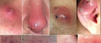
Any moles on a baby are classified according to the following criteria:
They have clear contours and a very dark color (black, brown, purple). The growths are located above the skin. If you press on them, no pain is felt. They can be up to 1 cm in size and have no hair. Sometimes they can grow up to 5 cm. The danger is possible malignancy.
- Intradermal moles
These are ordinary moles. They can be small or large. Moreover, such moles can also have different colors: flesh-colored, brown. There are children who have an intradermal nevus that has a characteristic stalk.
- Complex or mixed form
It is considered a transitional type between the first two. The neoplasm is dense, its shape is spherical, its color is dark, it can be either brown or red. Diameter - 1 cm. They can rarely degenerate into oncology, but they need to be observed. This species also has hairs on the surface.
- Congenital nevi
They are far from uncommon. Doctors explain their appearance by a violation of the mechanism of transformation of cells that were originally the skin of the embryo into melanin. Such moles cannot but be noticed already in the maternity hospital. Degeneration depends on their position relative to the epidermal layers.
Very large moles are rare for children. As a rule, moles are small and grow with the child’s body. As they grow older, their sizes stabilize.
But if you still suspect something is wrong, then to dispel doubts, contact a competent specialist.
Diagnosis of a growing nevus
Parents who notice that a dark and flat mole is growing in their child should definitely consult a pediatrician or dermatologist for advice. The doctor will conduct an examination and determine the type and danger of the stain. The degeneration of a benign lump into oncology in infancy is rare. It is necessary to observe the reaction and behavior of a child with marks.
Growths occur on skin areas with reduced immune defenses.
A weakened body is the main factor in the change in color and shape of the birthmark.
The doctor may additionally conduct an examination:
- dermatoscopy - examination of the spot on an enlarged scale, the method determines the malignant type of mole;
- Digital dermatoscopy – obtaining a clear image with a nevus approaching several times.
It is difficult to determine the type of growth on your own; you should visit a specialist in a timely manner.
Types of birthmarks
All pigmented neoplasms on the face or body of a child are divided into 2 main groups - hemangiomas and nevi. In the first case, we are talking about the accumulation of blood capillaries, which give the spot a reddish tint. Nevi are formed on the basis of melanin, so they can have a different color - from light golden to black.
Hemangiomas are divided into types:
- flat - considered the most common, characterized by a regular shape and smooth edges;
- star-shaped - they look like red dots with capillaries coming from them, disappear on their own after 1.5-2 years;
- capillary - look like wine-colored spots located in the deep layers of the epidermis;
- cavernous - when palpating such a formation, you can feel the pulsation of blood;
- strawberry (berry) - characterized by a bright scarlet hue and deformed edges.
A birthmark on the head or body of a child does not require special treatment and does not cause discomfort for the newborn. Damage or tearing off of a formation, as well as a sudden change in its structure, shape or size in newborns on the forehead, is considered a cause for alarm. In such cases, you should never hesitate to visit a doctor.
Is it necessary to remove a child's mole?
Small flat spots do not pose a threat to the life of a small person. Statistics show that in 40% of cases of tumor formation, oncology occurs. A small number of marks on the arm or back is not a cause for concern if the spots are not subject to friction.
If you constantly find new marks with a diameter of more than 5 mm, you should see a doctor. He will conduct a professional diagnosis and advise on possible treatment options. You shouldn’t delay going to the hospital when your baby accidentally rips off a bulging formation: the injury is the impetus for destructive phenomena in the body.
A special laser is used to remove dark spots in childhood on the temple, neck, and nose. After the manipulation, the baby may be left with a burn; it will take some time for rehabilitation.
You can remove spots on your leg using radio waves. The main advantage of this technique is that it does not affect healthy skin areas.
Removal takes place without pain or bleeding.
A reliable and safe method of getting rid of a mole is cutting it out with a surgical scalpel. There are no contraindications, the risk of relapse is reduced to a minimum level. The disadvantage is the presence of a scar. This method is not recommended for use in children. It is better to opt for laser removal
Localization of nevi in the area of the hands, fingers and forearms
If the nevus is located on the forearm, this indicates that this area of the body receives a large amount of sunlight. Thus, a large number of melanocytes are concentrated in this area, which provoke the appearance of moles. Over time, spots can change their appearance due to many factors.
Moles themselves are not dangerous . But if they are located on the hands, they can cause discomfort and are often injured. This will contribute to the degeneration of moles into malignant ones. The most dangerous disease that can arise in this regard is melanoma - skin cancer.
If the nevus is located on the hands, then the risk of damage increases . After all, we constantly perform actions with our hands. The hands can easily be rubbed, scratched or damaged, in addition to touching a mole.
If you have moles on your hands, it is better to consult a doctor and consult with him about removing such moles. In addition, the hands are open all the time, both in winter and summer. Therefore, the sun's rays fall on them, which can also contribute to the development of malignancy.
The same should be said if the mole is located on the fingers: middle, index, ring, thumb or little finger. With their help, we do cleaning, cooking, etc. every day. Therefore, the risk of injury to the mole increases significantly.
Important! A mole is a benign formation. As long as it is not injured, it is absolutely safe for health. But if a mole is constantly exposed to friction and other influences, this can cause it to degenerate into a tumor and cause melanoma.
In some people, nevi are located on the inside of the palms and wrists. This is also a very dangerous place, which is often injured while performing some everyday activities, so you need to monitor such moles very carefully and try not to injure them.
If the mole is located on any part of your arm and does not bother you, then there is no need to worry. The following changes and sensations may be a cause for concern :
- pain at the location of the mole;
- appearance of blurred outlines in the formation;
- swelling of the nevus;
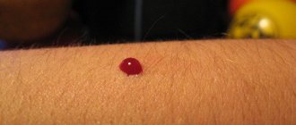
- discharge of blood or other unknown fluid from a mole.
In this case, you need to visit a doctor as soon as possible. Such symptoms are dangerous and may indicate that the mole is malignant.
Dangerous accompanying symptoms
Any spots on the baby's body should be controlled by parents. Periodic examination allows you to notice the first signs of transformation of moles into melanoma:
- Asymmetry: in its natural form, a nevus is an even geometric figure, an oval or a circle, the halves are symmetrical in relation to each other. The growth of one part of the spot is an important symptom for examination.
- A healthy mark has smooth edges, while a pathological mark has blurred borders with jagged sides.
- Change in color: uniform color and color is normal, the presence of inclusions or a change in tone is a sign of deformation of the spot.
- If the diameter exceeds 6 mm, you should visit a doctor.
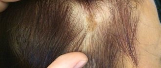
- Hair loss in the affected area.
If one of the above symptoms is detected, you must seek help from a medical facility to avoid dangerous consequences.
Possible complications and precautions
The main feature of a progressive pathological process is that the tumor grows rapidly in a short period of time (over the course of a month). When a white mark forms around a birthmark, do not be alarmed; this is a sign of Setton’s nevus. It occurs as a consequence of sunburn and disappears on its own after some time. A dangerous signal is the growth of moles throughout the body. This phenomenon requires specialist supervision.
Precautions to be taken:
- Limit children's exposure to the sun between 11 a.m. and 4 p.m.
- Protect your skin from ultraviolet rays with sunscreens and lotions.
- In hot weather, wear a hat and walk with your baby in the shade.
- If the tumor is injured, it is necessary to treat it with hydrogen peroxide and immediately contact a specialist.
Treating stains using folk remedies at home is prohibited due to the risk of complications.
Inspection of suspicious elements should be carried out at an early stage to avoid undesirable consequences.
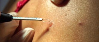
How does the removal take place?
Removal of skin tumors is carried out by a qualified specialist in the field of dermatology and oncology. If it is necessary to remove a mole for aesthetic purposes, no preliminary preparation is required. When removing a pathological mole, it is necessary to first undergo certain tests and conduct a specialized study. During the removal process, the specialist may suggest the use of local anesthesia. The most common methods for removing nevi are the following:
- using a laser;
- using nitrogen (cryodestruction);
- surgical intervention;
- using radio waves (radio wave removal);
- using high frequency current (electrocoagulation).
When a pathological mole is removed, its element must be sent for histological examination. Medical observation continues to monitor the patient's condition in the postoperative period.