Symptoms of pathology
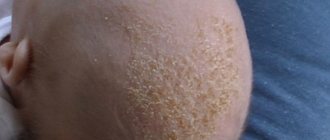
Externally, the growth looks like an oval or linear, waxy plaque with a clear contour; it has a soft, elastic structure and is absolutely painless. The color can range from flesh to yellow-brown. The surface is hairless, granular, covered with scales.
The nevus can reach 9 cm in diameter and appears mainly on the head, in the temporal region or on the forehead, eyelids, along the hairline on the neck, behind the ears.
In young children, the plaques are light, smooth, devoid of vegetation, the outer layer consists of small papillae. In adolescence, under the influence of a high level of gonads, the surface of the growth becomes coarser, covered with warty dark brown papules, the pathological tissues are equipped with blood vessels, so they are easily injured and bleed. The neoplasm increases in size and rises above the surrounding skin. Hair does not grow on its surface; when the nevus is localized on the head, alopecia areata is present.
After puberty and in older people, the disease in 5–30% of cases degenerates into a benign or cancerous tumor. Neoplastic changes, pronounced peeling, and hyperkeratosis are noted. With malignancy, the course of the disease is relatively favorable, it progresses slowly, and rarely metastasizes.
Clinical manifestations of the disease
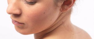
As you know, such a disease can manifest itself on the head. This nodular formation arises from the embryo after certain abnormalities have appeared in it. An alarming sign is the degeneration of papilloma into an oncological disease called adenoma of the sebaceous glands.
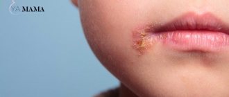
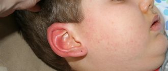
Chloasma very often appears on the surface of the face. What kind of disease is this and what does it represent? It looks like a single bald patch on the head. Their color range can be either yellow or dark brown. These formations have a velvety feel to the touch, and their appearance is the same as ordinary warts. The nevus visually resembles papillomas, measuring about 3 mm, but can reach 2 cm.
Chloasma may be light brown in color
Causes of formation of nevus of the sebaceous glands
The etiology of the disease is not fully understood. Moles usually appear in children in infancy or the first few years of life, becoming larger as the child grows older, and their structure changes. The pathology is diagnosed equally often in both sexes. The hereditary factor plays an important role; if the patient’s close relatives suffered from similar ailments, then there is a high probability of a nevus of the sebaceous glands appearing in the baby.
Active growth and malignancy of the node can be provoked by congenital hyperplasia of glandular tissue, concomitant chronic diseases of the gastrointestinal tract, hormonal disorders, the presence of rosacea on the skin, and constant trauma to the growth.
Stages of development
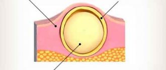
Nevus of the sebaceous glands of Jadassohn has a lobular structure, consists of cells of the sebaceous glands, grows from the middle and upper layers of the epidermis. Refers to harmartomas that arise as a result of pathologies of intrauterine development. The tissues are formed by the same cells as those of a healthy person, but differ in their dislocation and differentiation. Additionally, expansion of apocrine sweat glands and hair follicles is observed.
There are 3 stages of nevus development classified:
- At an early stage, hypoplasia of the follicles and sebaceous glands is noted.
- The mature stage is characterized by hyperpigmentation of the growth, superficial papillomatosis, and hyperplasia of the sebaceous glands.
- Stage of development of a benign or malignant tumor.
Various complications
Statistics say that in 15% of patients this disease develops into cancer (basal cell). It happens, less often, that an adenoma of the sebaceous glands is formed, consisting of epithelial cells. She is benign. Blepharitis or rhinophyma may appear, which impairs vision.
Unfortunately, the prognosis for a malignant tumor is unfavorable. It is not removed surgically, as they fear that malignant cells will multiply even faster. Doctors prescribe medications that keep the patient's condition stable. They give him painkillers. When the neoplasm is benign, the prognosis is favorable. It can be removed surgically or with laser. This is the best prevention of the development of a malignant tumor. It is best if at an early stage, when you notice a nevus, consult a doctor and he will prescribe histology. Some trigger the disease, and sebaceous gland adenoma and other nevus-based neoplasms occur. A sample will be taken from you and the tissue will be examined in the laboratory. If it is a benign neoplasm, then the oncologist or dermatologist will recommend removing it. If a scar remains, you can get rid of it.
Seborrheic nevus is dangerous. It can affect your eyes, spread to your bone tissue, penetrate your nerve endings and directly into your central nervous system or genitourinary system. Over time, Jadassohn's syndrome may occur. This is a linear nevus, the patient may experience intellectual retardation and epileptic seizures.
Diagnostic methods
The correct diagnosis can be made based on the results of histological examination and distinctive morphological features. Microscopic analysis reveals characteristic changes in soft tissue at the cellular level that correspond to the stage of development.
Modern clinics use videodermatoscopy, a non-invasive way to assess the condition of a pathological node. During the examination, the image increases in size, this allows the doctor to analyze the nature of changes in the structure of the epidermis and establish the correct diagnosis.
Nevus of the sebaceous gland is differentiated from aplasia of the skin, papillary nevus, juvenile xanthogranuloma, solitary mastocytoma, and a localized form of warty nevus.
Publications in the media
Nevi (moles, birthmarks) are hamartoma-like malformations of the skin that can develop both from elements of the epidermis and the dermis itself (connective tissue, vascular elements or melanocytes). Nevi are pigmented formations that usually protrude above the surface of the skin. Almost every person has moles; they can be congenital or occur throughout life, especially during puberty, in women during pregnancy, and hormonal dysfunctions. The clinical picture is characterized by extreme diversity in the number, size, morphological type and degree of pigmentation of individual elements - spots, nodules, plaques - up to subtotal damage to the skin with the so-called giant nevi. Large congenital nevi are considered to be elements >20 cm or occupying 2.5% of the body surface; they are considered a risk factor for malignancy.
• Epidermal nevi (warty or mole-like) are most often found at birth, although they can also develop in childhood (very rarely in adults). Externally they may be similar to papillomas (viral origin), but are usually represented by large linear plaques or an array of small papules. Epidermal nevi are often asymptomatic. Malignization is rare, with the exception of nevi of the sebaceous glands (basal cell carcinoma occurs in 5% of cases). Histologically they are characterized by acanthosis and hyperkeratosis. Nevus of the sebaceous glands is also characterized by the presence of a large number of sebaceous and apocrine glands. Cellular atypia is not observed. Treatment is mainly for cosmetic purposes. Preventive removal of sebaceous gland nevus is performed due to clinical manifestations (hair does not grow on it), and not the danger of malignancy.
• Dermal nevi •• Connective tissue nevi are predominantly congenital and are often represented by dense, single papules and flesh-colored plaques. The number of hair follicles in them can be significant (“pigskin”). This variant of nevi is idiopathic and is not associated with other diseases, with the exception of “shagreen skin” in tuberous sclerosis. Histologically, they consist of dense conglomerates of collagen and elastic fibers. Prognostically safe and removed solely for cosmetic reasons •• Vascular nevi (hemangiomas) are vascular formations lined with endothelium, mainly capillary, but cavernous structures can also occur, especially in large nevi. Three clinical types are considered typical: strawberry nevus, cherry nevus and cavernous hemangioma. Strawberry nevus and cavernous hemangiomas more often undergo spontaneous regression, while cherry nevus continues to grow throughout life. Surgical treatment is carried out for cosmetic reasons, for functional disorders (localized in the conjunctival area, lips) or thrombosis of the cherry nevus (due to suspicion of malignancy) •• Melanocytic nevi are of the greatest clinical importance, given the need for differential diagnosis with melanoma •• Pathohistology. Nevus cells are located in the form of nested clusters of various sizes and configurations in the dermis, subepidermally and between cell complexes. Sometimes the accumulations become diffuse and are located very close to the epidermis along the basement membrane, and atrophic changes in the epidermis are noted. The cells are large, of various shapes and sizes, with a clearly visible nucleus, sometimes several hyperchromatic nuclei, arranged in the form of “rosettes” or lumpy clusters. In some elements, the shape of the cells is spindle-shaped, which makes the nevus resemble a neurofibroma. Pigment content may vary. In “young” nevi, the stromal component is weakly expressed; over time, the connective tissue stroma begins to predominate; such a nevus is considered “fibroepithelial.” Localization of nevus cells among basal keratinocytes and in the zone of the basement membrane without its disruption is characteristic of a border nevus, and in combination with the intradermal component - of a mixed nevus.
Clinical types of nevi • Basal cell nevus - a hereditary disease (109400, 601309, BCNS gene, 9q22.3, Â) - characterized by (usually benign) skin lesions of the eyelids, nose, cheeks, neck, upper jaw, looks like a flesh-colored papule, without signs erosion, histologically does not differ from basal cell carcinoma. • Verrucous nevus is a skin-colored (or darker) lesion of the epidermis similar to a wart, often linear, appearing at birth or in early childhood; can be of different sizes and localization, single or multiple “verrucous nevus. • Hairy nevus is a mole covered with a large amount of growing hair. • Giant pigmented nevus is a large congenital, with pronounced hair growth, pigmented nevus with a predominant localization on the lower extremities “pigmented hairy nevus” .
• A blue nevus is a wide-based papule or plaque of varying shades of gray-blue color. Histologically, clusters of process melanocytes with a high pigment content are found in the dermis, located parallel to the epidermis, which determines the optical effect of blueness when incident rays of the visible spectrum “Jadassohn-Tische nevus”. • Intradermal nevus - a nevus with localization of nests of melanocytes in the dermis, and not at the border between the epidermis and dermis. • Ito nevus - pigmentation of the skin area innervated by the lateral branches of the supraclavicular nerve and the lateral cutaneous nerve of the shoulder; nevus cells lying randomly in the dermis.
• Strawberry nevus - a small vascular nevus similar in size, shape and color to strawberries - a bright red solitary formation of spongy density. It is observed in 3% of newborns and often spontaneously obliterates by 6-7 years of age. « cavernous hemangioma « cavernoma « cavernous hemangioma. • Unilateral nevus is a congenital linear nevus, which is located along the nerve either on one side of the body or on part of a limb also on one side linear nevus. • Ota nevus (oculodermal melanosis) - skin pigmentation in the area of innervation of the trigeminal nerve and conjunctiva of the eye in the form of bluish-gray spots; occurs in Asia, more often in women. The treatment is cosmetic. • Nevus dark blue orbital-maxillary .
• Borderline nevus - a nevus consisting of nests of nevus cells near the basal layer, the border of the epidermis and dermis, appears as a small, slightly raised, flat, non-hairy, pigmented (dark brown or black) tumor epidermal nevus. • Acquired nevus is a melanocytic nevus that is not detected at first after birth, but appears in childhood or in adults. • Flaming nevus ( port wine stain) is a large, vascularized nevus with a purple coloration, usually located on the head and neck. Unlike other vascular nevi, in this case there is no proliferation of a large number of vascular elements, but an expansion of the normal number of skin vessels. As the size of the vessels progresses, protrusion appears and the color of the nevus changes from pink to purple-red. See also Sturge-Weber syndrome in the appendix to this article. • Sebaceous nevus (Jadassohn) - congenital hyperplasia of the sebaceous glands with papillary acanthosis of the epidermis. • With thick curly hair, nevi are congenital foci of growth of spirally twisted hair in the scalp “limited [symmetrical] allotrichia.
• Atypical nevus syndrome (ICD-10: D22 Melanoform nevus; *155600, #155601 [mutation of the CDKN2A cyclin-dependent kinase inhibitor 2A gene, OMIM 600160.0001]) is an inherited disease (Â)) of pronounced familial nature. Prevalence: in the USA up to 5% of the general population; in the late 90s, about 40 thousand familial cases and 4.6 million sporadic forms were registered. Synonym: dysplastic melanocytic nevus •• Clinical picture. Usually spotted elements, but there are papular, plaque-like elements with a central papule or micropapules, oval or irregular in shape, sometimes with processes, 5 mm in size or more, with a finely scalloped clear outline, of varying intensity of brown color (up to black), with an erythematous rim along the periphery •• Pathohistology. There are no generally accepted criteria for diagnosing dysplastic nevus. The architecture of a dysplastic nevus suggests uneven hyperplasia of the epidermal processes with hypertrophy of the terminal sections. Melanocyte cells of adjacent processes are often connected. Among epidermal melanocytes, a spectrum of cytological changes is observed: from moderate pleomorphism to obvious atypia. The pronounced degree of the latter can be interpreted as melanoma in situ. • Red-blue vesicular nevus syndrome (*112200, Â)). Blistering hemangiomas of the skin, especially the trunk and upper extremities, night pain, regional hyperhidrosis, bleeding gastrointestinal hemangiomas, angiomatous gigantism, cerebellar medulloblastoma.
• Mucosal spongy white nevus is a congenital keratosis of the mucous membranes, manifested by a thickened white spongy fold of the oral mucosa. • Vascular nevus - congenital uneven red coloring of the skin due to the proliferation of skin capillaries “vascular nevus” hemangioma. • Spitz nevus is a benign juvenile melanoma (see below Juvenile nevus). • Elastic Levandowski nevus - clusters of smooth or bumpy papules of ivory or flesh color, found symmetrically on the trunk and limbs “collagenous nevus”.
• Juvenile nevus •• Spindle cell and/or epithelioid cell nevus (juvenile melanoma, benign juvenile melanoma, Spitz nevus) is a benign melanocytic nevus, usually acquired, having a characteristic histological structure different from other types of melanocytic nevi. Initially it was believed that it occurs mainly in children, but it has now been shown that juvenile nevus is more common (up to 60%) in adults •• Clinical picture. Usually the exophytic formation is hemispherical, less often flat; dense elastic consistency, with clear boundaries; color - from light red to dark brown, sometimes black; the surface is smooth or papillomatous •• Pathohistology. According to its architectonics, a nevus can be borderline, mixed (most often), intradermal. According to the cellular composition, it can be spindle-shaped, epithelioid cellular, and more often mixed. The pigment content in melanocytes is variable. Characteristic is the presence of eosinophilic accumulations - fragments of the basement membrane (the so-called Camino bodies). Spindle-shaped melanocytes are elongated with an elongated nucleus and a large eosinophilic nucleolus. In the epidermis, melanocytes have long cytoplasmic processes and form large elliptical nests, the long axis of which is usually perpendicular to the skin surface. In the dermis, spindle cells are often located parallel to each other in the form of bundles or cords. Polygonal or round epithelioid cells with abundant fine-grained, reticulate or vacuolated cytoplasm. Giant multinucleated cells are often found.
Treatment is indicated for dysplastic, nodular and giant pigmented nevi due to their possible malignancy • Indications for excision of any pigmented formation •• Change in color, size, shape and consistency •• Pain syndrome •• Regional lymphadenopathy • Further therapy after excisional biopsy of a healthy area skin are prescribed based on the results of histological examination and localization of the formation.
ICD-10 • D22 Melanoform nevus • I78.1 Non-tumor nevus • Q82.5 Congenital non-tumor nevus
Application. Sturge-Weber syndrome (185300) is classified as phakomatoses, characterized by a triad: congenital cutaneous angioma (flaming nevus), usually along the trigeminal nerve, often unilateral; homolateral meningeal angioma with calcification and neurological signs; angiomas of the choroid, often with secondary glaucoma; in incomplete forms, any two or more signs are possible, sometimes with angiomas of a different localization “encephalotrigeminal angiomatosis” Sturge-Weber disease “nevoid amentia.”
Treatment of Jadasson's nevus
In young children under 2 years of age, therapy is not carried out. All patients with congenital pathology are recommended to remove growths to prevent malignancy of the nevus of the sebaceous glands . Surgical excision must be performed before puberty.
Several methods are used to treat congenital moles:
- Cryodestruction is the effect of low temperatures on skin formations. The altered nevus tissue is frozen and gradually dies.
- Electroexcision is performed using an electric knife.
- Traditional surgical removal with a scalpel is performed for large nevus sizes.
When a large defect forms, skin grafting is performed at the site of the removed growth. After excision, the biopath is sent to the laboratory for pathomorphological examination. The analysis allows you to assess the nature of the tumor and identify cancer cells. If the malignant etiology of the nevus is confirmed, then a repeat operation is performed and the altered structures are excised within healthy tissues.
We treat
The tumor is removed only surgically under anesthesia. Unfortunately, if you perform electrocautery or cryodestruction, the tumor will grow again in the same place. The nevus is excised, and care is taken not to touch healthy tissue. If it is impossible to remove everything at once, then several operations are performed. The gap between them should be minimal.
Nevus in the sebaceous glands is often localized on the head or face. Its removal is considered a complex operation, but you need to get rid of the tumor as soon as possible. Do you want to delete? You need to go to a hospital that specializes in cancer surgery.
Anesthesia is given local or, more often, general anesthesia for children. This takes into account both the patient’s age, how large the tissue damage is, and how long the operation will last. If the nevus is somewhere on the head, then the edges of the wound can be sutured by tightening. When, after surgery, the wound remains on the facial part, then techniques inherent in plastic surgery are used. A flap of skin is applied to that place. Everything will heal soon.
For a whole week you need to frequently change dressings and lubricate the wounds with antiseptic agents. You will be prescribed all medications. Only sterile dressings are applied to avoid accidentally introducing infection. When the wound has healed, the stitches will be removed. After surgical removal of such a nevus, it heals and everything is fine. There were no relapses.
Probability of malignancy
Seborrheic nevus most often degenerates into basal cell carcinoma. This is a malignant tumor that can grow into surrounding tissues and recur even after complete removal. Malignancy can be triggered by exposure to ultraviolet rays, hormonal changes in the body, and frequent trauma to the node.
In some patients, the simultaneous presence of benign and cancerous tumors is detected. It is worth noting that when moles become malignant, the disease progresses slowly, and metastasis occurs very rarely.
Sebaceous nevus can also develop into the following types of skin cancer:
- hydradenoma;
- apocrine cystadenoma;
- apocrine gland carcinoma;
- keratoacanthoma;
- squamous cell carcinoma.
Cancerous nevi are removed using the classical method, laser or cryodestruction. The prognosis for treatment is favorable, most patients recover. Complications occur less frequently, metastases form, and tumor relapses occur.
Nevus of the sebaceous and apocrine glands of Jadassohn is a congenital skin disease. The size of the growth increases as the child grows older, and the structure of pathological tissues changes. With the onset of puberty, the risk of the tumor degenerating into a malignant form increases, so doctors recommend treating diseases before puberty.
Risks and consequences
Jadassohn's nevus is a rather dangerous health problem. This is due to the fact that processes of cell degeneration can occur in its structure. In some cases, everything is limited to a benign tumor. However, quite often a nevus degenerates into a malignant disease, which requires immediate medical intervention.
Important: basal cell carcinoma is the least dangerous, since it has a low degree of aggressiveness and does not metastasize to other tissues.
In general, the following pathologies can develop:
- hidradenoma;
- basal cell carcinoma;
- cystoadenoma;
- adenocarcinoma;
- common sebaceous nevus.
Against the background of the disease, an oncological process may develop
In rare cases, the disease provokes disorders in the central nervous system, visual organs, and vascular system. Signs of mental retardation and epileptic seizures may appear in the child.