What is it, features of Kaposi's eczema
Varioliform pustulosis of Juliusberg is another name for Kaposi's eczema herpetiformis. Pathology was discovered by various scientists. Eczema was first described by the Hungarian dermatologist Kaposi in 1887. The professor insisted on the fungal etiology of the disease. After 12 years, Juliusberg identified a disease with similar symptoms. In his opinion, the disease developed after infection with a herpes virus infection or smallpox. The disease on the forehead is caused by the herpes virus.
The disease affects weakened children. The infectious disease develops in adolescents and adults with HIV infection, malignant tumors, tuberculosis, syphilis, after radiation and chemotherapy.
Eczema is a contagious and dangerous pathological condition. If diagnosis and treatment are not timely, the disease progresses and causes serious complications.
Diagnosis of the disease
To diagnose Kaposi's eczema, a number of diagnostic measures are used. Each technique differs in some features and accuracy. To carry them out, a scraping is performed, blood, urine and saliva are taken for testing. The main diagnostic methods include:
| Cytological test for Tzanck cells | One of the most popular methods for studying the disease, however, it cannot be called the most reliable. |
| Virological method | Able to detect and identify a simple form of herpes. |
| Molecular biological method | One of the most reliable and fastest diagnostic methods. |
| Immunoenzyme method | The most reliable way to diagnose the disease. Based on the detection of antibodies to the virus. The accuracy of the method is about 90%. |
Causes of eczema herpetiformis
Kaposi-Juliusberg pustulosis occurs when infected with the herpes simplex virus. The causative agent of eczema is tropic to nerve cells. The virus circulates throughout the body, causing pathological inflammation in the internal organs. Transmitted by airborne droplets and household contact. The source of infection can be parents who are sick or have had herpes.
In adults, the pathology practically does not occur. The pathological condition develops in young children with signs of atopic dermatitis. The peak of the condition occurs during the cold periods of the year (winter, autumn). Children from 6 months to 2 years are affected. Until the age of six months, the baby is protected by maternal antibodies. From the age of 2, he begins to produce antibodies on his own. Atopic dermatitis affects children from 2 months of age. Both pathologies coincide in time period and state of immunity.
Among the reasons are:
- frequent hypothermia;
- violation of cellular and humoral immunity;
- long-term use of antibacterial drugs;
- recurrent colds.
The risk group includes infants who receive glucocorticosteroids, immunosuppressants or are bottle-fed. Premature/post-term children with intrauterine infection, congenital pathologies, birth injuries, and reduced immunity are prone to the disease.
What causes the disease
Eczema is called herpetiformis because the herpes viruses of the first and second types are responsible for its occurrence. They differ in the location of the lesion: the first type affects the head, neck, arms and provokes Kaposi's eczema, the second is manifested by rashes on the buttocks, thighs, and groin. If a two-month-old baby is diagnosed with atopic dermatitis, it is difficult to protect him from eczema. Those babies who are treated with glucocorticosteroids or immunosuppressive drugs or who are bottle-fed are at risk.
The infection enters the body through the saliva of an infected person, during contact with the skin and mucous membranes. The virus is viable for ten hours if the ambient temperature is 22-25 degrees Celsius.
The occurrence of Kaposi's eczema can be caused by diseases:
- lichen planus;
- pemphigus vulgaris and foliaceus;
- erythroderma;
- contact dermatitis;
- skin burns.
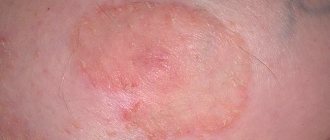
Lichen planus can cause Kaposi's eczema
The disease is activated when the body of a child or adult weakens under the influence of concomitant diseases.
Symptoms and diagnosis of viral course
The incubation period of the disease is 1-10 days from the moment the virus enters the child’s body. There is a prodromal period when herpetic eczema has not yet manifested itself, but there are prerequisites for the onset of the disease (weakness, loss of appetite, lethargy). The latent course is 3 days. Next, pronounced symptoms of the disease appear:
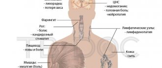
- Tachycardia, tachypnea, and shortness of breath are noted.
- Severe intoxication syndrome (fever, lethargy, weakness, decreased performance, drowsiness).
- Lymphadenopathy.
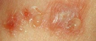
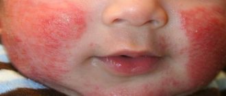
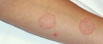
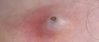
- 2-3 days after the temperature rises, a rash appears on the skin. Rashes appear in places of chronic dermatitis: on the arms, legs, lips, buttocks, neck, face. Skin lesions are represented by bubbles of different diameters, which are filled with clear liquid. Over time, pimples burst and erosion forms. There are dried crusts on the erosive surface. The rashes are painful.
- The child suffers from itching. Scratching the rash leads to infection with secondary microflora.
- After two weeks, the patient’s condition improves and the body temperature drops. Scars remain in place of the previous rashes.
- Viral eczema is often accompanied by severe diarrhea, prone to dehydration.
- With eczema herpetiformis, stomatitis develops. The disease manifests itself as painful rashes on the oral mucosa. The pathology is represented by aphthae in the form of small blisters that burst to form ulcers. The tongue is drawn into the pathological process. It is covered with a white coating with a small amount of bubbles. There is severe salivation and swelling of the gums.
- Sometimes viral conjunctivitis develops. The eyelids become swollen, the whites of the eyes turn red. There is constant lacrimation, photophobia, and intense itching.
Scientists presented several facts about the unusual disease:
- signs of atopic dermatitis immediately disappear after the appearance of eczema, but clinical manifestations of dermatitis may return;
- After illness, lifelong immunity is not formed. In adulthood, re-infection may occur due to reduced immunity or severe illness. It occurs again without complications. Recovery occurs in 5-10 days.
The diagnosis is made on the basis of complaints, anamnesis, examination, and additional research methods. A detailed questioning of relatives will help establish the fact of a herpes infection in the parents, frequent atopic/allergic dermatitis in the baby, and the use of immunosuppressants and glucocorticoids.
Upon examination, the doctor discovers a specific rash with a characteristic localization, lymphadenopathy, and an increase in the size of the liver and spleen.
A general blood test will help indicate the viral nature of the disease (leukocytosis/leukopenia, hypochromic anemia, lymphocytosis, eosinopenia). In some children, changes are observed in the general urine test (proteinuria, leukocyturia).
To confirm the diagnosis, an ELISA is performed, where a high titer of antibodies is determined. Using PCR, the DNA of the virus is detected.
In dermatovenerology, another examination is carried out called the Tzanck test. Using the study, cells are obtained from pustules. In case of herpetic infection, they contain herpesvirus inclusions.
Differential diagnosis of the disease is carried out with various pyoderma, chickenpox, and herpes zoster.
Clinical picture
Lesions spread to areas that have already been subject to inflammatory processes.
The incubation period for dyshidrosis herpetiformis is one week. At this time, there may be weakness, lethargy, and irritability.
After the specified time, there is a sharp deterioration in the child’s condition - the temperature rises, the pulse quickens, and rashes begin to appear.
A characteristic feature of this type of eczema is enlargement of the lymph nodes under the jaw, on the neck and in the back of the head. There may be tachycardia, difficulty breathing, and in severe cases, convulsive muscle contractions.
The appearance of bubbles filled with liquid is noted on the skin. Sizes vary from one to four millimeters. The formations grow in groups, often merging into large plaques. There is intense itching.
The blisters open on their own, and bleeding wounds form on the inflamed area, merging with each other. Gradually they become crusty.
After a couple of weeks, the crusts fall off and the body temperature returns to normal.
With the herpetiform type of eczema, patients in rare cases are diagnosed with intestinal disorders and dehydration.
Lack of treatment for eczema herpetiformis is fraught with adverse consequences in the form of purulent diseases of the epidermis and meningitis. Fatal outcomes are possible due to kidney and liver problems and problems with heart rhythm.
Treatment of Kaposi's eczema herpetiformis
Eczema is a contagious disease that is treated strictly in an infectious diseases hospital. The child and mother are placed in a separate room to avoid infecting other babies.
Herpetiformis is treated with the following drugs:
- Acyclovir effectively fights the herpes simplex virus. The drug destroys viral DNA, as a result of which the reproduction of viruses stops. Indications for the use of the drug include illnesses caused by Herpes simplex types 1, 2, Epstein-Barr viruses, and cytomegaloviruses. Contraindications for use include individual intolerance to the components of the drug and allergic reactions. The medicine is prescribed comprehensively in the form of tablets and ointments.
- Antiherpetic immunoglobulin is an antibody that acts against the herpes simplex virus. It is administered intramuscularly at 1.5 ml once a day. Course - 1 week.
Symptomatic therapy includes:
- antihistamines - prescribed to reduce symptoms of itching. Tavegil, suprastin are administered intramuscularly according to the child’s age;
- infusion therapy is carried out with saline, glucose, and Ringer's solution. Aimed at combating intoxication and dehydration;
- antibacterial drugs are prescribed to prevent the development of bacterial complications;
- antiseptics disinfect vesicles and prevent secondary infection. Erosion is treated with a solution of brilliant green, dermatolic, boric acid;
- To heal erosions, salicylic-zinc paste is used;
- antipyretics in syrup (Ibuprofen, Paracetamol) or injections (Diphenhydramine + Analgin + Papaverine) are used to reduce body temperature.
Prognosis and possible complications
A severe course is accompanied by serious disturbances in the functioning of internal organs. The circulation of the virus throughout the body leads to the following complications:
- meningitis, encephalitis;
- purulent otitis;
- keratoconjunctivitis;
- septic shock;
- development of secondary bacterial infection.
Complications are treated in the intensive care unit, where the patient is monitored around the clock.
The prognosis of the pathological condition directly depends on the resistance of the macroorganism, immunity, severity, and form of the disease. Death occurs in 20-30% of cases. According to statistics, an unfavorable outcome is observed with untimely antiviral treatment, late diagnosis, and non-compliance with doctor’s recommendations. With mild eczema, the prognosis is favorable.
There is no specific prevention for Kaposi's eczema.
If a child suffers from atopic dermatitis, avoid contact with possible carriers of the virus (patients with chickenpox, shingles, labial/genital herpes). The article has been verified by the editors