Almost every Caucasian person has age spots. Nevi appear on all parts of the body. They may vary in size, shape and color. A mole in the eye occurs in people of different age groups: elderly, adults and even young children. There are many factors due to which this formation is formed. Almost always, a nevus on the eyeball is benign.
Types of birthmarks
Nevi that form before the eyes are divided into two types based on their location. In accordance with this, if necessary, diagnosis and treatment are prescribed:
- A mole that appears on the front wall of the eye is called a conjunctival nevus. In this case, research procedures will not be needed, since the formation is detected visually without additional medical equipment.
- If a mole appears on the fundus, it is called a choroidal nevus. To identify such a pigment spot, it is necessary to use special tools.
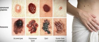
You need to know that any type of such formation does not pose a great threat to human health. But, like other moles, eye moles also require control. If it begins to transform, you need to undergo an examination immediately, otherwise melanoma may develop.
What to do with an ocular nevus?
If you suddenly notice a nevus appearing on your eye or your loved ones, then under no circumstances try to get rid of it using home methods. This can cause irreparable damage to your visual function!
You should contact an ophthalmologist and be registered with him at the dispensary. If the doctor has doubts about the benign quality of the formation or if it suddenly begins to change its color and size, the doctor will most likely recommend removing it, as this will prevent the development of melanoma.
Formation on the conjunctiva
The human eye is surrounded by a thin layer of mucous membrane, called conjunctival mucosa in medicine. It is so thin that the white of the eye and blood vessels can be seen through it. A person may develop a nevus at this site. Based on the findings of scientists, 5% of the entire earth's population faces such a problem.
It should be noted that the formation can appear not only on the outside of the conjunctiva, but also on the inside . There are several types of such nevus:
- Vascular. It is formed from a collection of capillaries that are contained in the mucous membrane of the eye. This nevus will be red or pink in color.
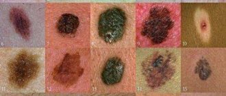
- Pigment formations. Melanin cells are concentrated in them. Such formations come in different colors, including black, they look like dots.
- Cystic nevus. In this case, the appearance of a mole was provoked by lymphatic vessels. It is noticeably different from other neoplasms; it looks like a bubble filled with liquid. If the cyst-like formation is greatly enlarged, then its surface resembles a honeycomb.
A formed nevus on the eye does not interfere with a person’s vision and does not cause discomfort. Even if it is located very close to the pupil, it does not block the light and does not affect the quality of vision. Treatment will be prescribed only if the neoplasm shows symptoms of malignant degeneration. According to the degree of development, a nevus on the conjunctiva can be stationary and progressive:
- The stationary one does not change its shape and location. It is absolutely safe.
- A progressive nevus changes its shape, size, and can quickly degenerate into a malignant tumor.
Such education needs to be given special attention and supervision. Medicine recommends getting rid of progressive moles using laser therapy.
Folk remedies
It is impossible to remove a mole at home, and it is very dangerous to engage in such self-medication. There are many methods for removing moles at home, some of which we inherited from our ancestors.
To remove moles, it is recommended to use:
lemon juice – the juice must be diluted with water and applied to the mole;- sea buckthorn oil – used to heal damaged moles and eliminate unpleasant sensations such as itching, peeling;
- castor oil – used to soften the skin;
- cauliflower juice – will help lighten pigmented skin;
- Celandine is a poisonous plant that will help disinfect a damaged mole;
- Chamomile decoction and lotions from it will help discolor the mole;
- grated potatoes and potato starch also help lighten the mole a little;
- pineapple juice - wipe moles with it in order to lighten them.
In fact, there are quite a lot of traditional methods for removing moles. The danger of such methods is that they can help, but they can also cause irreparable harm.
Some people remove moles on their own and then cauterize the removal site with brilliant green, alcohol, etc. but this is absolutely forbidden. Such mole removal can cost a person his life. The inflammatory process may not develop immediately after such removal.
No one can say exactly how a mole will behave after removal, but the doctor will conduct the necessary tests and be able to determine whether your formation is malignant or benign. If you have a malignant formation, then such a mole should be removed as quickly as possible, and the material should be sent to a specialized laboratory, where the necessary studies will be carried out.
These methods can help, but not remove, but slightly lighten the color of the mole. As for convex and hanging moles, these treatment methods will not bring any results. You won’t be able to remove a mole using these methods, but you can easily harm yourself, so you shouldn’t waste time looking for remedies for moles, but rather consult a doctor.
ATTENTION! You should be especially careful if you have thin, sensitive skin. Also, such procedures should not be performed on children.
Moles on the choroid
Such formations form inside the eye, and therefore are invisible to others. Choroidal nevus is formed from accumulations of vessels or pigment located on the inner surface of the eyeball, at the junction of the iris and the vessels. It can only be discovered through research. According to statistics, choroidal nevus is found in 2% of the population. Typically, such moles appear in adolescents subject to hormonal changes.
Choroidal nevi are small, dark or black in color. Very rarely they are colorless. In this case, it is impossible to recognize the birthmark until a certain point. Choroidal formation is usually progressive. As the nevus grows, it can impair vision. In addition, a person has complaints of pain in the eye, a feeling of the presence of a foreign object. If such symptoms appear, you should definitely consult an ophthalmologist.
The treatment prescribed by the doctor should be taken very seriously. He will choose the most effective methods, taking into account the condition of the nevus on the eyeball. Often in such cases, eye microsurgery is prescribed.
Is it possible to delete
If a nevus that appears on the lower or upper eyelid does not cause discomfort or interfere, there is no need to take action to remove it. If the mole is located on the eyelashes, in the corner of the eyes, or is painful, surgery should be performed to remove the growth. To remove a nevus on the eyelid, you must have experience and qualifications. Doctors involved in surgical intervention can excise the formation using the following methods:
- Radio wave excision is carried out using a high dose of ionizing radiation. The method is used for malignant growths. The advantage of the operation is that there is no scar or scar.
- To eliminate the risk of reappearance of the mole, a surgical method is used. Using a scalpel, deep layers of growth are removed. The disadvantage of the method is a scar at the excision site, which will ruin the person’s appearance.
- Laser removal prevents infection from entering the wound by cauterizing the blood vessels. The method is considered the best for getting rid of birthmarks on the eyelids. The operation lasts 3 minutes, there will be no scar left.
- Excision of nevus with high-frequency current. The benign formation is burned out with an electrocoagulator. After the operation, a barely noticeable scar may remain.
- Cauterization of the growth using liquid nitrogen is used for flat formations.
The doctor chooses which method is suitable for the patient based on the examination and test results.
Reasons for appearance
The etiology of the formation of a mole on the pupil or eyeball is always of the same origin. A mole in the eye means that the cells of the organ are oversaturated with a large amount of pigment - melanin. There are provoking reasons for the formation of a benign spot in the form of a nevus:
- Labor activity. Daily exposure to environmental factors during professional activities affects the pigmentation of any part of the organ of vision. Typically, an acquired benign nevus is found in hot shop workers - welders and metallurgists. In this case, the mucous membrane of the eye comes into contact with heated air and a bright source.
- Ultra-violet rays. Tanners who spend a lot of time in direct sunlight and do not wear sunglasses may develop excess pigmentation in the eyeball. This is how the body reacts to ultraviolet radiation, which has a negative effect on vision.
- Hereditary predisposition. Birthmarks are passed from parents to children with information in the DNA. In most cases, the dermatological defect passes through the male line, so the female half of the population almost does not suffer from this pathology.
- Frequent visits to the solarium. Artificial ultraviolet rays have a negative effect not only on the pupil, but also on the entire visual organ. Although protective glasses are used, the procedures may result in the formation of a benign mole.
- Chronic inflammatory processes caused by fungal or viral microorganisms can also lead to the appearance of a nevus. This provoking factor is the most dangerous, since a mole that arises due to chronic inflammation can transform into a malignant formation.
- Adolescence. During adolescence, the child's hormonal system is restructured. Increased secretion of hormones leads to excess accumulation of melanin both throughout the body and in the cells of the eye.
The meaning of nevus
On the upper eyelid
A rare occurrence is dark spots on the upper eyelid. People who interpret the origin of moles on the skin believe that such an arrangement indicates a person’s irritability and explosive nature. These people are critical, often dissatisfied with themselves and the world around them. Those with nevi on the eyelid are menacing in appearance, but easily vulnerable in their souls, they are emotional and take everything to heart. If a benign formation appears on the right eyelid, then most likely the person has poor resistance to stress and does not know how to control himself. A mole on the left eyelid means that a person is more frivolous, fickle in his thoughts, views and in love.
Do not pay special attention to the “magical” interpretation of moles on the lower eyelid and treat this with irony.
The meaning of nevus on the lower eyelid
What does a benign formation located at the bottom of the eye mean for women? We can immediately say that this sign does not bode well. Representatives of the fairer sex with such moles often have casual affairs and divorce their husbands. Their behavior is frivolous and family is far from the first place for them. A mole on the lower eyelid of any eye in representatives of the stronger sex indicates that the man is a supporter of a wandering lifestyle, and he can, under any circumstances, leave his wife, children, break off relationships with friends and change his place of residence.
Symptoms and diagnosis
The signs of a choroidal and conjunctival mole are almost the same. Their existence is usually not accompanied by any manifestations. Changes are detected only during an ophthalmological examination and then completely by accident. A nevus is dangerous in the eye when it degenerates into a malignant formation, then the following symptoms occur:
- the quality of vision deteriorates;
- the shapes of objects are distorted;
- sensation of a foreign body in the eye;
- limited field of view.
If such symptoms occur, you must follow all doctor’s recommendations to prevent dangerous consequences. The doctor gets acquainted with the medical history, clarifies the time of occurrence of the mole in the eye, and finds out about the presence of discomfort. Then he examines the mucous membrane of the organ and carries out diagnostic measures:
- Ophthalmoscopy. This procedure allows you to assess the condition of the retinal vessels and optic nerve using an ophthalmoscope. Using a red filter, you can easily detect a tumor. Using green, the doctor examines pathological changes in the structure of the eye that begin to progress as the nevus transforms.
- Echography. This diagnostic measure allows you to identify the focus of pigmentation.
- Ultrasound of the eye. The procedure examines the deep structures of the eyeball, which is necessary to monitor the development of a nevus.
- Fluorescein angiography. This test is designed to identify retinal angioma. The procedure is carried out by injecting a contrast agent (fluorescein), which illuminates the affected areas of the eye.
Usually, the presence of a mole in the eye can be detected independently if it is located on the outer layer of the mucous membrane. But it happens that the patient does not even suspect the existence of a nevus for quite a long period of time until symptoms arise. In this case, diagnosis of a mole is carried out only using comprehensive studies.
Types and forms of pathology
Choroidal nevus occurs:
Typical nevi:
Progressive nevi:
Atypical nevi
Treatment methods
If the mole is benign and does not cause inconvenience to its owner, treatment is not carried out. But the progressive formation should be under constant supervision. Control visits to a specialist are carried out twice a year. If the development of formation is detected, treatment is prescribed. The therapeutic complex is selected in accordance with the location of the spot and its size.
The developing form of pathology requires mandatory surgical intervention. First of all, an atypical mole is treated, since it has a high risk of transformation into a malignant form and a large cell nevus located in the area of the optic nerve. In medicine, there are several methods of surgical removal of moles on the eye organ:
- Electroexcision. The nevus is removed using an electric scalpel. This method is applicable for large malignant tumors with subsequent plastic surgery.
- Laser treatment. The mole is removed using a laser beam, which is effectively used to perform manipulations in hard-to-reach places.
- Cryodestruction is a frequently used method for removing a birthmark using liquid nitrogen.
- Brachytherapy. This method of nevus removal is considered simple and safe. Within an hour, the patient gets rid of a mole in the eye. Here, radio waves of various frequencies are used, with the help of which they not only remove the tumor, but also force the tissue of the eyeball to regenerate. Brachytherapy is used as the main procedure, but is also part of a treatment complex.
It is important to follow preventive recommendations to reduce the likelihood of developing a mole in the eye area. To avoid pathological changes in the eyes, it is necessary to eliminate infectious foci in the body. If there are hormonal imbalances during pregnancy or menopause, you should consult a doctor to restore hormone levels, if necessary. It should not be forgotten that the eyes should always be protected from the negative effects of ultraviolet rays, which contribute to the development of nevi.
When should you go to the doctor?
Any changes in the nevus, which is located on the eye, are very dangerous. Damage to such a mole is also dangerous.
You should consult a doctor if:
a mole on the eyelid increases in size. If we think logically, then the increase in such a nevus cannot in any way be related to your height. If this process is in no way connected with a person’s growth, then, consequently, some processes are occurring in the mole that can lead to the formation of a malignant tumor.- Itching, dryness, unpleasant tingling sensations and irritation on the mole should alert you. Painful and unpleasant sensations indicate an infection.
- Change in color of a mole on the eyelid. Moreover, the color of your mole may not change completely, but a spot on the nevus itself will be added to the main color.
- The appearance of pus, blood, and redness clearly indicate an inflammatory process, so you cannot postpone a visit to the doctor.
- The disappearance of the nevus and the appearance of a halo. The appearance of a halo and the disappearance of your mole on the eyelid are not always safe. Therefore, as soon as you notice changes, immediately consult a doctor, there is no time to hesitate.
- The owner of a mole should be alerted to changes in shape and the appearance of various growths on its surface.
- If damaged, it is also better not to self-medicate, but to seek help from specialists.
IMPORTANT! Damage to the nevus can cause complications and even lead to death.
Forms of the disease
Modern experts distinguish two types of ocular nevi:
- conjunctival moles;
- nevi of the eyeball.
Conjunctival nevi
The conjunctiva (iris) of the eye is the transparent mucous tissue.
Neoplasms on it can arise both from the outside and from the inside.
Depending on which cells take part in the formation and growth of the nevus, iris pathologies are divided into:
- Vascular. They occur in places where small blood vessels accumulate on the mucous membrane and have a red or pink tint.
- Pigmented. This form of pathology is characterized by a dark color, which is explained by the large amount of melanin in the tissues.
- Cyst nevi , as a rule, they are colorless, and in appearance they resemble small air bubbles. Cyst-like moles appear in places where lymphatic vessels connect.
Sometimes moles on the iris can change under the influence of hormones. In addition, some nevi may lighten over time and even fade completely.
Moles on the eyeball
Just like moles of the iris, nevi of the eyeball can be divided into several types depending on their variability:
- Stationary. This type of mole does not change its color or size. As a rule, no serious problems arise with such neoplasms. They are not dangerous to health and do not require surgical intervention, although examinations by an ophthalmologist should not be ignored.
- Progressive. This group of moles has the ability to change their shape, shade and size. Progressive nevi have a yellow border, which in some cases can cause worsening vision or a decrease in viewing angle.
IMPORTANT! If there is the slightest change in the appearance of the nevus or if a discomfort occurs in the eye, you should immediately consult a specialist.