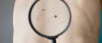
I thank Boris for the excellent opportunity to conduct an educational program on the issue of the connection between ordinary moles (nevi) and malignant skin tumors.
Read this short text to the end and your chances of encountering the serious consequences of these diseases will be close to zero. Here and in the future I will talk specifically about melanoma, because... It is the most dangerous among skin tumors.
Why chemotherapy doesn't help with melanoma
Melanoma is not sensitive to either chemotherapy or radiation therapy. The treatment of melanoma does not have an algorithm; it simply does not exist. For example, for lung cancer there are algorithms, there is an understanding of what drug regimens (protocols) to prescribe. In melanoma, the tumor can change the configuration of its proteins in a short time.
The proteins of melanoma cells are protected from the penetration of drugs, so the use of chemotherapy and interferons brings results in no more than 11% of cases and only for a short time - up to 6 months. Further, melanoma progresses significantly when immune function decreases. Treatment of melanoma with chemotherapy suppresses the immune system, stopping the synthesis of antibodies, monoclonal bodies, and lymphocytes. Lymphocytes' main function is to protect the body from microbes and foreign tumor cells. But if the patient suppresses the activity of lymphocytes using chemicals and prevents them from multiplying, then there is no one to protect the body.
Forecast and consequences
Major surgery carries a high risk of complications. If the form of the tumor is initial, then the consequence will be only scars or cicatrices. But you have to be careful. If an infection gets into the wound, a purulent complication may occur. This rarely happens in medical practice.
The prognosis of melanoma development plays an important role in successful treatment. It is worth calculating all the risks and possible consequences. Based on the information, the doctor prescribes treatment that is effective in a particular case. Talk to the doctor.
Forecast
It is good if the tumor can be found in the initial stage. The likelihood of a favorable treatment outcome is high. The main threat of melanoma is that during the first year of therapy there remains a high risk of relapse. Comprehensive treatment will eliminate melanocytes and reduce the likelihood of its development in the body.
For advanced stages of cancer, palliative therapy is carried out. This helps maintain the vitality of the body. The rate of spread of malignant cells directly affects life expectancy. The smaller the metastasis, the better.
The age of the patient is also important. A young body has more strength and a chance to recover quickly. After tumor removal, life expectancy varies among people. A person is capable of living to a ripe old age. You must follow your doctor's recommendations and follow his instructions.
Removal of lymph nodes contributes to the occurrence of lymphostasis. The movement of lymph will be disrupted. The result is swelling of the affected limb, which leads to an increase in its volume. There is also a high risk of developing seroma. This is a situation when lymph begins to accumulate in the area of the removed nodes. It is dangerous because it later begins to rot. And this excess liquid must be removed.
Possible complications
Any method, both surgical and laser, carries a risk of complications. If you have any concerns about your condition getting worse, you should tell your doctor.
Laser removal of melanoma
This situation needs to be discussed and a further solution must be reached. There are 4 groups of complications:
- Getting infected. This situation occurs when treatment is carried out under unsterile conditions or wounds are poorly treated. As a result, the patient experiences burning and itching around the wound.
- The appearance of a new tumor. It may occur near the site of a previous excision. This is the result of an incorrect procedure to eliminate melanoma. As a result, the neoplasm is also removed along with healthy epidermal cells.
- The appearance of scars. They appear when large tumors are eliminated. The areas need to be sutured, which is why visible scars remain.
- Long wound healing and bleeding. It depends on the individual abilities of the body. Or it is a sign of dysfunction of the lymphatic system.
Melanoma can only be removed in a medical facility. Other methods pose a danger to the health and life of the patient. The tumor is eliminated by removing the root; special equipment is required. An incorrect operation will cause the cancer cell to grow at a faster rate.
Melanoma surgery
Surgery is performed in the presence of a primary lesion. If necessary, lymph node dissection of the sentinel lymph node to prevent the spread of cancer cells along the lymph flow paths. Surgery as the only method of treating melanoma is used in the early stages of melanoma development without metastasis.
In this case, the tumor is excised along with the surrounding skin (at least 3-5 cm away from the edge of the tumor on the trunk and limbs, 2-3 cm on the face), subcutaneous tissue, fascia or aponeurosis. Therefore, plastic surgery has to be performed frequently to close the wound defect.
Indications for melanoma removal
If you suspect a malignant tumor, you should consult a doctor. After a complete examination, the doctor will be able to prescribe the correct tests and treatment. Removal of melanoma is carried out at an early stage of development. This will prevent it from further increasing.
The method for removing melanoma depends on a number of factors. Size, degree of germination, individual parameters of the body - all this influences the choice of treatment. The matter will not be limited to one operation. Later, you will need to consolidate the result with the help of postoperative treatment - you will have to take medications and undergo chemotherapy courses.
In advanced stages of tumor development, cancer cells actively multiply and penetrate into tissues. During this time, treatment is carried out using intensive immunotherapy. To avoid such situations, familiarize yourself with the first symptoms of the disease:
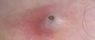
- The mole changes shape, and its boundaries become fuzzy and blurred.
- The size of the tumors increases.
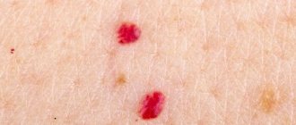
- The pigment changes color - it becomes bright, red dots appear.
- These areas are itchy and itchy.
- Places with moles bleed.
If you notice a similar symptom, consult a doctor as soon as possible.
Modern treatment of melanoma
Long-term observation has shown that the spread of metastases occurs in a period of 6 to 12 months, the average life expectancy of patients with metastases in one organ is 7 months, in two organs - 4 months, and in three or more organs is 2 months. Dacarbozine, other chemotherapy drugs and interferons increase life expectancy by only 9%, but they do not help everyone.
What modern drugs can patients pay attention to?
1. T-lymphocytes (TIL therapy), 2. Biovaccine based on dendritic cells, 3. Activated NK cells.
We invite patients to take part in new methods of treating melanoma, as well as in clinical trials of new drugs
Removal methods
There are two ways to eliminate a malignant tumor: using a laser or a scalpel. Removing melanoma with a laser is painless and is performed without anesthesia. The procedure does not leave large scars or complications as a result. This method only affects malignant epidermis. Healthy areas are not affected. The laser is prescribed exclusively for small tumor sizes - 5-8 mm in diameter. And the device does not have the ability to reach the deep layers of the epidermis.
And using a scalpel allows you to do this. This is the only way to penetrate deep into the skin. Although the scalpel is effective, it is not without its drawbacks. The instrument leaves a large scar. This occurs as a result of the process creating a large, slow-healing wound. It will need to be processed periodically.
There is a risk of bleeding during surgery. Since this is a serious intervention in the body, infection can be introduced through the wound. Special care is prescribed. Plus, incomplete removal of melanoma does not guarantee recovery - the risk of recurrence of the formation remains high.
The second option is popular. Doctors prefer to work with a scalpel. In their opinion, this is an extremely effective way to eliminate melanoma.
Contraindications to laser use
Both methods are used in medical practice. But laser melanoma removal is not for everyone. This is the penetration and influence of rays. You should be careful. The use of laser is prohibited:
- During pregnancy and during breastfeeding.
- The presence of chronic heart and vascular diseases, pre-stroke and pre-infarction conditions.
- Very sensitive skin.
- New occurrence of melanoma after elimination.
Although the procedure is painless, side effects are possible. Doctors do not prescribe it to all patients.
Specifics and methods of surgical intervention
The essence of surgical removal of melanoma is that the cancer cell is removed along with surrounding healthy cells. This prevents the spread of malignant cells to nearby areas of the skin. The depth of the cut depends on the size of the tumor. In medicine, 4 methods of eliminating tumors have been created:
Melanoma on the hand
- Easy removal. Painkillers are injected into the area around the tumor, then the melanoma is cut out with a scalpel. And the wound is sewn up with threads and then removed. A scar remains on the area of skin.
- Wide excision. The method is carried out in advanced stages of melanoma. A marker is used to outline the area around the tumor with a margin of 2 cm in each direction. This helps reduce the possibility of a new formation appearing. The resulting wound is sutured.
- Amputation. This method is used in cases where melanoma appears on the extremities. At an early stage, parts of the fingers or toes are removed. If the tumor form is neglected, you can lose a limb.
- Removal of lymph nodes. Due to melanoma, metastases quickly appear in the lymph nodes nearby. Getting rid of them involves removing the lymph nodes. This reduces the risk of their distribution throughout the lymphatic structure.
For a routine operation, anesthesia is used. Sometimes the procedure is performed under local anesthesia. Amputation of limbs and lymph nodes is performed under general anesthesia. This will help to overcome the painful shock.
Therapy
- • Biovaccines as the future of oncology
- • Cellular immunotherapy of melanoma
- • Therapy of melanoma at different stages
- • Melanoma with metastases to lymph nodes
- • New drugs
- • We treat metastatic melanoma with a T-lymphocyte-based vaccine
- • TIL program
- • Treatment of the third stage of the disease
- • Treatment of stage 4 melanoma
MYTH #1: Moles cannot be removed - it leads to death from skin cancer
There are currently no scientific studies that show that removal of benign nevi is associated with an increased risk of melanoma. The scenario “removed a mole and died” is possible only in two versions.
– removal of a malignant tumor without histological examination, “diamond by eye”
- error of the doctor who conducts the histological examination
Other scenarios are not possible . The risk of developing melanoma at the site of removal of a benign mole is the same as at any other part of the body.
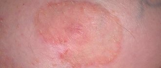
Superficial spreading melanoma, stage I.
Removal of benign moles (nevi) with histological examination does not increase the risk of developing melanoma.
Life expectancy with new drugs
- • Zelboraf
- • Ipilimumab
- • Keytruda
- • Opdivo
- • Tafinlar
- • Dacarbazine
- • Tumor necrosis factor
General principles
If melanoma is not removed, it will continue to grow and metastasize, so if left untreated, the process will most likely be fatal. Therefore, surgical intervention is contraindicated only in two cases:
- the patient’s general condition is so unstable that he will most likely not tolerate anesthesia;
- the prevalence of the tumor is such that it is impossible to remove it: conglomerates of lymph nodes are fused with the surrounding tissues, there are multiple distant metastases.
Now that non-invasive methods for diagnosing melanoma have become widespread, such as dermatoscopy, when a doctor can examine a suspicious tumor under high magnification and make a preliminary diagnosis, the first stage of the operation to remove melanoma takes place even before histological examination. The neoplasm is excised to the entire depth of the skin with an indentation of about 5 mm and sent for examination under a microscope. This procedure is called an excisional (stab) biopsy.
There is still no consensus on under what anesthesia to perform an excisional biopsy: some experts believe that local anesthesia promotes the spread of malignant cells, while others believe that it does not worsen the course of the disease. In practice, a suspicious tumor is excised on an outpatient basis and under local anesthesia.
Moles and melanoma
In no case should moles be removed (even if they do not appear malignant) using a laser, cryo- or electrical destruction, or other methods that do not leave material for histological examination. Practice shows that “ugly” moles, which are usually removed without medical indications, often have areas of malignant degeneration. Therefore, any removed nevus should be considered as material for histological examination.
If the biopsy confirmed the malignancy of the tumor, the thickness of the tumor should be indicated in the histological report. Next, the suture after removal is resected again, capturing the surrounding skin:
- 0.5 cm, if the neoplasm is located within the epidermis and does not grow into the basement membrane;
- 1 cm for tumor thickness up to 2 mm;
- 2 cm if the thickness of the neoplasm is more than 2 mm.
The skin is removed to its full depth, fatty tissue down to the fascia (a thin connective tissue membrane that covers the entire body like a stocking).
Since during such operations the skin defect becomes quite significant, they are supplemented with pedunculated skin grafts or other plastic methods.
If the primary tumor is thicker than 0.75 mm, the nearest lymph node is removed under anesthesia, which drains lymph from the affected area. He is also sent for histology. If malignant cells are detected, the entire complex of lymph nodes with surrounding tissue is excised, and conservative therapy is additionally prescribed.
Surgical tactics for progression
Local relapses of melanoma are also subject to removal, when tumor foci reappear in the area of the postoperative scar or excised lymph nodes.
Advanced forms of melanoblastoma, when distant metastases are clearly identified, are subject to surgical treatment only if they can be removed. For example: the initial lesion has not grown into the muscles, and there is a single metastasis to the brain that does not affect vital centers, or a single metastasis to the liver, when it is possible to remove part of it, and so on. In each case, the issue is resolved individually. Treatment must be supplemented with medicinal methods.
Debriefing
Now that we have a specific reason for panic in our hands after removing a mole with good histology, let’s analyze it in detail. There are many questions for the author that cannot be answered based only on the message itself:
- What is your name, where do you live? Are you a real person?
- Are there medical documents to support your words?
- Where was the histological examination of the mole performed and was it done at all?
- Are you sure that melanoma developed in the exact same place where the mole was removed?
- Was a histological examination of the removed tumor performed? Where are his results?
- How did the doctors at the clinic find out that melanoma developed from “cells left behind in the dermis”?
- Tell us more about the innovative method of removing melanoma, which leaves a hole and not a surgical suture?
- Are you by any chance an employee of a clinic that decided to advertise itself in such a clumsy way?
The absence of an answer to even one of these questions casts serious doubt on the reliability of the entire post as a whole.
Stage 3
At the time of diagnosis, melanoma had already spread to the nearest lymph nodes. Therefore, during surgery they are also removed. The cost of surgery for melanoma will be higher. The person may need more time to recover. In addition, additional treatment methods are always used after melanoma removal; the cost of the course of therapy increases as a result.
After surgery use:
- targeted therapy;
- immunotherapy;
- radiation therapy.
Pathological areas of tumor tissue can be found not only in the lymph nodes, but also under the skin, inside the lymphatic vessels. All of them are removed if possible. But sometimes at stage 3 melanoma cannot be cured completely. In this case, to increase the patient’s life expectancy, additional regional chemotherapy, therapeutic vaccines, and external irradiation are used.