What it is
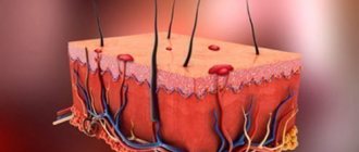
Moles (nevi) are clusters of pigment cells. They look like spots or compactions of varying sizes and shades. Pigmented ones begin to appear at the age of 5-10 years.
There are also vascular formations that arise due to the proliferation of endothelial cells lining the inner surface of blood vessels. They are called angiomatous. They are often congenital, but they also occur with some somatic diseases, for example, pancreatitis.
Warts are raised plaques that rise above the surface of the skin and resemble cauliflower. They are considered dangerous. With regular injury or exposure to direct sunlight, they can turn into a cancerous tumor.
Accumulations of pigment have different shades.
- Red (vascular). They are rarely reborn. They are a collection of blood vessel cells.
- Brown, resulting from the accumulation of melanin. They require special attention, as they most often turn into melanoma.
- Blue and bluish. Located in the deep layers of the dermis. They often appear as a result of hormonal changes in the body. They almost never degenerate into cancerous tumors.
According to their shape, they are classified as convex and flat. Convex ones are more dangerous, since they are easier to injure, for example, by hitting clothes or jewelry. Their condition must be monitored especially carefully.
Features of nevi (moles)
Moles are skin formations consisting of melanocytes (pigment cells) involved in the production of melanin. There is a varied number of nevi, which are characterized by the following features:
- The process of mole formation begins at birth and lasts on average up to 10 years. In later life, the appearance of new moles or changes in existing ones may also be observed.
- Pigment formations can have different sizes and diameters: from small 1-2 mm to large ones - 10-20 cm.
- The base color of moles varies from light beige to dark brown. Red moles can also occur, particularly during pregnancy - angiomas, which are benign vascular tumors.
- The surface of the formations is often flat, but bumpy nevi are also found.
- Some moles are accompanied by hair growth (usually from the center of formation), which is not considered a pathology.
Initially, all moles are benign formations and are not considered as a skin pathology.
Only in some cases, under the influence of various factors can provoke the development of a malignant process.
(Genetic predisposition, nevus injury, increased exposure to UV).
Causes
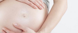
Normally, pigmented nevi appear when exposed to ultraviolet radiation or as a result of changes in hormonal levels. The latter reason leads to the appearance of a large number of brown spots in expectant mothers.
An increase in moles during pregnancy is also considered normal. After childbirth they disappear on their own. If this does not happen, there is no need to be upset. If the stain does not change color or size, it is not dangerous.
A sharp increase in moles during pregnancy should be a reason to consult a doctor. This indicates a sharp change in hormonal status and problems with the endocrine system.
Myths
The formation of moles during gestation is a common phenomenon. Due to the lack of knowledge on this issue, women began to come up with their own explanations for this process. Most of them have not been scientifically confirmed, but are still popular among the population.
Such myths include:
- If, while carrying a child, a mole forms on the body of the expectant mother, then the baby will also have one, and in the same area. There is no evidence to support this opinion. Most children emerge with clear skin without any growths. They begin to form by adolescence, and their location can be any. Although, if there is a genetic predisposition, a similar outcome is possible, and if the mother had many moles, they may appear in the child.
- Based on the location of the nevi, predictions can be made regarding the fate of the mother and baby. This myth is also not confirmed. There is no relationship between skin spots and life events.
- A pregnant woman can cause nevi to appear on the baby's body. This is supposedly possible if, in severe fright, she grabs some part of her body. According to myth, the baby will form a birthmark in this place. But this assumption is wrong. Expectant mothers should not worry about this; in their condition, it is harmful to worry.
Moles on the body are a harmless phenomenon, even if they form in a pregnant woman. This is due to changes in hormonal levels. When it returns to normal, most of the tumors will disappear. Those nevi that remain are also not dangerous if they are not accompanied by pathological symptoms. Removing moles during pregnancy is rarely practiced if there is a threat to the health of the mother and baby.
What is the danger
Nevi are benign formations. They do not pose a danger to the expectant mother and child. But doctors consider clusters of melanocytes to be an optional precancerous condition.
Under certain conditions, they degenerate into a malignant tumor that can grow into surrounding tissues and cause metastases.
Hanging moles during pregnancy require special attention. They are easily injured and even torn off, so it is advisable to remove them before the intended conception.
More often, nevi degenerate into melanoma, a malignant tumor of pigment cells. It can quickly metastasize to distant organs.
Because of this, her treatment is successful only in the first and second stages of development. During this period, metastases are absent or appear only in nearby tissues.
Melanoma cells are small. Through blood and lymphatic vessels they enter any organs and tissues, including the brain.
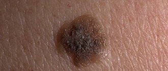
Changes in a pigment spot that has developed into a tumor may be minimal. Therefore, melanoma is often diagnosed only by the presence of metastases in the liver, brain and even in the heart.
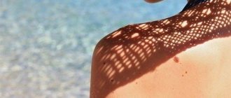
Particularly dangerous are formations located in areas subject to constant trauma (on the scalp, neck), as well as in open areas of the skin exposed to sunlight.
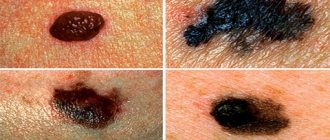
The following changes should be alarming:
- increase in size;
- the appearance of itching or burning;
- redness of the skin around;
- bleeding, the appearance of small ulcers, discharge of pus;
- the appearance of several new spots next to the nevus.
A malignant tumor affects the condition of the entire body as a whole. The following symptoms are observed:
- increased fatigue;
- drowsiness, irritability;
- decreased appetite;
- losing weight while maintaining the same diet.
These signs may accompany normal pregnancy. You should consult a doctor if your condition worsens suddenly and unexpectedly.
How to prevent the formation of moles during pregnancy
In order to once again protect your body from the appearance of neoplasms, pregnant women are advised to do the following. Necessary:
- Try to stay in the open sun as little as possible or wear clothes that cover your body as much as possible. It is necessary to forget about visiting a solarium during pregnancy, because this procedure dries out the skin, which also stimulates the activity of melanocytes.
- Take proper care of your skin. During gestation, the female body increases sharply in size, which can cause itching of the skin and scratching of existing moles. To prevent your skin from itching, you should moisturize it regularly and choose soft bathing products.
- Take enough vitamins. For example, a lack of folic acid in a pregnant woman’s body can lead to the appearance of new moles.
- Wear clothes that are pleasant to the body and do not restrict movement. Synthetic, rough material can injure the skin, which will lead to the formation of neoplasms. Try to wear clothes made primarily of natural fibers, with minimal addition of synthetics.
Having discovered a new mole on your body, you should not immediately worry and run to the doctor. A pregnant woman should be less nervous and listen to her body, which itself will tell her if everything is okay.
Which doctor should I contact?
If the formation has changed color or size, you should consult a dermatologist. He will prescribe additional tests (biopsy, blood test for tumor markers).
If a dangerous neoplasm is detected, the woman will be sent to an oncodermatologist (a specialist in malignant and benign tumors). He will conduct additional examinations and choose future treatment tactics.
If it turns out that a process of malignancy has occurred, that is, degeneration into a malignant tumor, examinations such as MRI are recommended to identify metastases in organs and lymph nodes.
Removal
New moles are rarely removed during pregnancy. This is due to the fact that any intervention poses a potential danger to the health of the fetus.
Removal is recommended only if the risk of developing melanoma is high. The nevus is removed under local anesthesia using a special scalpel.
The removed tissue is sent for biopsy. Doctors must evaluate the condition of the cells that make up the tumor to determine whether it is malignant or benign.
Removing moles on the stomach during pregnancy in beauty salons is strictly prohibited. The surgical method must be chosen by an oncodermatologist. The decision to intervene is made if the benefit of the operation for the mother significantly outweighs the potential risk to the fetus.
Diagnosis of moles during pregnancy
What to do if you notice the appearance of new age spots on the skin or an old mole has enlarged during pregnancy?
It is necessary to make an appointment for an in-person consultation with a dermatologist.
The doctor will conduct a visual examination of the pigment formation, assess its condition and appearance.
Dermoscopy is mandatory to determine the possible pathogenicity of the nevus.
Dermatoscopy is a non-invasive method for the differential diagnosis of pigmented skin lesions and early diagnosis of malignant melanoma.
This technique is also known as dermoscopy, reflected light microscopy, and epiluminescence microscopy.
Dermatoscopy is an integral part of the dermatological examination of the skin.
Today, the dermatoscope is considered the “dermatologist’s stethoscope.”
In addition to the diagnosis of pigmented and non-pigmented skin tumors, the method will be able to establish the diagnosis of infectious, parasitic and inflammatory lesions of the dermis.
In the field of dermato-oncology, dermatoscopy contributes to preoperative planning for the removal of pigmented skin lesions.
Helps you choose the best treatment option and subsequent care.
The main task of a dermatological examination is to find out the following points:
- whether the lesion is melanocytic or non-melanocytic;
- whether the formation is benign, suspicious or malignant.
In modern dermatology, digital dermatoscopy is more often used.
Where diagnostics displays the results of the procedure on a computer monitor.
This allows you to create reports with patient data and images of his pigmented lesions.
This makes it easier to make a diagnosis and makes it possible to consult with other specialists.
Another advantage of this method is that the images can be printed and given to the patient.
Alternatively, the digital images can be transferred to a CD and sent to the appropriate physician.
Prevention
To avoid degeneration of education, you should:
- at the planning stage, remove dangerous nevi that are subject to constant trauma and are located on open areas of the body;
- do not be exposed to direct sunlight without using sunscreen;
- consult a doctor if you notice changes in the shape, size or shade of a mole during pregnancy;
- take medications prescribed by your doctor;
- independently examine nevi in order to track their pathological changes in time;
- regularly take blood tests and check the level of hormones in the blood.
Only attention to your health and monitoring the condition of formations will help avoid the development of cancer.
The appearance of moles during pregnancy is normal. If they are small, round in shape and do not rise above the surface of the skin, there is no need to worry. Bleeding, increase in size, shape - a reason to immediately consult a doctor.
The material was prepared specifically for the website kakrodit.ru, edited by diagnostician O.S. Vasilyeva.
Symptoms of dangerous moles
Moles that appear during breastfeeding are most often the result of hormonal imbalances. If nevi don’t hurt, don’t grow, and don’t bother you at all, then in principle you can forget about their existence.
However, there may be situations when moles cause a lot of problems. Mothers should know the symptoms that indicate degeneration into melanoma:
- The nevus began to change its color.
- Education began to grow rapidly.
- The boundaries are not clear.
- The development of the inflammatory process is observed.
- Feeling of discomfort in the form of pain and itching.
A young mother can independently see these signs in herself, but only a doctor can determine the true nature of the changes.
Pros and cons of a significant number of moles on the body
If a person has many moles, is it dangerous? Most often, moles do not pose any danger. When nevi do not change over many years, this is not a disease. Sometimes it can happen that flat nevi rise above the surface of the skin (the neoplasm becomes convex). Such nevi are called dermal nevi.
Important! If moles change, you should consult a doctor. This will help rule out melanoma at an early stage. For this, patients should consult a dermatologist. A qualified specialist diagnoses the process of degeneration that has begun.
If the skin has many moles, then ultraviolet radiation under the open rays of the sun becomes dangerous, and human health is susceptible to the negative consequences of tanning.
The rapid formation of melanoma occurs not only under the influence of the sun, but also when the mole is injured. The cellular melanocyte begins to multiply rapidly to heal the injury.
If a person is predisposed to cancer, the mole develops into melanoma.
Therefore, doctors warn about the dangers of tanning to those patients who have a hereditary predisposition to the occurrence of a tumor process. Tanning is allowed under the influence of scattered rays. After swimming in a pond, and especially in sea water, you need to dry yourself.
This is due to the fact that moisture tends to accumulate on convex nevi. Under the influence of drops, a magnifying glass effect is obtained. When the sun's rays pass through moisture, the effect increases several times.
For some people, tanning in the open sun is generally contraindicated. This category includes people with white skin.
Those representatives who have more than 40 moles on their body are also prohibited from sunbathing.
be careful
The presence of papillomas, warts, condylomas, moles and spines on the body is the first sign of malignant melanoma!
We hasten to warn you that most medications “treat” warts, papillomas, moles, etc. - this is a complete deception of marketers who make hundreds of percentage points on drugs whose effectiveness is zero. They do not cure the disease, but only mask the symptoms.
The pharmacy mafia makes huge money by deceiving sick people.