Angioma is a neoplasm that is classified by doctors as a benign tumor. This pathology is a collection of blood and lymphatic vessels. Outwardly, most neoplasms resemble moles, and therefore are popularly called “red moles” or “port-wine stains.” In most cases, angiomas occur during fetal development, but sometimes they appear in older people or can be an external manifestation of a number of diseases.
What is angioma
Angioma is a benign vascular tumor that appears as a result of the transformation of blood vessels. The neoplasm can be of different sizes and shapes; its appearance resembles a regular mole, but is red in color. The localization of angiomas also differs and depends on the individual characteristics of the woman’s body.
Angiomas on the body
In addition, the mole may be flat or raised above the surface of the skin.
Complications
When such neoplasms appear on the surface of the skin, there is no need to panic, since they do not pose a danger to human health and life. Neoplasms that are located in deeper layers and on internal organs are considered more dangerous, since they often provoke the development of internal bleeding.
The following complications may occur:
- Formation of thrombosis.
- Penetration of infection.
- Deterioration of respiratory function.
- impairment .
- Complication of urination and defecation processes.
- Impaired functioning of the gastrointestinal tract.
The appearance of such symptoms requires an urgent visit to a specialist in order to diagnose and prescribe adequate treatment.
Reasons for appearance
Angiomas on the body (the causes in women may be related to physiological and pathological factors) appear in patients of different ages. The greatest number of cases of their occurrence was recorded among people from 20 to 45 years old.
The main causes of tumor formation are the following:
- Disturbances in the functioning of the vascular system, when small capillaries in the skin change and form a nodule.
- Diseases of the lymphatic system that provoke the accumulation of lymphatic vessels under the skin.
- Hormonal imbalance as a result of stress, overwork, treatment with potent drugs, during pregnancy and breastfeeding.
- Liver diseases of varying severity, provoking changes in blood vessels.
- Genetic predisposition to the appearance of such neoplasms.
- Self-administration of hormonal drugs.
- The presence of malignant tumors in the body becomes a cause in exceptional cases.
In women during menopause, all processes in the body change. This can also lead to angioma, but the likelihood is not as high.
Causes
Various factors can provoke the formation of red growths, and they are somewhat different in adults and children. Therefore, when talking about the causes of angioma, it is necessary to take into account the age of the patient.
Causes of pathology in adults
Tumors can appear on any part of the body, but the most “favorite” places are the back, arms, neck, and chest.
On this topic
- Skin covering
Treatment methods for atheroma without surgery
- Natalya Gennadievna Butsyk
- December 9, 2020
The reasons due to which angioma forms are not fully understood, but medical scientists put forward several theories on this matter, according to which the following factors provoke the disease:
- Thinning vessels , which occurs as a result of vitamin deficiency in an adult.
- Inflammatory reactions occurring in the digestive organs.
- Concomitant diseases of the cardiovascular system.
- The development of hemophilia is a rare hereditary disease associated with a violation of the blood clotting process.
- Pyelonephritis is an inflammatory pathology of the kidneys, which is characterized by damage to the kidney parenchyma, calyces, and renal pelvis.
- Bad heredity.
- Disturbances at the hormonal level as a result of diseases (diabetes, thyroid pathologies, etc.), as well as during pregnancy, at the onset of menopause, and when using contraceptives.
- Liver diseases - in this case the tumor acquires a rich burgundy color.
- Regular injuries , for example, this is often observed during shaving, when capillaries are damaged.
- Chronic infectious pathologies.
- Malignant tumors.
- Regular exposure to direct sunlight - such neoplasms are the result of ultraviolet radiation. At the same time, others prove that such a factor is not capable of causing the appearance of angioma, since they do not contain melanin.
A distinctive feature of a red mole on the skin is that the color disappears when pressed, but after a few seconds the color returns.
If a person observes a large number of neoplasms, then an urgent need to visit a dermatologist, since often such a phenomenon can indicate the formation of complications, including the development of cancer pathology.
Angiomas can grow as a result of inflammatory reactions occurring in the stomach and intestines.
Reasons for formation in children
Sometimes neoplasms are diagnosed in newborns in the first days of life. This indicates a disruption in the development of the child’s circulatory system. The main reasons for this pathology are the following factors:
- The growth of capillaries into regional tissues is observed with abnormal development of blood vessels in the womb.
- Abuse of alcoholic beverages and tobacco products by the expectant mother during pregnancy.
- Uncontrolled use of medications during pregnancy.
- Infectious experienced by the mother during the intrauterine development of the child.
- Acute deficiency of vitamins in the body of a pregnant woman.
- Progression of chronic pathologies during pregnancy.
On this topic
- Skin covering
Why does the wen itch?
- Olga Vladimirovna Khazova
- December 5, 2020
If single neoplasms are observed that are not large in size, then specific therapy should not be carried out.
However, constant monitoring is necessary to ensure that the pathology does not increase in size, does not change color, and does not cause painful discomfort. If such symptoms do not appear, then most often after a few years the angiomas disappear on their own.
Types of angiomas and locations in women
Experts divide angiomas into several types. The first classification involves the identification of 2 species, taking into account the structure, into monomorphic and polymorphic. The first type is a tumor formed as a result of mutation and accumulation of one type of cell. The second type is a tumor formed by several types of cells, usually growing to large sizes.
Taking into account the form of the neoplasm, doctors also distinguish several varieties:
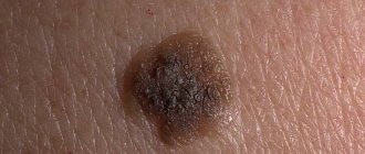
- A simple one is formed as a result of the accumulation of capillaries. Externally, such a mole looks like a small red spot, located on the body or mucous membranes; when pressed, its color becomes less bright. Usually the patient does not pay attention to the stain, since it does not bother and is not damaged by clothing.
- Cavernous angioma is usually medium or large in size, raised above the surface of the skin, and burgundy in color. When you touch it, you can feel a local increase in temperature. In most cases, inside the formation there are cavities filled with thrombotic masses, so when damaged they protrude out and provoke bleeding.
- The branched type of tumor is considered dangerous. It consists of a plexus of blood vessels; upon close examination, you can see a barely noticeable pulsation. Around the neoplasm there are vessels located close to the surface layer of the skin; they visually increase its size and nourish the changed cells.
The location of angiomas on the body also differs. In women, they can be localized on the back, inner and outer surfaces of the shoulders and forearms, abdomen, chest, and neck. Less commonly, neoplasms appear on the legs, head, and genital area.
Symptoms
The manifestation of symptoms directly depends on the location of the formation and its size. More often, disorders are diagnosed in newborns in the first days after birth.
A characteristic feature of this phenomenon is the fact that girls suffer from neoplasms much more often than boys.
Rapid growth of angiomas is often observed; literally within three months, the neoplasm can increase from one millimeter to several centimeters at once.
On this topic
- Skin covering
What does a wen look like on the body?
- Olga Vladimirovna Khazova
- December 5, 2020
Localization can be observed, for example, vascular forms can form on any part of the body:
- Bone elements, muscle fibers (damage to the spinal trunk is more often observed).
- Leather.
- Mucous epithelium (oral cavity, genital area).
- Internal organs (most often pathology manifests itself in the brain, head, liver).
The cavernous and venous form, which is localized on the surface of the skin, does not hide any special problems and is considered only a cosmetic defect that does not require specific therapy and does not cause other problems for the patient.
However, it should be remembered that pathologies located on the internal organs can provoke a number of serious complications.
When bone structures are damaged, patients note the following symptoms:
- Severe pain discomfort.
- Deformation of the bone skeleton in the affected area.
- Radicular syndrome.
On this topic
- Skin covering
Treatment methods for lumps in the armpit
- Olga Vladimirovna Khazova
- December 4, 2020
In places where the tumor has appeared, repeating pathological fractures are possible.
Pathologies of the brain are considered the most dangerous to health, and at the initial stages they do not manifest certain signs.
As the disease progresses, the following series of neurological symptoms appear:
- Deterioration of vision.
- Speech dysfunction .
- Sensitive headaches.
- Impaired motor function, deterioration of motor coordination.
- Increased blood pressure.
- Damage to intracranial nerve fibers.
- Hemiparesis is a neurological disorder in which there is a restriction in the movement of muscle fibers on the right or left side of the body when the opposite hemisphere of the brain is affected.
Often, an angioma located on the brain can cause impairment of consciousness and paralysis of certain parts of the body, which also directly depends on the location of the tumor.
The most dangerous complication in such situations is considered to be hemorrhages in the brain tissue, in which there is a sharp deterioration in health, and death is not excluded.
A number of general symptoms that are characteristic of all types of pathology have also been identified:
- Feeling of heaviness in legs and arms.
- Hyperhidrosis is a sweating disorder, manifested by increased sweat production as a result of dysfunction of the autonomic nervous system.
On this topic
- Skin covering
Lump behind the ear
- Natalya Gennadievna Butsyk
- December 4, 2020
If the tumor is located in the oral cavity, the patient suffers from difficulty swallowing, and is also plagued by pain in the mouth, directly in the place where the tumor is located.
Brief manifestations of neurological signs cannot be ruled out; this applies to situations where the tumor spreads to areas of the brain.
The appearance of signs of the disease directly depends on the location, size of the tumor and the characteristics of its structure.
Are red moles on women's bodies dangerous?
Angiomas on the body (causes in women can provoke the formation of several moles at once) are benign growths, and therefore in most cases do not pose a threat to a woman’s health. However, this is a tumor that, under certain circumstances, can develop into a malignant one.
Red moles become dangerous when they are constantly damaged during hygiene procedures or when injured by clothing. In addition, there is a high risk of cell degeneration with regular exposure to chemical compounds present in cosmetics. Sun rays also have a negative effect on angiomas, so they can provoke cell degeneration.
What it is?
skin angioma in the photo
Angiomas are considered benign neoplasms, and they appear on the skin of the body of both women and men. Many experts associate their appearance with disturbances in the normal functioning of the circulatory system or lymphatic system. Until the age of seven, red moles can disappear from the surface of the skin on their own, without special treatment.
It is believed that the occurrence of these growths does not pose any danger to humans, but it is important to know that any neoplasm on the surface of the skin should be carefully monitored, since cases of degeneration of benign growths into a cancerous tumor are not uncommon. Therefore, if an increase in size of red moles or a change in their color was noticed, then this is a good enough reason to seek medical help.
Do red formations go away on their own?
When a single and small formation appears on the body, there is a possibility that it will disappear on its own after a certain period of time. However, this doesn't happen very often. In most cases, angioma remains on a woman’s body for life or until it is removed using one of the known methods.
If the tumor is large and bothers the patient, you should not expect it to disappear on its own.
Characteristics of education
Liver hemangioma (angioma made up of blood vessels) is quite common. In the vast majority of cases, this formation is found in female patients. All the causes of liver hemangioma have not been reliably identified, but it is often congenital. The tumor grows quite slowly. But in some cases it begins to actively grow, threatening the development of complications dangerous for the patient. An angioma can reach a size of 20 cm. A hemangioma can be capillary (consist of one cavity filled with blood) or tricky (have several cavities fenced off from each other). The tumor can be single or affect an organ multiple times. Most often, right-sided liver damage occurs. Treatment of angioma is not always required; in some cases, it resolves on its own. Treatment is carried out only on the recommendation of a doctor. Self-medication will not be effective.
Is it possible to remove angiomas, in what cases?
Red growths are allowed to be removed, but the decision is always made by a dermatologist during examination and examination. If the mole does not bother the patient and is located on an inconspicuous or hidden area of the body, it is not removed, but the patient must regularly visit a doctor who will monitor the condition of the angioma.
If the patient regularly feels discomfort, the mole hurts, bleeds, quickly increases in size, is constantly injured by clothing, its removal using one of the hardware methods or surgery is indicated.
Preventive measures
The expectant mother can prevent the onset of the disease. It is necessary to approach the process of conception and pregnancy competently. Having decided to become parents, a couple should undergo a medical examination, identify and get rid of existing diseases, and eliminate cigarettes and alcohol-containing drinks from their lives.
Particular attention is paid to diseases of the cardiovascular system. Angioma is on the border between a benign tumor and a disorder in the formation of vascular tissue. A woman should identify vascular problems and begin treatment before conception.
Expectant parents need to maintain healthy reproductive organs and normal hormonal levels. A woman should stop taking contraceptives for a long time.
During pregnancy, it is not recommended to stay in the sun for a long time. Daily walks are required, but direct sunlight should be avoided.
If the disease is present, the patient must regularly measure blood pressure, stop smoking and alcohol, carefully take medications that affect the blood, follow a sleep and rest schedule, and not force the body through physical and mental labor.
Diagnosis of angiomatosis
If a single angioma or multiple neoplasms are detected on the body, a woman is advised to consult a specialist. He will conduct a full examination and determine whether the mole needs to be removed.
| Method | Description |
| General examination and interview of the patient | The specialist examines the neoplasm, the skin around it, and determines the time of appearance of the mole |
| Clinical blood test, biochemical blood test | Analyzes make it possible to identify abnormalities in the internal organ systems that could cause angiomas |
| MRI | This type of diagnosis is not always indicated; it is most often required when identifying signs of a tumor of internal organs. During the procedure, the specialist can see even a small tumor |
| Biopsy | It is prescribed for pain, discomfort and rapid growth of a mole. The doctor takes a sample of material that is examined in the laboratory |
After receiving the results of the examination, the doctor will determine whether removal is necessary and the best method suitable for the patient. Sometimes an additional ultrasound examination is required to identify diseases of the internal organs, as well as studying the woman’s hormonal levels by taking a blood sample and sending it to the laboratory.
Diagnostics
Diagnosis of angiomas occurs already at the stage of visual examination by a doctor who can use special equipment. If necessary, the specialist prescribes a number of additional tests, for example, an ultrasound or a blood test.
You might be interested! Blood spots on the body: why do they occur and how to treat?
In cases where neoplasms entail changes in surrounding tissues, tend to bleed, hurt or itch, a number of additional studies will be required. Your doctor may order a full examination, including a biopsy of the lesion, an MRI, or a CT scan, to make an accurate diagnosis and, if necessary, remove the angioma.
In addition to angiomas, other red moles on the body are also diagnosed, which are found especially in women. On our website you can read about the reasons for the appearance of red moles on the body in women.
Surgical removal
Angiomas on the body (the causes in women in many cases are related to internal processes in the body) are successfully removed surgically in a hospital. Typically, this method is used when moles are located in closed areas of the body and are small in size. Neoplasms on the face are not excised in this way; large angiomas are also removed using another method.
The procedure can be carried out on an outpatient or inpatient basis and takes place in several stages:
- Medical staff prepares the patient and clears the area of skin where the excision will be performed. Usually the patient is positioned horizontally.
- The mole site is treated with antiseptic solutions several times.
- After this, a local anesthetic is administered, for example, Novocaine 0.5%. The amount of product is determined individually.
- Using a disposable scalpel, the surgeon excises the angioma within the healthy tissue, at the same time washes the wound and stops the bleeding.
- After excision, the wound is treated several times and packed with gauze soaked in 10% saline solution.
- A sterile bandage is applied on top.
If the manipulation is successful, the patient is allowed to go home after 3-4 hours. She will have to come in every day for dressing changes. For the first 2-3 days, do not apply ointment to the wound, but change the bandage, and apply a tampon with saline solution directly to the affected tissue.
After this, the specialist prescribes a healing and antibacterial ointment, for example, Levomekol. It is also applied under a bandage, which is changed daily. The patient is prohibited from removing the bandage on her own and lubricating the wound with anything. The bandage is removed when the edges of the wound are pulled together and a thick crust appears on its surface.
The patient is then allowed not to come for a dressing, but to independently treat the area around the wound with a solution of brilliant green until it is completely healed and the crust is removed. Healing usually takes from 7 to 14 days, depending on the characteristics of the woman’s body. The cost of the procedure in clinics starts from 1000 rubles.
Laser removal
The laser removal procedure is considered the most preferable option for the formation of angiomas. It is suitable for excision of moles on the face and other parts of the body, and is used for tumors of various sizes and shapes. The manipulation is carried out on an outpatient or inpatient basis.
The patient is placed on the couch, the part of the body with the mole is freed from clothing, and the site of treatment is numbed. Lidocaine or Novocaine is used for pain relief. After this, the specialist takes the handle of the laser device and directs the beam to the tumor.
To remove a medium-sized mole, you will need from 2 to 5 sessions, depending on the depth of its germination. This is due to the fact that the laser beam excises the angioma layer by layer and is not able to immediately affect all layers. If the tumor is located deeply, a scar remains after the manipulation.
After the last session and complete excision of the mole, a wound remains, but it does not bleed, since the laser immediately cauterizes the vessels.
The wound can be lubricated with Solcoseryl or Methyluracil ointment. These drugs stimulate healing. Typically it will take 7-10 days for complete recovery. The price of the procedure depends on the size of the tumor, starting from 2000 rubles. per session. Healing ointments may only be used after consulting a doctor.
Treatment
If the tumor does not cause discomfort, it is not necessary to remove it. The tumor may disappear without treatment. Treatment is required if:
- Intensive tumor growth;
- Bleeding;
- Localization of the tumor in the neck and head;
- Tissue damage around the formation.
Modern medicine offers the following treatment methods:
- Surgery – used to remove large and deep growths on the skin;
- Electrocoagulation – a bulging tumor is cauterized with electric current;
- Laser removal – suitable for excision of superficial tumors;
- Radiation therapy - with its help, flat subcutaneous or hard-to-reach angioma is removed;
- Cryodestruction - the tumor is removed by exposing it to low temperatures for 30 seconds.
Cauterization
Angiomas on the body often cause serious discomfort to the patient. The reasons for women may be different, but only a doctor can tell you the best method to combat the problem. Often the cauterization procedure is effective. For this purpose, a high-frequency electric current is used, which is directed to the formation zone using a special apparatus.
The process of preparing for manipulation is standard. The patient undergoes an examination, the doctor appoints a day for the manipulation. The patient must be in a horizontal position and standard local anesthesia is used. The specialist directs the handle of the device to the area of the mole, the electric current acts on it layer by layer, which leads to tissue necrosis and simultaneous coagulation of blood vessels.
1-2 procedures are enough to completely get rid of the formation. Sometimes more sessions are needed if the angioma is large and deep. After removal, a small wound remains at the site of the mole. Experts recommend not to damage delicate tissues and not to use drugs for healing. After 3-5 days, a brown crust forms on the wound site.
After this, it is allowed to treat the affected area with brilliant green. After 10 days, a small trace of removal remains. Sometimes a scar forms if the angioma was deep. The cost of removal in different clinics is approximately 2000-3500 rubles. depending on the size of the mole.
What is the difference between fibroids and angiomyomas?
Uterine fibroids are the general name for any benign tumor of the reproductive organ. But these neoplasms may differ in their structure.
There are several types of fibroids:
- leiomyoma - a tumor consisting of muscle fibers;
- fibroma is a neoplasm formed by connective tissue;
- rhabdomyoma - emerging from skeletal muscles;
- angiomyoma is a neoplasm that has a highly developed network of blood vessels.
Depending on which vessel predominates in the structure of the neoplasm, angiomyoma can be:
- venous;
- arterial;
- mixed;
- poorly differentiated.
Cryodestruction method
Cryodestruction is a procedure in which the neoplasm is exposed to liquid nitrogen at the lowest possible temperature; it is approximately -170-1900C. This allows you to freeze the changed cells and provoke their death.
Preparation for manipulation is standard; the procedure is performed in several stages:
- The patient sits on the couch and removes clothing from the area where the mole is located.
- The nurse prepares all the instruments for the doctor. He anesthetizes the affected area with Novocaine and waits a while for the medicine to take effect.
- The next step will be exposure to liquid nitrogen. The device has a special handle that allows nitrogen to act only on the angioma. Within a few seconds, the substance penetrates into the depths of the tumor and affects the cells, coagulating the blood vessels.
- After the manipulation, it is better for the woman to stay in the office for 30-40 minutes, since unpleasant sensations can cause loss of consciousness.
The angioma does not disappear immediately after the manipulation. It changes color, becoming dark brown or black. After 5-7 days, the neoplasm disappears on its own, leaving in its place a small depression covered with delicate, renewed skin. There is no need to lubricate the affected area, as this may slow down the healing process.
In some cases, only part of the angioma becomes necrotic, while the deeper layers remain intact. Then the doctor performs the procedure again to remove the remaining changed cells. The price of manipulation in different clinics across the country ranges from 2500-5000 rubles. depending on the number of angiomas and the depth of tissue damage.
X-ray treatment
Angiomas on the body (the causes in women are often associated with hormonal disorders) are quite successfully treated with x-rays, but this method is used very rarely today. This is due to the occurrence of complications as a result of exposure to rays on the body.
The essence of the method is to carry out several procedures during which the angioma is exposed to rays using a special apparatus. Under their influence, the neoplasm brightens, decreases in size and gradually disappears. It is worth noting that the method is not so reliable, it can lead to the opposite effect and provoke complications.
The advantage of the method is the absence of pain and the need to violate the integrity of the skin. In addition, after the manipulation there are no scars left, and there is no need to use products to speed up healing.
The price of a course of treatment with X-rays ranges from 3000-5000 rubles. depending on the size, depth and number of tumors. After any removal procedure, experts recommend excluding the sauna, bathhouse, solarium for 4 weeks and avoiding direct sunlight on the delicate skin in the area where the angioma was.
What is angiomyoma?
Angiomyoma
Angiomyoma is a benign tumor that can form anywhere; it grows slowly and rarely degenerates into a malignant neoplasm. It consists of fat and muscle tissue, mutated blood vessels and epithelial cells. Another name is vascular leiomyoma and angiomyolipoma.
Most often, the pathology is diagnosed in patients aged 30-60 years. This is due to high levels of female sex hormones estrogen and progesterone.
Traditional medicine methods
Alternative medicine recipes are often used to eliminate angiomas or reduce their size and lighten them. This eliminates discomfort, especially if the mole is located on an open part of the body. One of the popular recipes is castor oil purchased at a pharmacy.
It helps to reduce angioma, lighten it, make it less noticeable and eliminate the likelihood of cells degenerating into malignant ones. If the tumor is small, the oil can completely eliminate it. It is easy to use; you should treat the mole every day before going to bed, do not wash it off. Continue use for 2 weeks.
Flat and small angiomas can be lightened with black radish. For the procedure, you only need crushed root pulp. It must be applied to the mole area for 30 minutes, repeated 3 times a day. The duration of use depends on the color of the angioma.
If it is bright red, it will take 5-7 days. When a burgundy mole forms, the course lasts from 7 to 10 days. During the procedure, a slight burning sensation may occur in the area of application. For patients with sensitive skin, it is better to use a different recipe. Crushed dandelion root is also used to lighten and get rid of small angiomas.
The recipe for use is as follows:
- Collect raw materials and rinse thoroughly with running water.
- Grind using a meat grinder.
- Apply the resulting paste to the mole for 1 hour.
The procedure should be repeated daily, the course duration is 10 days. To achieve a better result, after spreading the pulp, it is allowed to fix it with polyethylene. Lubricating angioma with regular honey is also considered an effective way to eliminate or lighten it.
The method involves daily treatment of the mole 3 times a day for 2 weeks. This recipe is strictly contraindicated for patients with allergies to bee products. It is important to remember that traditional medicine recipes are allowed to be used only in the absence of negative manifestations from the angioma and after consulting a doctor.
If a mole hurts, increases in size, becomes heterogeneous, changes color and shape, provokes inflammation of the surrounding skin, it is strictly contraindicated to use any remedies at home. This can lead to a worsening of the condition, introduction of infection into the blood, its spread throughout the body and the development of diseases.
Treatment of skin angioma
Treatment of angiomas is carried out only after diagnosis and recommendation of the attending physician. Treatment can be carried out using a conservative method or urgent intervention to remove the formation. The choice of treatment will depend on the rate of growth of the compaction and unpleasant symptoms.
Conservative treatment
The use of this treatment method is carried out in cases where the growth is small in size and located on a visible area of the body. The use of special medications can reduce the growth of education and prevent further development of the disease.
The following drugs can be used:
- Hormonal drugs are used to normalize the structure of blood vessels, thereby reducing their damage.
- Products containing interferons reduce inflammation and restore damaged cells.
- Cytostatic agents – reduce the formation of connective tissue cells and reduce tumor growth.
- Sbeta blockers - normalize blood flow and reduce the manifestation of angiomas.
Let's celebrate! The drugs are prescribed by the attending physician only after diagnosis; the types of drugs are selected individually for each patient.
Surgical removal
Used for cases where conventional methods do not work. Used for large formations that quickly increase in size. In the procedure, a specialist makes an incision and removes the entire lump.
After this type of removal, marks and scars remain on the skin. Before removal, local anesthesia is performed. After treatment, the specialist prescribes additional medications to restore the skin after surgery.
Laser removal
Laser removal is used for angiomas that are located on the surface of the skin in single quantities. For large formations, long-term laser treatment should be carried out, up to 6 sessions.
Before starting the procedure, the specialist numbs the area of skin using a special cream. A laser beam destroys cells and removes formations. Most often, after such treatment, no traces remain at the site of the angioma. In rare cases, burns to healthy areas of the skin may occur.
Cauterization
This type of treatment is most often carried out on open areas of the body; after the procedure there are no visible marks left on the skin. The formation can be cauterized using liquid nitrogen and low-intensity electric currents. The type of treatment method is selected individually for each patient, depending on the size of the angioma.
Before starting treatment, a special preparation is applied to the skin in the area of the seal to reduce discomfort. After removal, a crust appears at the site of the angioma, which disappears on its own after 7-10 days.
Traditional methods
Small growths on the skin can be eliminated with the help of traditional treatment, for this you can use the following methods:
- Kalanchoe juice - for preparation, you need to pour half a glass of plant leaves with a glass of water. Place in a dark place for 6 days. Strain the resulting mixture and wipe the growth twice a day. Duration until the defect disappears.
- Kombucha - apply a piece of kombucha to the growth every day for a week and fix it with a band-aid.
- Onion infusion - mix onion pulp, soda and copper sulfate in equal quantities. Apply the resulting composition to the growth and fix it with a band-aid before going to bed. Duration of treatment until the lump disappears.
- Apple cider vinegar and lemon oil - helps eliminate the red color and make the formation invisible. Mix vinegar and lemon juice in equal proportions, wipe the resulting mixture twice a day for 5-7 days.
Good to know! Before starting treatment, it is necessary to check that the body does not react to the ingredients that are part of the recipe for traditional treatment.
Is it necessary and how to get rid of angioma during pregnancy?
During pregnancy, the appearance of angioma is considered normal, since the lymphatic and vascular network becomes especially vulnerable as a result of changes in hormonal levels and increased load on all systems. This leads to the formation of red moles.
Experts believe that it is extremely undesirable to remove any tumors in the 1st trimester of pregnancy. If in the later stages the tumor does not bother the woman and is small in size, it is also not removed. After childbirth, angioma may disappear on its own.
In the 2nd trimester of pregnancy, neoplasms are removed when pain, bleeding appears, growth and changes in structure and color are observed. For removal, the laser method is predominantly used, since it is the safest and in some cases you can do without anesthesia. Other methods are not so harmless, so they are used extremely rarely.
The procedure is carried out in a standard manner, the healing period does not last longer than with removal for other patients. Angiomas on the body are neoplasms of different shapes and sizes that cause aesthetic and sometimes physical discomfort. The reasons for women may vary, but if the condition worsens, it is recommended to immediately consult a doctor.
What is the danger of the disease?
The appearance of angiomas on the skin can contribute to the manifestation of cancer cells, so the appearance of any change in the structure and color of the growth requires diagnosis. Also, depending on the location of the growths, bleeding may occur, which provokes the development of complex diseases.
Especially if such disorders occur in the spinal cord or in the head area. Therefore, if unpleasant symptoms occur, you should contact a medical facility.
The formation of angiomas can disrupt the normal course of a person’s life, especially if such lumps appear on open areas of the skin.
Let's celebrate! A small number of formations tend to disappear on their own, but compactions may occur that require urgent treatment. A medical specialist will help determine the correct treatment and duration of therapy after diagnosis.