An interesting fact was recorded by anthropologist E. Belts. His attention was drawn to a dark spot on the newborn’s buttocks. His discovery was continued by O. Finsch and B. Adahi. Analyzing the frequency of cases when a pigmented spot in the sacral area appeared at the birth of a child, scientists came to the conclusion that this is the prerogative of people of the yellow race. This phenomenon is called the Mongolian spot.
What does Dr. Komarovsky say about the red rash?
The reasons for the appearance of such anomalies, and what determines a certain area of their localization, have not been precisely determined. Scientists associate such darkening of the skin in infants - representatives of the Mongoloid race - with genetic characteristics that cause a disruption in the production of pigment and its movement to different layers of the skin.
The fact is that the skin consists of two layers. The superficial layer is the epidermis, and the deeper layer is the dermis. The stratified epithelium, which makes up the epidermis, produces the pigment melanin. Skin color depends on the activity of melanocytes - cells that produce melanin. In Caucasians, pigment production occurs only under the influence of ultraviolet radiation and manifests itself in the form of tanning.
During development in the womb, the fetus undergoes migration of melanocytes from the ectoderm to the surface of the epidermis. Modern dermatologists believe that the appearance of a Mongoloid spot in a newborn is due to the incompleteness of this process, so a certain number of melanocytes do not have time to move to the upper layers and remain in the deep layers of the dermis. The melanin they produce is most likely what causes the frightening darkening.
If the baby’s general condition is normal, appetite and sleep are preserved, then you can cope with the rashes on your own. The following tips will help with this:
- First of all, try to wear diapers on your baby as little as possible, especially in the warm season. Using them too often leads to diaper dermatitis, prickly heat, and allergic reactions to diaper filler.
- Organize proper care for your baby: regular washing with baby soap, as well as hygienic baths will help not only get rid of rashes, but also prevent their occurrence. After bathing, dry your skin with a clean towel or napkin.
- After each bowel movement, toilet the baby's perineum. Find out how to properly wash your baby.
- Buy zinc ointment or Bepanten cream at the pharmacy and lubricate the rash on your butt after washing. Apply creams only to clean and dry skin, after washing your hands. This is important to prevent infection.
- Buy only high-quality clothes for your baby, made from natural fabrics. All diapers, onesies and baby vests must be clean and ironed on both sides.
- If a rash on the butt and other parts of the body appeared after the introduction of complementary foods, as well as after the nursing mother consumed an allergen product, this food should be removed from the baby’s portion.
You can add decoctions of chamomile, thyme, and other herbs to the baths. Remember that keeping your baby clean is an integral part of the treatment and prevention of skin rashes.
If you don’t know how to get rid of rashes on the butt of a newborn or baby, you can contact a pediatrician who will give recommendations on caring for the baby. A visit to the doctor is required in the following situations:
- General condition disorders, fever, severe accompanying symptoms (difficulty breathing, cough).
- Ulceration of the elements of the rash with the formation of weeping surfaces, which are the entrance gate for infection.
- With enlarged lymph nodes in the occipital and cervical region, as well as in other parts of the body.
- A progressive rash with a star-shaped appearance, which may indicate meningococcemia.
- Suppuration of pimples, discharge of blood, pus, and fluid from them.
Do not forget that in difficult situations, self-medication is very dangerous, because it can cause significant harm to children's health.
Natalya Korol, pediatrician, especially for Mirmam.pro
- Change the brand of diaper, give your baby's bottom a rest more often, using regular diapers instead of a diaper.
- Bathe your child 2 times a day, in chamomile, calendula, thyme. You may also be interested in the article: Herbs for bathing a newborn{amp}gt;{amp}gt;{amp}gt;.
- If the child behaves nervously (when he experiences severe discomfort), dust the newborn’s bottom with starch or powder.
- If thrush occurs, treat the child's mouth with a soda solution (1 teaspoon of soda for 1 glass of water).
Mild redness of the skin after birth should not be treated in any way; the child is examined by an obstetrician, and after discharge by a pediatrician. If doctors do not see the need for therapy, then the baby should not be treated.
https://www.youtube.com/watch?v=sIvzxGyvyFw
For mild diaper dermatitis, specific therapy is also not required. It is enough to follow the preventive measures described below.
Severe redness requires contacting a specialist who will determine further treatment tactics.
Local medicines
In children, the use of any medications is limited. Use one of the following medications:
- Grinder with zinc oxide, talc, glycerin. It is prepared in a pharmacy according to a doctor's prescription.
- Bepanten - contains a precursor to vitamin B, helps restore damaged skin.
- 1–2% tannin solution or 0.25% silver nitrate solution. They have an antiseptic effect.
- Zinc paste or syntomycin emulsion in the phase when there are no weeping phenomena.
System Tools
Diaper dermatitis can only be treated topically. And the use of drugs orally or in injections is required for viral, bacterial and autoimmune diseases. The following drugs can be used:
- Antibiotics of various chemical groups.
- Antiviral drugs.
- Detoxification solutions.
- Cytostatics and glucocorticosteroids.
- Antihistamines.
Of course, the use of these drugs without the advice of a doctor is prohibited. Each of them has a number of side effects.
Folk recipes
You can also relieve inflammation using folk remedies. Use one of the following recipes:
- Apply a thin layer of Vaseline before putting on the diaper. It protects the baby's skin from moisture.
- Baking soda in the amount of two tablespoons is diluted in two glasses of water. The baby's bottom is washed with this solution and then wiped dry. Repeated up to 5 times a day.
- One teaspoon of vinegar is dissolved in two glasses of water. This weak solution is used to wipe the skin on the baby’s bottom to achieve an antiseptic effect.
- Rub the irritation with coconut oil three times a day. This product accelerates skin healing. Can be used under a diaper.
- Other oils, such as shea or plantain, also help.
- Oatmeal flakes can also soothe the skin by adding water, straining, and then rinsing the baby’s skin with the solution 3 times a day.
- A decoction of the string can be used as a bath for the butt. The product is brewed in the ratio: 2 tablespoons per glass of water.
The famous doctor E. O. Komarovsky did not ignore the problem of the appearance of rashes in children. He devoted an entire TV show to a topic of interest to many parents.
According to Dr. Komarovsky, there are two main reasons for the appearance of a rash on a child’s bottom:
- allergies to external or internal irritants;
- the body's response to a viral infection.
In the case of infection, the patient is simultaneously prescribed antiviral and antihistamine drugs aimed at suppressing the allergic reaction in the body.
Dr. Komarovsky recommends adhering to the following rules in the fight against allergies:
- limit skin contact with external allergens;
- prevent allergens from entering the body;
- provide conditions to reduce the child’s sweating;
- maintain an optimal air temperature in the room of 18-22 degrees and humidity at 60%;
- Provide the child with plenty of fluids.
Following these recommendations will reduce the manifestations of allergies or eliminate them altogether.
Who has it?
It is no coincidence that this mark is called the “Mongolian spot.” Most often it occurs in infants of the Mongols, Buryats, Yakuts, Koreans, Chinese, Japanese, Khakassians and other representatives of the Mongoloid race. The appearance of such a congenital spot has been noted among North American Indians, Ainu and Eskimos. It is extremely rare that the “Genghis Khan spot” can be seen in infants of other races, and, as a rule, its appearance means that there were Mongoloids in the family. In this case, the spot occurs in both newborn boys and girls.
Causes of congenital nevus
The German physician and anthropologist Erwin Baltz, while in Japan in 1883, noticed blue spots that stood out on the surface of the skin of newborn babies. They looked like bruises on a child's bottom, but were not painful or sensitive to the touch. Erwin called them “Mongolian blue spots,” considering them characteristic of the Mongoloid race. The reason for their formation is proliferation (increase in the number of cells, growth) of melanin in a specific area of the dermis.
Patients with dark skin, including people of African, East Indian, and Asian descent, tend to be more susceptible to this pigmentation. For example, among Asians they are as common as freckles. Approximately 10% of white children are diagnosed with dermal melanocytosis.
The sacral spot can have a gray-blue, cyanotic, blue-black, greenish, and sometimes bluish-brown tint. Usually the color of such pigmentation is uniform over the entire surface. The neoplasm can be a regular oval or round shape, but darkening of irregular shapes is often found.
Typically, the Mongoloid spot in infants is located in the sacral area. However, there are cases when darkening is localized on the skin of the back or lower leg. Sometimes migration of a skin defect is observed when the spot moves from its original position (for example, from the buttock to the lumbar region).
Immediately after birth, the pigment in the child is weakly expressed; the spot has a pale blue tint, but under the influence of sunlight it acquires a dark blue color. Doctors do not consider a nevus that formed in the first days of a baby’s life to be a deviation.
A dark spot on the butt of a long-awaited baby often frightens loved ones, but among the indigenous peoples of Central Asia, pigmentation on the baby’s tailbone is of particular importance. Buryats believe that a child born with such a mark will be happy, since higher powers will patronize him.
The Kirghiz still rejoice when a baby with a bluish spot appears in the family, because they believe that Genghis Khan himself protects the baby. A similar interpretation is found in the myths and legends of the Kazakhs, Turkmens, and Uzbeks.
Features of the meaning of Mongoloid spots
With the exception of the yellow race, other colored races are less likely to have congenital nevus. The white race is practically not susceptible to blue birthmarks. Hematologist B. N. Vishnevsky attributed the Mongolian spot on the tailbone to a phenomenon that could reveal the secrets of the evolution of the yellow race and signs of pathology in the development of the embryo. Now the birth of a child with a blue pigment spot is not considered an emergency by dermatologists.
According to statistics, the presence of Mongoloid-type birthmarks is observed:
- 90% of babies born are of the Mongoloid race;
- in 80% of East African children;
- in 46% of Hispanic children;
- in 1−9% in children of the Caucasus and Europe.
As for the mythology of different nations, they explained the Mongoloid spot on a newborn in a completely different way. The meaning of this phenomenon was interpreted as the child’s chosenness for “God’s purposes .
European countries with developed medicine explain it from a purely scientific point of view. According to them, the essence of blue pigmentation lies in the oversaturation of epithelial cells with melanin, which is responsible for skin color.
What is
The Mongoloid spot does not form in an adult or a schoolchild, but appears in a baby at birth on the buttocks, on the back, and on the legs. Sometimes the pigmentation shifts to another place. Over time, the rich shade fades, the nevus decreases in size and disappears completely. The spot can be colored blue, black, gray and even green, and have an oval, round, blurry shape.
A single formation disappears in childhood, multiple pigmentation sometimes persists in an adult. The spot, which forms when the baby is in the mother’s womb, does not cause itching or pain, does not rise above the skin, can be a completely crumbled formation or reach a radius of 50 mm.
Color change
In the first months after the birth of the baby, the shade of the neoplasm can be quite saturated. However, over time, its color begins to gradually fade and the darkening decreases in size. Mongoloid spots on the butt usually disappear by age 4 or 5. Although there are some cases where they lasted longer.
To find out the origins of pathology, you need to understand what it is and how the process of skin pigmentation occurs. Color is influenced by melanins - pigments of complex structure and chemical composition. They are divided into
- Pheomelanins have a red tint and impart it to organs: lips, nipples, genitals. Hair saturated with pheomelanin pigment will have a red color.
- Neuromelanin is found in brain tissue. Its purpose has not been revealed.
- Eumelanins are predominantly brownish or black in color.
Melanins create our appearance, saturating it with different colors. They are also found in other parts of the body. This pigment is produced by specialized cells - melanocytes.
The human skin consists of two layers:
- upper (epidermis, consisting of squamous epithelium);
- internal (dermis - a porous area created by connective collagen tissue).
These are the causes of Mongolian spots in newborns.
Symptomatic picture of a small citizen
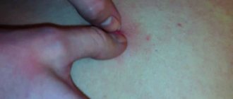
During the initial diagnosis, the doctor pays attention to the location and appearance of redness and irritation in the butt.
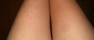
The photo shows what pronounced red spots on a child’s butt may look like on all or some part of the butt:
diaper rash
allergy
There are different types of redness on the bottom of a newborn:
- Redness, pronounced in and around the skin folds, causes a burning sensation - these are signs of prickly heat or irritation during dysbacteriosis.
- Extensive areas covered with spots with blisters, which are localized over the entire area of the buttocks and anus. Signal about allergies.
- Red small spots and pimples indicate external manifestations of dermatitis.
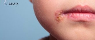
- Watery blisters can be a manifestation of the initial stages of chickenpox or herpes.
- Rashes in the form of spots covered with scales that constantly itch. Indicate the first manifestations of psoriasis.
- A large patch extending to the genitals, in which the inflamed skin becomes rough and rough. Signals seborrheic eczema.
- Confluent pustules, localized on the buttocks. Indicate infection with streptococci and staphylococci.
Redness and rough skin on a child’s bottom is divided into 3 degrees:
- 1st degree - mild. Slight redness without compromising the integrity of the integument.
- 2nd degree - average. Bright red redness, accompanied by microcracks, sometimes pustular rashes.
- Grade 3 - severe. Weeping cracks appear, detachment of the epidermis begins, at this stage microbial eczema may appear due to infection with bacterial and fungal flora.
At grades 2 and 3, accompanying signs are:
- itching;
- pain;
- burning.
As a result of these signs, the child becomes restless and feels unwell. If infection does occur, these signs are added to:
- fever;
- worsening sleep;
- loss of appetite.
Red spots and crust on the child’s bottom and legs:
hives
allergy to candles
Appearance
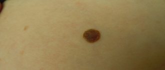
The dark mark is a congenital nevus. In most cases, the Mongoloid spot in a newborn has a blue-gray color, reminiscent of a bruise. Sometimes these spots are blue-black or blue-brown. A distinctive feature of these particular spots is considered to be uniform coloring throughout the entire area with altered pigmentation.
The shape of the spot can be completely different, mostly irregular in shape. Sizes also do not have standards - they range from specks no larger than a coin to large spots covering the entire back.
The Mongoloid spot in a newborn is most often concentrated on the lower back or sacrum. But other places of manifestation are also quite likely: spots are known to appear on the legs, back, forearms and even hands. Even migrating spots are very rare, gradually moving, for example, from the buttocks to the lower back and back.
Most often, the spot is present in a single copy, but there are also manifestations of multiple marks.
Immediately after birth, the “blobs” darken, but over time they become paler and smaller. In almost all children, by the age of 5, the skin acquires a uniform color. Rarely, marks can be found in adolescents. Mongoloid spots remain in an adult only if there were a lot of them in childhood, and in atypical places.
Attitude
The Mongoloid spot, a photo of which accompanies this article, has different meanings among different peoples. For example, in Brazil they consider it a shame to have such marks; parents carefully hide this fact even from their closest relatives, not to mention strangers. In addition, the color of the spot among the inhabitants of Brazil is close to greenish, therefore, if a nevus is suddenly discovered in an adult, he will be teased as “green-backed.”
For most peoples, a stain is a “slap from Buddha”, “a kiss from God”. It is believed that a child with such a mark will be happy, since God (Buddha, Allah) is looking after him. And, of course, this is another opportunity to make sure that the child is a representative of a certain people.
Is the Mongolian spot dangerous for children?
The spots form in the fetus and can be detected immediately at birth or several weeks after. In the course of research, it turned out that these are not vascular skin lesions. They are benign and harmless. They are produced by the same cells in the body that are responsible for eye color. The closer the cells are to the surface of the skin, the more noticeable the Mongolian spot is. No special tests are required to diagnose a skin pathology: a qualified doctor can identify it simply by examining the stigma.
Blue-gray Mongolian spots can be single or multiple, ranging in size from a few millimeters to 10 centimeters or more in diameter. Just look at the photo.
As a rule, they appear on the back, shoulders, buttocks, legs, arms, face of the child and can be either very tiny or large formations of irregular shape, without a specific texture. They affect both girls and boys equally, although it is believed that dermal melanocytosis is more common in boys.
Some types of congenital nevi can degenerate into a malignant neoplasm - melonoma. True, not a single case has yet been recorded in which a Mongoloid spot has transformed into a cancerous disease. Therefore, such a pathology, as well as manifestations of papillomatous or verrucous nevus, intradermal pigmented nevus and Setton’s nevus, is considered a melanoma-free nevus and does not pose a threat to life.
However, patients who have certain types of pigmentary manifestations should be registered with a dermatologist and oncologist. Nevus of Ota, borderline and blue nevi are particularly dangerous. Only a highly specialized specialist can determine the exact diagnoses of these pathologies after conducting the necessary research.
Diagnostics
If you find an unknown spot on your child’s skin, you should consult a dermatologist. The doctor will conduct a special examination to make sure that these are not pathological pigmented nevi, since some of their varieties can be melanoma-dangerous. If one of these options is detected, it is necessary to be constantly monitored by a dermatologist and oncologist.
To distinguish the Mongoloid spot from other types of nevus, siacopy and dermatoscopy are performed. If the diagnosis needs clarification, the doctor may prescribe a biopsy of the pigmented area.
Diagnostic methods
The newborn is examined by a pediatrician before discharge. If there is a suspicion that the spot may be a sign of a dermatological disease, a fragment of the epithelium is taken from the baby for examination. This is done in order to distinguish Mongoloid formation:
- from Ota syndrome;
- blue mole;
- giant nevus;
- hemangiomas.
Such abnormalities also appear with the birth of a child, and although they look like benign formations, they sometimes turn into melanoma.
Differential diagnosis includes several examination methods, including:
- Dermatoscopy. The non-invasive procedure is carried out using a special device that allows the doctor to examine the structure of the formation, determine its nature, and understand its nature.
- Siascopy. Using a special device that directs light rays into the deep layers of the epidermis and displays a three-dimensional image on the monitor screen, you can not only examine skin formation, but also find out the concentration of melanin and identify even a slight deviation.
- Biopsy. During the manipulation, a section of the epidermis is cut off and sent for histology. The study helps to identify the structure of the tumor and determine the autoimmune disorder. During the procedure, the affected area of skin is removed, and medication intake is monitored.
Mongolian formation does not pose a threat to life, lightens and disappears before the age of 5, in rare cases it disappears in adolescence in the presence of multiple spots. Congenital pigment formation never develops into melanoma.
However, some types of nevi, similar to the Mongolian spot and appearing at birth, are not so harmless and can develop into a cancerous tumor. This means that if pigmentation is detected, the baby must be shown to a doctor.
Those birthmarks that are localized in places of constant irritation (on the neck, scalp, inside natural folds) are subject to removal.
Indications for surgical removal:
- rapid growth of the birthmark;
- the appearance of small bleeding cracks on the surface of the mark;
- statement;
- pain when touching the mark.
To excise the spot, laser therapy, cryodestruction and the use of medications for external use (for older children) can be used.
The choice of remedy depends on various factors: the types of formations, the age of the baby, and the results of a diagnostic study are taken into account. In the absence of suspicion of the presence of malignancy processes, preference is given to laser treatment.
But if there are symptoms of malignancy, the birthmark is removed using the traditional surgical method. Only excision of the tumor with a scalpel will completely remove the formation and obtain biological material suitable for histology.
If any pigment darkening is detected in a baby, it is advisable to immediately contact a dermatologist. Of course, a child born in a maternity hospital should be immediately examined by a specialized specialist. If this does not happen, you should visit a doctor in the first days after discharge. An examination by a specialist will determine the nature of the pathology and eliminate the likelihood of dangerous diseases.
Accurate diagnosis is of great importance. Mongoloid spots in a newborn do not require any treatment, however, there are other dangerous diseases that masquerade as harmless pigmentation. If necessary, the dermatologist may prescribe certain laboratory tests of the skin in the area of pigmentation.
Only a dermatologist should determine the origin of the spots.
If there are pigment defects on a child’s skin, he should be examined by a dermatologist in order to make an accurate diagnosis and establish the type of nevus present. There are a number of ways to conduct research and diagnose tumors:
- Dermatoscopy. Multiple enlargement of the skin area with pigment pathology.
- Siacopy. The pigmented area of skin is subjected to spectrophotometric scanning.
- Biopsy. However, this method is more often used in the study of warts, pruritus and other similar diseases; it can be prescribed rather to clarify the diagnosis.
This formation on the skin does not turn into cancer, does not cause discomfort to the child and is rather a cosmetic problem in life and in photos. However, in most cases, it goes away with age and the skin color returns to normal.
The need to diagnose nevi
The necessity of a thorough diagnosis is associated with the differentiation of congenital nevi with different etiologies of Mongoloid spots . Nevi of other types are capable of degenerating into a malignant skin tumor.
For diagnosis, a sample of epithelium from the focus of the anomaly is examined. The analysis allows us to exclude dermatological congenital pathologies:
- Otha syndrome;
- congenital giant nevus;
- blue nevus;
- borderline pigmented nevus.
Despite the symptoms, like the Mongolian spot, ailments can turn into skin cancer, which requires doctor's observation and treatment. Dermatology has laboratory tests and instrumental examinations at its disposal.
Common stain research technologies:
- siascopy;
- dermatoscopy;
- biopsy of skin fragments;
- histology of deep layers of epidermal tissues.
The presence of an excessive amount of melanin at the site of localization of the Mongolian spot in the superficial layer of the skin is considered normal. The prognosis is much worse if the concentration of melanin is in the deep layers of the dermis. This is evidence of the presence of a nevus for a long time.
Possible studies
To confirm an accurate diagnosis and determine the nature of the tumor in modern dermatology, studies such as:
- Dermatoscopy. Carrying out this procedure involves studying the pigment spot under a powerful microscope.
- Siascopy. This procedure is carried out using spectrophotometric scanning of areas of pigmentation. For these purposes, special modern devices are used. The procedure itself is absolutely painless and safe, and gives an accurate result even in the early stages of serious diseases.
- Skin biopsy of a pigmented neoplasm. Carrying out histological studies makes it possible to detect or exclude the presence of dendritic cells, which should not be present in the area of analysis (between the elastic and collagen fibers of the skin tissue).
Reasons for appearance
In the old days, body pigmentation was viewed more from the mystical side, rather than from the scientific side. Some Tatar-Mongolian peoples classified children with blue Mongolian spots as God's messengers, others, on the contrary, as servants of evil. In any case, such children were considered special. As for the Slavs, they were of the opinion that such defects are the result of the mother’s fright during pregnancy, and the form and localization of the pathology depend on the cause of the fright.
In the modern world, there is a scientifically based theory why a newborn develops a Mongolian spot. Such defects are formed during gestation, and not at the time of birth, and this is due to the incomplete movement of pigment cells into different layers of the skin. When the embryo is still in the womb, melanocytes migrate from the ectoderm to the epidermis.
In European countries, where medicine is at a high level, there is a scientific explanation for the unusual formation on the child’s body. It is based on the excessive saturation of skin cells with natural pigment.
Epidermis
The spot, which appears only at birth, is benign in nature; not a single case of its transformation into a cancerous tumor has been recorded. Darkening, very similar to a bruise, is formed due to a disruption in the production of pigment in the epidermis.
In a baby who is born with darkening on the buttocks, in the sacral area, melatonin accumulates in the cells of the upper layer of skin during development in the womb. Among the peoples of the Caucasian race, the substance is produced under the influence of sunlight; among the Mongols, Chinese, Japanese, and Buryats, synthesis occurs constantly, and the pigment begins to accumulate in the body.
The deep layer of skin consists of fibers and blood vessels. Collagen, which is formed in the fibroblast, gives it elasticity. Protein compounds retain water in the dermis, which provides sensitivity and thermoregulation of the skin.
Melanins
High-molecular pigments determine the shade of the hair and iris, absorbing ultraviolet radiation, they protect the deep layers of the skin from damage by rays. The concentration of melanins increases when their synthesis is disrupted, which occurs during the development of the human embryo. The most common pigments are black and brown in color.
Melanocytes
Cells that are localized in the epidermis, in connective tissue and have long processes, protect the skin from exposure to ultraviolet radiation and synthesize melanin. A healthy person who is exposed to the sun develops a tan. If a malfunction occurs in the body, the formation of pigment is disrupted, and spots form on the body.
Melanocytes are responsible for the color of hair, skin, and determine the color of the iris of the eyes. As the pigment concentration increases, the shade becomes darker. In Europeans, melanocytes are located in the outer layer of the epidermis; in Mongoloids and blacks, these cells are distributed over all layers of the skin, and in such peoples it has a dark color.
Birthmarks, the structure of which contains pigment cells
There are several types of pigmented formations, which are most often detected in infants. The reasons for the formation of such birthmarks are a high concentration of melanin in a small area of skin.
| Type name | Description |
| Mongolian spot | Appears on the body of infants with Mongoloid genes from birth on the lower back, in the sacrum area. Outwardly it looks like a bruise and disappears completely on its own by the age of five. The health and development of the child is not threatened |
| Dysplastic nevi | These markings are similar to irregularly shaped marks. They have different color intensities and vary in size. There are cases when dysplastic formations are formed from individual point-type elements |
| Moles | They resemble small spots of different colors and sizes and appear on any part of the body |
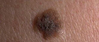
| Congenital nevus formed from pigment cells | Large markings. Each baby has a different shape of this spot; the color can be light, coffee or blue-black. Hair grows on the surface. This is the main diagnostic sign |
If there are several nevi on the child’s body at once, parents should observe them, control their size and color. If the shape changes, a sharp increase in the size of the formation, or asymmetry appears, you must immediately seek medical help.
You need to start sounding the alarm if the birthmark begins to change color and the skin next to it becomes swollen, red, begins to hurt or itch.
Diagnosis, treatment and prevention
Since God's Mark is not a disease, there is no cure for it. The prognosis for such a nevus is positive. During the entire period of observation of these spots, not a single case of its degeneration into melanoma was recorded. For this reason, there is no need for medical supervision.
In most cases, the spot disappears on its own by age five. But even in those rare cases when it remains for life, it does not have any effect on the health or functions of the body.
After differentiating the pathology and excluding congenital melanoma-dangerous nevi, no treatment is required. Children who have sacral bluish-gray spots on their bodies are not registered. Pigmentation does not cause any inconvenience to the baby: it does not hurt or itch. Such a cosmetic defect can bother loving parents, and some of them, in the hope of making the spot less pronounced, begin to use various ointments, creams and carry out some kind of cosmetic procedures.
Treatment
Having discovered a dark-colored formation on the baby’s body, parents should show the baby to a dermatologist. If a specialist confirms the presence of a Mongolian spot, no medications are prescribed, since the pigmentation goes away after a year, two or five and does not cause any inconvenience to the child.
Do not bleach a dark area with peroxide or treat it with bleaching ointments or gels. The stain cannot be removed this way, and cosmetics and medications irritate the baby’s delicate skin and often cause allergies.
No treatment is required. Mongolian spots do not have a tendency to malignant degeneration and gradually disappear, so many doctors do not even classify them as birthmarks. In children they go away at the age of 3-5 years. In very rare cases, they persist throughout school years and into adulthood.
Some people use a cream to even out skin tone to hide a pigmented nevus, others turn to aesthetic medicine, insisting on removing the spot using a laser, which inhibits the formation of melanin. In any case, you cannot do without consulting a dermatologist in this matter.
How to act in case of danger
For any skin rash, the child should be shown to the pediatrician. This symptom is an indicator of the internal state of the body. If necessary, the pediatrician will refer you to an allergist or other specialist.
The doctor prescribes a series of tests to identify the cause of redness:
- skin scraping for fungi;
- bacteriological culture;
- urinalysis and urinalysis;
- clinical blood test to identify the allergen.
Based on these studies, the specialist prescribes the correct treatment with the dynamics of a speedy recovery.
It is necessary to call an emergency ambulance if the redness on the child’s bottom is combined with the following dangerous symptoms:
- the child is lethargic and sleepy;
- there were complaints of headache;
- vomiting and signs of indigestion appeared;
- increase in the area of irritation or rash;
- The temperature has risen to 38-39oC and is not brought down by antipyretics.
If the situation is not critical, the following steps should be taken to alleviate the child’s condition before being examined by a qualified specialist:
- Do a wet cleaning of the room and ventilate it well.
- If possible, remove pets from the house due to possible allergies to wool.
- Remove all possible allergens that came into contact with the child, i.e. remove synthetic clothing, change hygiene products to hypoallergenic ones.
- Do not give your child obviously allergenic foods (chocolate, red fruits and vegetables, smoked sausages, canned food, foods with a high chemical content).
- Bath in water with the addition of herbs (chamomile, string, oak bark).
- Give the child antihistamines (Fenistil, Zodak or Suprastin) or use non-hormonal ointments, but before taking, consult a doctor by telephone.
Neonatal hemangioma
Another neoplasm that can be mistaken for a bruise without a contusion on the tailbone or back of a newborn is hemangioma. It is a formation consisting of cells of the vascular walls. It has uneven borders and texture, and may have a reddish, less often bluish or purple tint.
Hemangioma is a vascular neoplasm
The exact causes of vascular tumors have not been fully established. Presumably predisposing factors to the formation of hemangiomas are:
- viral diseases suffered by the mother in the first trimester of pregnancy;
- burdened heredity;
- unfavorable environmental situation;
- hormonal disorders;
- bad habits of the expectant mother.
Hemangioma is a benign neoplasm that has no tendency to become malignant. However, this type of vascular tumor is characterized by aggressive growth and development, as well as the ability to grow into nearby organs and tissues, disrupting their function.
The risk may be associated with increased pressure from the hemangioma on surrounding tissues and organs. The tumor can compress blood vessels and nerve endings, thereby causing disruption of the innervation and blood supply to nearby organs.
In some cases, the tumor goes away on its own without any treatment. If the tumor is not prone to progression, does not disrupt the functioning of organs, and does not cause discomfort to the child, a wait-and-see approach with constant monitoring of the condition of the tumor may be chosen.
Removing tumors with cold
If the hemangioma does not go away by the age of 1.5-2 years of the child’s life and tends to grow aggressively, a decision is made to remove it. Depending on the size of the tumor and the depth of its occurrence in the tissues, various techniques can be used:
- small hemangiomas located on the surface of the skin are removed by electrocoagulation, laser coagulation or cryodestruction;
- cavernous hemangiomas are removed using the scleroderma method, which makes it possible to “glue” the walls of blood vessels and avoid bleeding;
- if the tumor is deep and it is impossible to remove it by other means, surgical excision is performed, followed by suturing of the surrounding tissues or replacing the skin flap with an autograft.