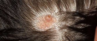
Setton's nevus (halonevus) is a pigmented nevus, the distinctive feature of which is the presence of a discolored skin area along the periphery of the formation. Externally, the area with the neoplasm is similar to vitiligo. There may be several such elements on the body. Setton's nevus is not prone to malignant transformation, but it is still worth keeping a close eye on. It is necessary to differentiate halonevus from melanoma, which also has foci of depigmentation along the periphery. For diagnostic purposes, a biopsy of the formation, dermatoscopy and siascopy are performed. If the diagnosis is confirmed, then specific treatment is not required. But in the case when the doctor suspects melanoma, surgical removal of the tumor is performed.
…
Setton's nevus itself does not degenerate into melanoma or skin cancer, and even better - it disappears on its own after a while. But this diagnosis must be made by a dermatologist who specializes in skin cancer processes: several more dangerous diseases, including melanoma itself, can be disguised as halonevus. The doctor will conduct several painless examinations, and at the slightest suspicion of a malignant formation, he will remove the halonevus itself and the depigmented skin around it.
Short description
The medical reference book contains information that Setton's nevus is a specific benign formation. During the disease, a tiny mole appears on the skin, which is surrounded by a discolored area of the epithelium, which is why the pathology is often confused with vitiligo. Doctors say that halonevus is one of the common manifestations of increased pigmentation of the epidermis, which many people are accustomed to calling a mole.
Experts began to refer to this disease as halonevus due to the fact that the neoplasm is surrounded by skin without pigmentation, and also resembles a specific glow around the object. Most often, this pathology occurs among young people. Nevus affects not only the torso, but also the limbs (in rare cases, the face).
What is considered surprising is that this type of formation disappears just as it appears. At the initial stage, slight pigmentation appears, which within a short period of time takes on the appearance and shape of a regular mole. After some time, a circle of discolored skin appears around the growth. The final characteristics of a nevus can differ significantly, since it all depends on the skin type and immunity of a particular person. A small tumor may not change its parameters for several years, after which it will resolve on its own.
Signs of halonevus
Education is a combination of two factors:
- an ordinary mole of brown color with a reddish tint, moderately protruding above the surface of the body, with clear edges,
- a white spot around a mole, which has all the signs of vitiligo; it is discolored skin in which there are no melanocytes.
Nevi most often have an oval shape and multiple distribution; they practically never occur alone. The localization of halonevus varies: any part of the torso, arms, legs, but they rarely appear on the face.
Setton's nevus differs from ordinary moles by a light rim of discolored skin
Characteristics
Research into this pathology began several years ago. Setton's nevus is a rather rare pathology, as it is diagnosed in only 1−2% of the entire population of the planet. Dermatologists most often diagnose growths in children and young people; a person’s gender predisposition does not matter. Many patients mistakenly confuse this disease with vitiligo (a chronic pathology that affects the scalp, meninges and retina).
People who have previously been diagnosed with autoimmune diseases are at risk. Of course, the nevus itself is not dangerous, but the patient must avoid various risk factors. Some experts note that the growth is often a reaction to increased exposure to ultraviolet rays. When the protective function of the immune system is reduced, cytotoxic antibodies are detected in the blood of patients.
Who gets sick more often?
A halonevus is a formation that is not present in a child from birth, but appears later - in children and people under 30 years of age. Both men and women suffer from it equally. All races are susceptible to the disease, and it affects 1% of the world's population.
People most susceptible to Setton's nevus are:
- who has vitiligo (patches of discolored skin) or immediately before its appearance: every fourth such person is sick;
- those suffering from autoimmune (when one’s own immune system attacks its own tissues) diseases: autoimmune thyroiditis, autoimmune hepatitis and others;
- siblings of a person who has vitiligo;
- living in stressful conditions;
- having sunburned skin;
- who have or have had skin melanoma;
- in women with Shereshevsky-Turner syndrome, who have short stature and underdeveloped genitals due to the fact that they have only 1 sex chromosome - X (X), and not XX, as it should be.
Setton's nevus in a child and adolescent is rarely single: more often than not, several such formations appear throughout the body (the maximum number of such formations has been 100 in one person).
Most of these diseases (autoimmune processes, stress, skin burns) also accompany vitiligo. However, Setton's nevus and vitiligo are different diseases with different causes, and not "vitiligo around the mole" as it is sometimes called.
Differences between other growths
Each nevus has its own characteristics. If the patient knows all the characteristics of neoplasms, he will be able to timely determine the type of disease in order to seek help from a doctor. The experts themselves note that each neoplasm has its own characteristics:
- Flame nevus. This is a flat pink spot that most often affects the back of the head, the bridge of the nose and the eyelids. In 98% of all cases it goes away on its own, but sometimes it can recur when the patient reaches middle age.
- Spider-like neoplasm. Located in the neck, face, shoulders and upper third of the body. The disease itself is presented in the form of a red, weakly pulsating vascular network.
- Nevus of the sebaceous glands. This is a congenital pathology that can remain in a latent form until adolescence. The growth appears as a flat solitary plaque.
- Hairy growth. An ordinary mole that is abundantly covered with hair. Most often it is congenital in nature, which can have a negative impact on the health of the infant.
- Specific hemangioma. Education is benign. The growth can affect not only the skin, but also various organs. The disease is represented by a small thickening with uneven edges, consisting of pathogenic and healthy cells.
Reasons for development
Most often, the disease appears after severe sunburn. Under the direct influence of ultraviolet radiation, the likelihood of a benign tumor significantly increases. Most cases of the development of Setton's nevus have been registered by dermatologists due to autoimmune problems. In addition, the disease can occur as a result of exposure to the following unfavorable factors:
- destruction of melanocytes by immune cells;
- violation of skin pigmentation;
- sunburn;
- genetic predisposition;
- frequent visits to the solarium;
- the presence of chronic autoimmune diseases (for example: thyroiditis);
- long-term stress, depression.
In some cases, the disease may precede the appearance of a more dangerous pathology, vitiligo. If there are too many of these moles on a person’s body, this may indicate the development of malignant melanoma inside the eyeball.
What is a dysplastic nevus?
A dysplastic nevus is a type of pigmented growth on the skin that is often called a birthmark or mole. This is an intradermal or subepidermal accumulation of pigment cells (melanocytes) that enter the skin during intrauterine development. These pigment cells are actually a subtype of nerve cells.
Nevi do not immediately begin to appear as brown spots on the skin. This process takes a long time and depends on many endogenous and exogenous factors. Dysplastic nevus is a dangerous neoplasm, as it is a precursor to the malignant disease melanoma (a particularly aggressive form of skin cancer). It is the term “dysplastic” that suggests that a nevus is not a typical mole, but has a predisposition to be malignant.
Causes of dysplastic nevus
The causes of dysplastic nevus and its degeneration into melanoma are as follows:
- hereditary factor;
- disruptions and changes in hormonal levels (during pregnancy, menopause, during puberty);
- excessive ultraviolet exposure;
- excessive passion for sunbathing.
Most often, genetic predisposition is the main reason for the appearance of dysplastic nevus. Often, 90% of a person with a dysplastic nevus has a family history of melanoma. For this reason, dysplastic nevus is a congenital hereditary factor.
Symptoms of the disease
- The surface of a dysplastic nevus is finely lumpy.
- It does not have a clear shape or boundaries.
- Heterogeneous color over the entire surface of the dysplastic nevus.
- It can be either single or multiple.
- Hair growth from a nevus is possible.
- A dysplastic nevus may not rise above the skin or rise only slightly.
Treatment of dysplastic nevus
Treatment of a dysplastic nevus consists of its surgical removal if there is a suspicion that the nevus has degenerated into malignant melanoma. This applies to both new formations and older ones that have changed their clinical characteristics.
Multiple dysplastic nevi, widespread on the body, are not removed for prophylactic purposes. Only those nevi that are located in places that are difficult to access for control and inspection, as well as nevi with severe dysplasia, are removed surgically.
A dysplastic nevus is surgically removed at a distance of several millimeters from the edge of the neoplasm towards healthy tissue. When removing a nevus, a Wood's lamp is used, which helps to accurately determine the outline of the neoplasm and completely eliminate it. Otherwise, there is a high probability of recurrence of the nevus, which can give rise to even greater growth of atypical cells.
Methods for removing dysplastic nevus
The most popular method of nevus removal is laser. The laser removal method is the safest, as it does not require anesthesia and does not leave scars or scars. But this method still has contraindications. Large nevi larger than 1 cm are not removed. In this case, there is a possibility of incomplete tissue removal, and as a result, new tissue growth and the occurrence of melanoma.
It is possible to remove the nevus using electrocoagulation (electric current), cryodestruction (liquid nitrogen) and a surgical scalpel (tissue excision). Removing a nevus by surgical method is the most reliable, as it makes it possible to completely remove the affected tissue.
Disease prevention
To avoid the development of melanoma, it is necessary to conduct a preventive examination every six months, depending on the growth of atypical cells and the rate of appearance of new formations. Unfortunately, dysplastic nevus does not have any effective ways to prevent its development.
The only way to reduce the likelihood of a nevus degenerating into malignant melanoma is to avoid exposing it to direct sunlight. You should protect yourself from UV radiation, especially if you have one or more birthmarks on your body. You can be in the sun from 8 a.m. to lunch and after 5 p.m., preferably not in direct sunlight.
You should protect your skin from the sun's rays in all possible ways: use sunscreens with a high protection factor, wear light-colored, lightweight clothing that covers most of the body, wear wide-brimmed hats, glasses and umbrellas.
In addition, the owner of a dysplastic nevus must be attentive to himself, monitor birthmarks, and pay attention to their condition. Contact a doctor immediately if the nevi suddenly change color, size, surface or structure, since early diagnosis of melanoma can help save and prolong a person’s life.
Something to remember!
- That a dysplastic nevus in itself is not a pathology.
- Most often it is hereditary and appears in individuals with a family history.
- This is an individual feature of the body that requires careful attention to birthmarks, as it has the ability to degenerate into malignant melanoma.
- Only single dysplastic nevi are removed surgically.
- Multiple dysplastic nevi require regular and careful monitoring.
Common symptoms
The pathology begins to develop from an ordinary mole, around which the pigment elements completely disappear. After 2-3 weeks, a white rim of discolored epithelium begins to grow around the growth. Dermatologists distinguish several main stages in the development of a neoplasm:
- Nevus stage. A small mole grows on any area of the skin.
- Forming the rim. The growth and the skin around it begins to turn red; this process may resemble normal inflammation. After 4-7 days, the skin around the mole completely fades, and a rim forms.
- The final stage of recovery. Numerous studies have proven that regression is very spontaneous. Both the mole and the rim itself can disappear at any time. Most often, the patient’s recovery occurs after 3 months. During this time, the mole resolves and the skin acquires its natural color.
Can Setton's nevus develop into cancer?
This is one of the main questions that worries patients after diagnosing halonevus. Doctors almost completely exclude the possibility of malignancy of a true Setton’s spot, but correct diagnosis and timely consultation with a doctor are of great importance in the future prognosis. To exclude melanoma and other malignant skin pathologies, a biopsy of the formation can be performed, followed by histological examination of the material. This procedure is quite painful, but you should not be afraid of it: the sooner existing deviations are identified, the greater the chance of a favorable outcome and preservation of life and health.
In some cases, a nevus biopsy is performed
It is necessary to consult a doctor without waiting for further progression of the spot if the following symptoms occur:
- itching and burning in or around the nevus;
- change in color of the formation;
- bleeding mole;
- severe hyperemia of the skin around the spot and its hyperthermia;
- pain when pressed.
Important! Some types of melanoma may have discolored areas of skin along the periphery of the formation, so self-diagnosis for any skin rash is unacceptable.
Diagnosis should be entrusted to a qualified doctor
Diagnosis of pathology
Setton's nevus is always accompanied by clear signs, on the basis of which it is not at all difficult to make the correct diagnosis. But in some cases, even an experienced dermatologist cannot determine the real nature of the origin of the problem without special devices. For example: if a halonevus has formed against the background of vitiligo, then you simply cannot do without universal diagnostic procedures.
The patient must undergo a complete blood count, which will allow dermatologists to understand the exact specifics of the immune response. The analysis shows the presence of inflammatory processes in the body. Specialists necessarily study the quality of blood clotting; for this purpose, liquid biomaterial is taken.
To eliminate the likelihood of developing dangerous diseases, experts use instrumental diagnostic procedures:
- Skiascopy. A universal procedure during which you can determine not only the structure of the growth, but also the presence/absence of dermal melanin cells.
- Biopsy. The doctor takes part of the tumor to determine its nature.
- Dermatoscopy. Innovative hardware research. A dermatoscope is used during the procedure. This procedure is especially important if the mole has changed color and shape.
It is worth noting that a biopsy is performed in every case when the dermatologist suspects that the growth is malignant. Setton's nevus itself never degenerates into melanoma.
But it is very important to promptly differentiate a normal growth from a malignant neoplasm. The danger stems from the fact that at the initial stage, melanoma can also look like an ordinary mole with a discolored halo.
How is the diagnosis made?
To determine whether treatment is needed in a particular case, you need to be examined by a doctor called a dermatologist. Such specialists work in oncology centers and dispensaries, and conduct consultations in private centers. Only they can distinguish halonevus from neurofibroma, pemphigus vulgaris, and most importantly, from the initial stage of melanoma (an extremely malignant formation of cells with pigment).
To do this, he uses the following methods:
- Dermatoscopy. A method that allows you to examine a neoplasm under high magnification, evaluate the structure of the nevus and the structure of the cells - to diagnose its malignancy. The study reveals an accumulation of melanocytes in the mole itself, a shaft of lymphocytes along the periphery of the nevus and the absence of melanocytes in the rim area.
- SIAScopy is a method recently developed by scientists at the University of Cambridge. The name is based on the abbreviation SIA, which stands for spectrophotometric intracutaneous (that is, intradermal) analysis. The method is based on the penetration of a light wave deep into the skin up to 2 mm, where light beams of different lengths will interact with melanin, collagen and hemoglobin. Using the SIAscope device, a three-dimensional multispectral image of the studied halonevus is received on the monitor, which allows you to evaluate its structure, color, and areas rich in melanin or hemoglobin. The probability that the SIAscope will “see” malignant cells (if any) inside Setton’s nevus is 96%.
- Examination of regional lymph nodes using ultrasound, and, if necessary, their biopsy. This way you can notice the presence of metastases in them, if it is not a halonevus, but a melanoma or other malignant neoplasm of the skin.
The possible cause of Setton's nevus is also being investigated. For this person, specialists such as a rheumatologist, endocrinologist, and medical geneticist examine.
Effective treatment
Most often, specialists do not prescribe drug therapy to combat Setton’s tumor. If the growth was diagnosed in a teenager or a small child, then the nevus will resolve in just a few months, leaving no trace on the skin. In older people, it takes up to 5 years of life to combat this formation.
A nevus is removed only in the most extreme cases, if the growth is often injured by clothing or during hygiene procedures. If even the diagnostic procedures performed were unable to determine the nature of the skin tumor, then it is treated as a malignant pathology. Excision is performed surgically. The laser procedure is not relevant in this case.
During surgery, the mole is cut out to healthy tissue. The thinnest cosmetic sutures must be applied to the wound. If the procedure is performed by an experienced surgeon, the wound will be as neat and small as possible. After six months, the scar becomes thinner, lighter and gradually dissolves.
Those patients who have Setton's nevus and vitiligo are at risk. They must continuously monitor their health. Several times a year you need to visit a dermatologist and monitor the condition of the skin.
Symptoms of seton nevus in children and adults
Symptoms of Galonevus:
- The first manifestation of Setton's nevus is a pigmented nevus, around which an area of depigmented skin forms. In some cases, redness may be observed before depigmentation appears.
- The neoplasm itself resembles a nodule of brown, red-brown or milky-brown color. The nevus rises slightly above the skin. It has clear boundaries and a rounded shape. As a rule, the depigmented rim around the formation is twice as wide as the nevus itself.
- Mostly, these formations affect the skin of the upper extremities and torso, and the face in rare cases. Basically these are multiple formations. The patient can count about a hundred nevi on his body, having a similar shape and approximately the same size. Setton's nevus is very rarely isolated.
- Setton's nevus is prone to gradual regression; it is classified as a group of melanoma-dangerous nevi. True, it has many common features with such formations as melanoma and neurofibromatosis. Setton's nevus may also be associated with malignant tumors of internal organs.
- Setton's nevus is characterized by staged development, which allows us to call it the most distinctive symptom. From the very beginning, pigment formations appear. An area of hypopigmentation, or discoloration, appears around it after a few months.
Only after a few years regression of the pigment part of the formation is observed. After this, the color of the depigmented rim gradually returns to its natural color.
In the photo, Setton's nevus on a woman's face
Can halonevus develop into cancer?
- There is no need to fear that Setton's nevus will degenerate into an oncological tumor; malignancy is not inherent in it. Moreover, it often disappears on its own without any therapy.
- However, if halonevus affects a large surface area of the skin, then the risk of developing a cancerous process on it or in internal organs increases.
- Since the likelihood of a nevus degenerating into melanoma is initially minimal, to reduce the risk to zero it is enough to follow medical recommendations.
Summarizing what was said earlier, we note that the prognosis of halonevus is good, subject to systematic monitoring of the dynamics of visual changes.
ethnoscience
It has long been believed that regular use of decoctions, infusions and other non-traditional therapeutic agents can speed up the healing process. The main thing is to use only proven recipes . The most effective folk remedies include:
- The mole should be treated daily with fresh celandine herb juice.
- At home, you can prepare the following recipe: pour three tablespoons of herbs into a liter of boiling water. The product is allowed to brew for several hours. Only after complete cooling should the healing mixture be filtered through cheesecloth. To achieve the desired result, the patient should take a tablespoon of tincture three times a day before meals.
- Add a spoonful of apple cider vinegar to 150 ml of warm water. The drug is drunk in one gulp on an empty stomach. If the patient does not have chronic diseases of the digestive system, then this folk medicine can be taken every day.
Prevention measures
Experts assigned ICD-10 code D22 to halonevus. This means that the growth belongs to the category of melanoform nevi. Under the influence of unfavorable factors, a mole can degenerate into a malignant neoplasm. To avoid negative consequences, experts recommend adhering to the following preventive measures:
- Strong exposure to ultraviolet radiation should be avoided.
- It is forbidden to miss scheduled visits to the dermatologist (at least 2 times a year). If any problems arise, the growth will change its color, structure and begin to grow. In such a situation, seeking help should be immediate.
- If the growth is located on an open part of the body and looks aesthetically unattractive, it can be masked with a high-quality foundation with a high SPF rating. If the cream contains an insufficient amount of valuable elements, then Sanskrin must be applied as a base.
- It is strictly forbidden to visit solariums.
- The neoplasm itself must be protected from exposure to direct sunlight.
- When using cosmetic products you need to be as careful as possible. If the skin begins to itch, an inflammatory process or swelling begins to develop, then you need to wash off the applied product with warm water. Negative manifestations can be minimized if you take high-quality antihistamines. If even after this the symptoms do not disappear, then you should immediately seek qualified help from a dermatologist.
It is worth noting that such a nevus in a child requires constant monitoring. To minimize the likelihood of developing adverse reactions, you need to show your baby to a specialist in a timely manner and undergo a high-quality examination.
Complications and consequences
Complications with Setton's nevus can only occur if the spot is damaged - for example, if it is scratched.
When microbes enter a damaged nevus, an inflammatory process begins in it, pus accumulates in the wound, and toxins penetrate into the blood, leading to intoxication.
To avoid such consequences, it is important to explain to the child that moles cannot be touched. When going outside in sunny weather, you should use sunscreen or cover the site of the nevus with a band-aid.