Why do blue spots appear?
Many researchers agree that melanocytic nevi appear due to mutations in pigment cells. And they, in turn, can be provoked by various factors:
- Hormonal imbalances.
- Excessive insolation.
- Dermatological pathology.
Against the background of a hereditary predisposition, adverse effects from the outside or from the body itself lead to genetic damage in melanocytes, which becomes a source of tumor growth. This is how blue pigment spots appear.
Blue nevi appear due to genetic damage in melanocytes, which occurs when exposed to external or internal factors.
Causes of blue nevus formation
A blue nevus is one of the types of birthmarks, so it is formed in a child while still in the womb.
The following are the reasons for the appearance of this mole:
- abnormal fetal development,
- infectious diseases of the genitourinary system of a pregnant woman,
- hormonal imbalances associated with pregnancy,
- the influence of harmful factors, these can be radiation or toxic substances.
In addition, one of the common reasons for the appearance of such a formation is the pathological development or improper placement of melanoblasts.
But a blue mole can increase as a result of the following reasons:
- regular use of hormonal methods of contraception,
- physiological changes in the body - pregnancy, lactation, transition to adolescence,
- various allergic and inflammatory skin diseases,
- the influence of ultraviolet rays - sunbathing at lunchtime, going to the solarium.
Tanning in the middle of the day provokes the growth of moles
Classification
Blue nevi come in several types. According to the existing classification, which takes into account morphological and histological features, there are three types of moles:
- Simple.
- Cellular.
- Combined.
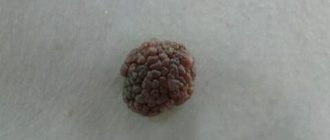
The first variety is small in size, has a smooth surface, and is mainly localized on the neck, face and hands. A cellular mole grows up to 3 cm, is dark in color, and lumpy. The combined type is a combination of a blue nevus with an intraepidermal, borderline or complex pigmented nevus.
Methods for removing pigmented nevi
- Small nevi are removed using a laser. Local anesthesia is used here and many small nevi can be removed at once in one visit. A crust appears at the site of removal, which will disappear in seven days. The marks are practically invisible, which is why the laser method is used to remove facial moles.
- For large nevi, surgical excision is used. The operation is performed under local or general anesthesia with suturing.
The decision on the treatment method is made as a result of examining the nevus. In some cases, consultation with an oncologist is required. When surgical excision is used, the tumor is sent for histology to determine whether it is melanoma.
You should never remove moles and birthmarks yourself. This is fraught with infection and other troubles.
Conventionally, pigmented nevi can be divided into four categories according to their size:
- small (up to 1.5 cm)
- medium (up to 10 cm)
- large (up to 20 cm)
- giant (more than 20 cm)
Multiple pigmented nevi are quite rare (5%). This nevus consists of a large lesion and small pigment spots and affects the lower body, limbs, back and chest.
How do they appear on the body?
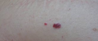
A blue mole looks like a small (up to 10 mm in diameter) round or oval formation located deep in the skin. This is a nodule of dense consistency with clear edges, slightly protruding above the surface of the skin. The color of the nevus varies from blue to blue-black, its surface is smooth, devoid of hair, but with a preserved epithelial pattern.
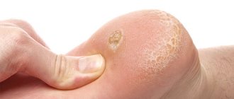
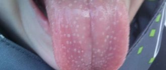
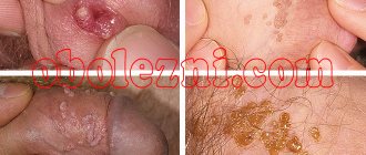
The formation can be located on almost any part of the skin, but in half of the cases it is found on the dorsum of the hands and feet. Sometimes it is found on the mucous membrane of the oral cavity (soft and hard palate). Cases of multiple nevus are rarely described, when pigmented formations are located in different parts of the body.
The mole does not bring any subjective sensations and grows slowly, and therefore can remain unnoticed for a long time. Gradually, the blue nevus may become pale and flatten. Very rarely does it degenerate into melanoma, a malignant skin tumor. But even a benign process can be accompanied by the spread of pigment cells to the lymph nodes (cases of familial multiple nevus).
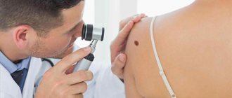
The symptoms of a blue mole are revealed during a medical examination and form the basis of a preliminary conclusion about the nature of the pathology.
Is blue nevus dangerous?
It is extremely rare for a blue mole to transform into a malignant tumor. However, medical practice confirms such cases. The danger of degeneration threatens a person with cancer, which in later stages is difficult to treat and can cause negative health consequences.
In order to promptly recognize the transformation of a nevus into an oncological disease, it is necessary to remember the following symptoms that are inherent in this process:
- The mole changes its size to a much larger one.
- Change in color.
- The appearance of hairs on the birthmark.
- Painful sensations occur on palpation.
- The nevus itches, itches and burns.
- The surface of the growth becomes bumpy and rough.
- Pus mixed with blood is released from the growth.
The most dangerous is the blue nevus, which is located in places subject to constant mechanical stress. Namely: face, back, neck, arms. When a nevus in such a localization is injured, there is a risk of introducing an infection into it, which can provoke its change into a cancerous formation.
To avoid negative consequences, you should take hormonal and contraceptive medications with caution, and do not stay in direct sunlight for a long time between 10 and 17.00. This especially applies to people with a light skin tone and the presence of multiple blue, brown or red birthmarks on the body. And also, to avoid consequences, you should limit your visits to the solarium and monitor the hormonal balance in the body.
If there are suspicious signs when a mole turns blue, you should urgently consult a doctor who specializes in moles, a dermatologist or oncologist. He will conduct a personal examination, a number of other examinations, and determine what actions should be taken in relation to the blue nevus.
Diagnostics
A typical skin lesion makes it possible to make a correct diagnosis with a high degree of probability. It is based not only on data from a routine examination, but also on the results of additional methods:
- Dermatoscopy.
- Skiascopies.
- Histological analysis.
Tissue examination is performed after the mole is removed. Histological analysis allows us to determine the structure of the tumor and its nature. A simple nevus contains melanocytes with a large number of granules, and in the cellular form they take on large sizes and are depleted in pigment. The tumor is located in the dermis, between the fibers or goes down to the subcutaneous tissue.
Jadasson-Tiche's nevus often requires differential diagnosis. Education must be distinguished from the following states:
- Melanoma.
- Dermatofibroma.
- Venous hemangioma.
Unlike melanoma, a blue mole retains its skin pattern and has clear edges. Dermatofibroma is mainly located in the lower legs and shoulders, and venous hemangioma is soft and turns pale when pressed.
Description
The mole is a round shape of a bluish-blue hue and is located in the lower layers of the epidermis. The congenital appearance protrudes above the surface of the skin. Usually located singly, less often in colonies. The size in diameter does not exceed 1 cm, but there are also up to 3 cm in diameter. The blue mole is smooth, without hair, elastic and thickened. Most often, localization occurs in the area:
- Shins;
- Buttocks;
- Forearms;
- Back of the foot and hand.
Rarely, similar growths appear on the face and head.
The neoplasm does not cause any discomfort, does not provoke itching or pain. If it is located in the groin or on the joint folds, it can become irritated during physical activity or walking. In medical practice, the appearance of such skin pigmentation is not associated with the aging of the body. Typically, blue moles can appear during puberty and are more common among women.
Treatment of a blue mole
If the nevus does not exceed 10 mm in diameter and has a low risk of malignant transformation, then patients are only recommended to undergo regular monitoring by a dermatologist. In other cases, surgical correction is necessary.
How is the removal done?
It is recommended to remove a blue mole if it is subject to constant trauma (razor, clothing, jewelry), which increases the risk of its malignancy. Manipulation is carried out in several ways:
- Instrumental excision.
- Electrocoagulation.
- Cryodestruction.
- Radio wave method.
- Laser removal.
Surgical tactics are determined based on the characteristics of the pigment formation. If the nevus shows signs of degeneration into melanoma, its wide excision along with subcutaneous tissue and a strip of healthy skin is indicated. In other cases, minimally invasive techniques can be used.
In most cases, blue nevi are removed surgically. The doctor will tell you what is the best way to do this.
Treatment of blue nevus
If transformation of the nevus is not observed and is not predicted, then removal of the formation can be completely avoided. However, the patient is obliged to regularly contact specialists in order to constantly monitor the condition of the pigment spot. The patient himself should be interested in this and carefully monitor the nevus. At the slightest sign of transformation of the birthmark, you should contact a dermatologist.
If the nevus is located in an area of the body that is subject to regular mechanical stress (trauma or friction), the doctor may conclude that removal is necessary. In some cases, such treatment is prescribed to prevent the occurrence of melanoma.
Blue nevus can be removed in several ways: laser or radio wave exposure, electrocoagulation and cryodestruction. The most delicate surgical work is performed in the case of removal of a blue nevus from the face. Surgery is required in cases of suspicion of transformation of a nevus into a malignant formation. The damaged area of skin is deeply excised using subcutaneous tissue. Excision is carried out taking into account the necessary capture of healthy skin around the borders of the nevus in order to avoid the risk of further changes in cellular tissue near the removed formation.
Thanks to timely research, the doctor prescribes adequate treatment to the patient in the early stages. Therefore, a person who has a blue nevus on his body can live a long time and remain almost completely healthy.
Symptoms
There are 4 symptoms by which you can judge a blue mole:
- less infiltration;
- greater isolation;
- solitary lesion with a mole;
- color (blue).
| By its nature, a blue mole, even immediately after damage, is extremely rare, but can degenerate into melanoma. In the history of medicine, there have been cases when a blue mole degenerated into malignant melanoma. | |
Types of blue moles
There are 3 types of blue moles:
Common blue mole
Melanocytic mole
Cellular blue mole
The blue mole first described by the dermatologist Tiche in 1906. When studying these neoplasms in the future, the German dermatologist Jadasson discovered many new things. The blue mole was named after two dermatologists - blue nevus of Jadasonna-Tiche .
Basically, a blue mole protrudes above the skin, but sometimes it can appear in the form of an infiltrated nodule located directly under the skin itself. The appearance of a blue mole has been recorded in all age categories of people. Most often, carriers of blue moles are women.
A blue mole inherently congenital ; it usually appears after puberty.
Prevention
There are no specific methods for preventing neoplasms - this also applies to blue nevus. But the risk of its development can be reduced - the recommendations are as follows:
avoiding exposure to aggressive chemical and physical factors on the skin;- prevention of the development of dermatoses and dermatitis, and if they occur, timely detection and prescription of adequate treatment;
- avoiding injury to the skin, and if this does happen, proper treatment of the wound surface;
- prevention, diagnosis and treatment of infectious skin pathologies, identification in the body and elimination of chronic foci of infection - first of all, they are detected in the presence of chronic tonsillitis (inflammation of the tonsils and arches) and caries (damage to hard dental tissues with their inevitable destruction);
- prevention, detection and treatment of chronic somatic pathologies;
- rejection of bad habits;
- avoiding the influence of negative environmental factors; if this is not possible, change your place of residence.
Clinical manifestations
There are several signs according to which it will not be difficult to understand that you have had to deal with this particular disease:
- the presence of clear boundaries of the neoplasm,
- shape – oval, circle, hourglass,
- homogeneity of the structure of the mole,
- high density,
- lack of hair,
- variable sizes (minimum 1-2 mm, maximum 3 cm),
- dependence of color characteristics on pigment content and depth of location,
- the likelihood of elevation above the skin,
- slight infiltration,
- presence of blue shades.
Traditionally, a blue nevus is a harmless type of mole with a risk of degeneration into a cancerous tumor. Outwardly, it is similar to other growths and pigment spots, which requires differential diagnosis with Dubreuil's melanoma, pigmented nevus, and borderline moles.
It is important to know! Often the patient does not detect clinical manifestations of the disease. Due to the insignificant growth rate of the formation, it remains unattended and is treated in the later stages.
Causes and development of pathology
As with most cases of cancer, the causes of this tumor are unknown. It is assumed that the immediate cause is a violation of the growth and development of melanocytes (cells that produce the pigment melanin), but why this process occurs is currently unknown. Factors that may contribute to disorders of melanin formation have been identified (some of them are debated in dermatology). This:
- physical impact;
- dermatitis;
- dermatoses;
- injuries;
- skin infection;
- chemical factors;
- a number of chronic diseases;
- bad habits;
- environmental factors;
- hereditary predisposition.
The physical factors that contribute to the development of blue nevus are those against which many other skin cancers develop. This:
- ultraviolet irradiation;
- radioactive exposure;
- temperatures are too high or low.
Ultraviolet irradiation is one of the most significant factors that can lead to the restructuring of skin tissue and start the process of neoplasm formation. This threat exists when:
- frequent visits to the solarium;
- prolonged exposure to direct sunlight;
- the combined effect of these two factors.
Radioactive exposure to the skin followed by the formation of a blue nevus is observed less frequently. It is exposed under such circumstances as:
- diagnostic examination - namely radiography, fluorography, different types of tomography (computer, multispiral computer);
- radiation therapy for malignant pathologies (not only the skin, but also other structures of the body);
- work activity with the need to come into contact with radioactive substances and/or equipment that generates radioactive radiation;
- accidental or unauthorized contact with radioactive elements.
Too low or high temperatures play a role in the occurrence of blue nevus when exposed locally to the skin. The thermal effect is of practical importance - for example, the described neoplasm often appeared after skin burns (most often under the influence of sunlight, which is a reason to doubt the effectiveness of the thermal factor).
The role of dermatitis and dermatoses is the occurrence of inflammatory and degenerative-dystrophic processes in the skin, respectively, due to which its structure is disrupted - preconditions arise for the start of the oncological process. These factors are among the most significant in the development of blue nevus.
The body can react by launching an oncological process in response not only to inflammatory or degenerative-dystrophic skin lesions, but also to its injury. In case of regular trauma, the risk of the formation of neoplasms in general and blue nevus in particular increases - for example, with constant rubbing of the skin with clothing or jewelry (tight narrow bracelets, watch straps).
In addition, more pronounced trauma to the skin may also play a role - a violation of their integrity of a non-medical and medical nature. In the first case, these are injuries received, as a rule, at home, at work, during physical education and sports - namely wounds:
- cut;
- torn;
- bitten.
In the second case, this is a violation of the integrity of the skin associated with invasive manipulations, which can be:
- diagnostic (biopsy of the skin or underlying tissues);
- therapeutic (opening purulent formations, removing small tumors of the skin and subcutaneous fat, cosmetic surgeries, and so on).
Infectious skin lesions, against which a blue nevus may occur, are most often associated with inflammatory processes. Basically, a nonspecific infection plays a role - staphylococci, streptococci, Escherichia coli, Proteus and some others (sometimes their associations). It is assumed that infectious diseases caused by specific pathogens are also involved in the appearance of the described pathology, but they themselves are less common, so they do not play a special role in the occurrence of blue nevus.
Any aggressive chemical factors when exposed to the skin can trigger the oncological process - this also applies to the occurrence of a blue nevus. There are a lot of such factors - first of all, these are:
- chemicals that are used in everyday life, in production and in agriculture;
- toxins produced by the body itself;
- toxic substances produced by an infectious pathogen that has settled in the skin;
- poisons secreted by some plants.
note
Household products are the largest group of chemical aggressors that contribute to the development of blue nevus. These could be seemingly harmless and certified washing and cleaning products, varnishes and paints used in repairs, and even decorative cosmetics.
Toxic substances that are produced in the body and can contribute to the start of the oncological process are pus and products of necrotic decay that occurs in a number of diseases.
The infectious agent is also involved in an aggressive chemical effect on skin tissue with the subsequent formation of a blue nevus - the role played by:
- exogenous toxins of microorganisms (those that are produced and released into the external environment);
- substances produced during their life activity;
- decay products of dead bacterial and viral bodies.
Somatic pathologies play an indirect role in the occurrence of the described tumor. As a rule, these are protracted chronic diseases, against the background of which the body becomes depleted, and it is no longer able to regulate tissue and cellular processes. Most often these are violations:
- respiratory – mainly of an inflammatory-allergic nature. Most often these are diseases of the bronchi: bronchitis (inflammation of the mucous membrane), bronchial asthma (spasm of the bronchi with the occurrence of suffocation);
- cardiac - myocarditis (inflammation of the heart muscle), previous myocardial infarction (death of a section of the heart muscle due to impaired blood flow through the coronary vessels);
- vascular - as a rule, this is a pathology of microcirculation (blood circulation at the tissue level), as well as hypertension (regular increase in blood pressure);
- metabolic – obesity;
- hormonal - mainly pathologies of thyroid hormones (hyper- and hypothyroidism - respectively, an increase and decrease in their quantity) and, possibly, sex hormones;
- gastrointestinal – gastric ulcer (deep defects of the mucous membrane), colitis (inflammation of the mucous membrane of the large intestine).
Bad habits are one of the most dangerous factors against which the oncological process of the skin starts very often. First of all, this concerns smoking. Nicotine has a dual effect because it:
- itself is an oncogene - a substance that can lead to the start of the oncological process;
- constricts blood vessels, causing blood flow to suffer, tissues to starve and, in the form of a “protest,” react by forming a tumor—in this case, a blue nevus.
Smokers, trying to “soften the sentence” for themselves, believe that the occurrence of a tumor can only be provoked by long-term smoking. In fact, the oncological process is an uncontrollable phenomenon, so it can develop under the influence of provoking factors (in this case, nicotine) at any time.
Unfavorable environmental factors affect not directly the skin, but the entire body, causing a disruption of homeostasis (stable internal environment) and thereby triggering the oncological process – in this case, the formation of a blue nevus. Most often this is the influence of factors such as:
- atmospheric air polluted by combustion products of automobile engines and harmful industrial emissions;
- water and food that contain harmful substances (nitrates, nitrites, heavy metal salts, etc.).
The hereditary predisposition to the occurrence of blue nevus is being studied. Cases of detection of this neoplasm in several family members have been described, but the involvement of burdened heredity is controversial. The argument is that:
- different forms of blue nevus were detected in relatives;
- the neoplasm could arise in them as an acquired pathology under the influence of negative factors that simultaneously affected all family members.