Hemangioma, or benign tumor, is formed from several blood vessels. The main reason for its appearance is a violation of intrauterine development. Most often, hemangioma can be seen on the skin of a baby, less often on internal organs (bones, muscles). In appearance, such a tumor is represented by a small thickening with uneven edges. It is better to know in advance how to deal with it and what actions to take. We will talk about this further.
What it is?
Hemangioma is a benign tumor that develops from a vascular structure, and the causes of which have not been fully investigated. Typically, hemangioma forms in children and newborns from the moment of birth or some time after it. Growth in size does not depend on increasing age in children; heredity and hormonal changes are more important.
The most natural place of occurrence is subcutaneous fatty tissue; sometimes formation occurs in the liver or kidneys; in some cases, the vertebrae of the spine or other bone elements are affected.
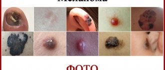
Photo 1. What different types of hemangioma look like
Symptoms appear after the tumor has gained a significant size ; before that, they do not appear.
Treatment is carried out using surgery, sometimes using a laser. In some situations, the hemangioma itself regresses and disappears, this often happens in children under 6 years of age.
Where does hemangioma occur in adults?
In four out of five cases, tumor pathology manifests itself as a single tumor; multiplicity is a rarer occurrence. The most common sites of localization in adults differ little from those in children, usually the head :
- Forehead
- Eyelids
- Scalp
- Above and around the nose
- Cheeks, including from the inside
In adults, swelling rarely appears around the ears.
After the head, the most common place of occurrence is the external genitalia . Due to this location, the hemangioma often becomes infected, ulcerates, and bleeds. The tumor does not bypass the extremities; lesions of the arms and legs are also not uncommon. In some cases, pathology occurs in the tissues of internal organs and bone structures. In such a situation, various methods are used for diagnosis, from ultrasound and x-rays to MRI.
In general, cutaneous hemangioma in most cases occurs in childhood; it is not a common occurrence in adults. Let's look at the reasons why it occurs.
Diagnosis of oral tumors
There are no difficulties in diagnosing the disease. A visual examination by a surgeon and dermatologist and anamnesis are sufficient. When planning surgical treatment, additional studies are carried out:
- Local thermometry (measure the temperature of the tumor).
- Thermography (thermal video shows in infrared rays a picture of the distribution of temperature fields).
- Ultrasound (ultrasound examination).
- CT (computed tomography) if the jaw bones are affected.
- MRI (magnetic resonance imaging).
- Biopsy (a piece of tumor tissue is excised for microscopic diagnosis).
- Angiography (studying the condition of blood vessels). The research is based on the properties of X-rays. A contrast agent (iodine) is injected into the vessel. It is impenetrable to X-rays. If you inject it into the blood and “enlighten” the body, the vascular tree will be visible in the image.
- Tissue puncture (the tumor is punctured and the material is sent for cytological examination under a microscope of the structure of the tumor).
Small oral tumors can be accidentally diagnosed by a dentist during a routine examination, during the treatment of caries and other dental procedures. An accurate determination of the type of tumor is possible only after a histological study of its structure. Such a study can be carried out on material taken during a tumor biopsy or after its removal.
Ultrasound of the tumor is used to determine the depth of tumor growth in the oral cavity, and X-ray examination is used to assess the condition of bone structures. For gingival fibromatosis, an orthopantomogram is performed, which often reveals areas of destruction of the alveolar process. Angiography is often used in the diagnosis of vascular tumors.
Causes
Why hemangioma appears in children and adults has not been fully established . This pathology is usually considered congenital; it occurs due to abnormalities in the development of the vascular system of the embryo, when the vascular tissue grows excessively. It is important to remember that the formation of the vascular system occurs in the first trimester, so the expectant mother should monitor her health and well-being from the very beginning of pregnancy.
Medicine is not able to more accurately determine the period at which the causes leading to hemangioma in the future arise.
To summarize, the reasons for the formation of hemangioma in adults are as follows:
- Diseases of internal organs leading to vascular dysfunction
- Factor of heredity and congenitality
- Increased exposure to ultraviolet rays
Adviсe
Before starting treatment, you should familiarize yourself with two tips that will help you cope with the pathology:
- If a new growth appears, monitor it closely and visit your doctor immediately if it has accelerated its growth. You should also go to the hospital if the swelling darkens.
- When treating neonatal hemangioma, do not use folk remedies. Decoctions with lotions will not help get rid of tumors and can worsen the baby’s condition.
Symptoms in adults
As we indicated above, hemangioma in an adult mainly manifests itself on the skin. As a rule, it initially looks like a red or bluish spot, which has an irregular shape and is located slightly above the level of the skin. Sometimes a network of diverging small vessels can be observed from the center.
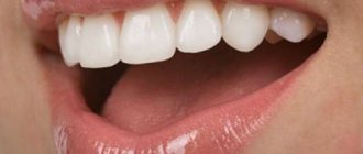
Photo 2. Hemangioma on a man’s face
Often, a hemangioma very quickly progresses in size, occupying an increasingly larger area, which causes significant cosmetic discomfort in the patient.
Symptoms and what a hemangioma looks like in adults depends on its type and location, the main ones being:
- A simple or capillary tumor is a tumor consisting of many small capillaries located on the surface of the skin.
- Cavernous is a hemangioma consisting of a set of vascular cavities located subcutaneously
- Combined consists of both supracutaneous and subcutaneous elements
- Mixed differs in that it contains dissimilar fabrics.
Let us consider in more detail the various structures of hemangioma
The basis of capillary formations is a network of capillary vessels, which are lined with a single layer of cellular structure (which forms the inner choroid). The state of some capillary groups may be normal, while others may be collapsed. In appearance, hemangioma is a soft tumor with a red or blue-purple tint and clear boundaries. Possible germination into tissue under pathology. When pressed, the color fades; if you release it further, the color returns to its original color. Capillary hemangioma in children goes away on its own in every twentieth case.
Cavernous hemangioma in adults is based on vascular cavities of various configurations, separated from each other by septa. They are often filled with clotted blood and a blood clot that has grown into the connective tissue. In this case, the color of the pathology will not be pale, but red. The general appearance is “multi-lobed”, the most common localization is the head and neck. With various kinds of stress, for example, as a result of screaming or coughing, the tumor increases and further returns to its original form after the stress is relieved.
Cellular neoplasms are called neoplasms, the basis of which are stem cells. Such a hemangioma is immature and has a tendency to grow deeper, as well as to grow peripherally. It is soft to the touch and the color is red. In addition to the skin, it also affects the mucous membranes.
Combined ones contain structures of capillary and cavernous pathologies. The basis is cavernous cavities in which underdeveloped capillary structures are observed.
A muscle vascular tumor is called a hemangioma in adults, of various types, initially formed in the cells of a muscle or tendon. However, then it grows from the depths and manifests itself on the skin.
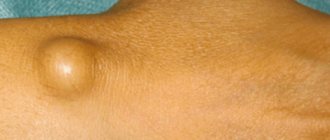
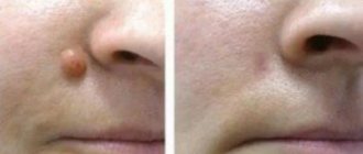
Photo 3. Manifestation on the face
In addition to skin manifestations, formations on the mucous membranes are also found. They are able to grow deep into tissues and increase in volume. There are known cases of hemangioma of the tongue, when the tumor has grown so large that the tongue does not fit in the oral cavity. At the same time, naturally, there were a fair number of accompanying symptoms, for example, the inability to swallow normally, breathe, and bleeding.
If hemangioma in adults affects internal organs and bones , then most often the disease proceeds secretly. It is usually discovered by chance, or if the tumor is aggressive and has led to the appearance of symptoms. In the case of the spine, this will be pain in the vertebrae as a result of compression of the spinal canal, damage to the rectum may begin to bleed through the anus, and the stomach may cause vomiting with red blood impurities.
It is not so rare for adults to develop liver hemangioma; in such a situation, due to internal bleeding, signs of compression of the bile ducts appear, which will cause symptoms of cholelithiasis or cholecystitis in the patient. Discovery of a tumor usually occurs when a person goes to the doctor, suspecting a completely different disease.
Why is hemangioma dangerous?
Hemangioma in adults or children, although it is a tumor, the causes of which are not fully understood, is not dangerous in itself . It does not have the ability to form metastases and does not degenerate into cancerous tumors. However, a significant increase in the size of the pathology may have health consequences. The specific complications will depend on the location where the occurrence occurs.
Let's consider the main danger factors:
- While inside, pressure on neighboring organs . For example, if a hemangioma in an adult is located on the neck and has grown quite deeply, this can lead to difficulty breathing. The same is with the eyes; if you are in the eyelid area, vision deterioration is likely. If we consider the internal localization, then the occurrence on the bones or blood vessels can, respectively, threaten bone fractures due to the weakening and deterioration of blood flow, respectively. In the latter case, hemangioma can even threaten life.
- If the tumor is located in a place that often comes into contact with something, for example, the edge of clothing or household items, when placed in the palm area, then there is a high probability of infection due to damage to its integrity. In addition, their independent ulceration periodically occurs, for example, in immunodeficiency states or in diabetes mellitus. As a result, an infectious agent is introduced and the course of the disease becomes more complicated.
- The development of the previous factor is a serious injury to the hemangioma, resulting in large blood loss . This situation is most dangerous during internal placement, when it is very difficult to identify and promptly stop bleeding. The most severe case in this case will be damage to the liver tumor. Such bleeding will be very profuse, and it can only be stopped by treatment through surgery.
- Weak blood clotting occurs due to the body’s constant struggle with hemangioma. To do this, it regularly supplies the damaged area with platelets and proteins that ensure coagulation. If the affected area is large enough, then there is a risk of platelet deficiency, which can result in poor blood clotting.
Question answer
Does neonatal hemangioma go away? Hemangioma, of course, is not the most pleasant of diseases. However, in most cases it does not require any intervention. It disappears on its own, on average, by the age of nine years of a child’s life. Once it disappears, it cannot reappear.
Is it possible to get vaccinated for hemangioma in newborns? You can vaccinate, but not during its active growth phase. Then vaccinating a child is contraindicated.
Do newborns with liver hemangioma survive? It is believed that such a tumor on the liver is very dangerous for the life and health of the baby. They carry a huge risk of bleeding, since even with the slightest impact on the liver area, the tumor is injured and ruptured. If symptoms are present, action must be taken immediately, otherwise it will be fatal. After a series of studies (ultrasound, MRI), the causes of the tumor are identified and treatment is prescribed. Subsequently, if there are no indications for surgical intervention, the hemangioma is simply observed to prevent its growth.
Is it possible to massage newborns with hemangioma? Most doctors agree that massage with hemangioma is contraindicated. This is explained by the fact that during its implementation, increased blood circulation begins, and this can lead to tumor growth.
Treatment of hemangioma in adults
Various surgical techniques are used for treatment in adults. In this case, the indication for removal is a neoplasm in the genital area or face. In addition, removal is carried out when there is a significant increase in size in a short period of time, for example, doubling in 10-15 days .
The main methods of treatment and removal of hemangioma in adults are:
- Sclerosing treatment . It is prescribed if the tumor is cavernous, located in the mucous membrane of the mouth or near the ears. The essence of the method is to introduce a special solution, sometimes based on ethyl alcohol, into the tumor, as a result of which a burn is provoked and the inflammatory process gradually subsides and leads to healing of the lesion.
- Cryodestruction , as freezing with liquid nitrogen is called, is used to remove capillary pathologies.
- Diathermocoagulation , or cauterization using an electric current and an electrode, removes small tumors
- , low-voltage X-ray therapy is used to remove large hemagiomas in adults, mainly located on the face.
- X-ray endovascular occlusion is a technique when the hemangioma is deprived of blood supply due to blockage of the vessel responsible for it.
- Hyperthermia by an electromagnetic field involves the rupture of molecular bonds inside tumor cells, which changes their structure and makes it impossible to synthesize new tissues.
Author: site editor, date July 01, 2017
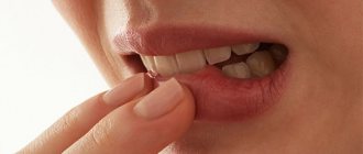
Hemangioma on the lip is a formation of a benign nature, which is formed due to excessive proliferation of vascular cells. It looks like a red nodule or a convex spot of a purplish-bluish color. This disease is often diagnosed in newborns, but can also develop in adults.
Features of the occurrence of hemangioma on the lips
Scientists suggest that congenital hemangioma is a consequence of improper development of the circulatory system during the embryonic period. Doctors note that there is a direct relationship between the appearance of characteristic spots on the baby’s lip and infectious diseases suffered by the mother during pregnancy. The circulatory system of an embryo is formed in the first three months of its life. If the expectant mother suffers a viral or fungal disease at this time, the fetus is likely to have the described defect on the body or lip after birth.
Causes of hemangioma in adults:
- hereditary predisposition;
- vascular disorders;
- excessive passion for tanning.
Depending on what form of the disease develops, certain symptoms appear.
Main types of hemangioma
Vascular formations have different structures. Different vessels (capillaries, veins) can participate in their formation. Tumors have different depths of damage. Taking these criteria into account, doctors classify pathologies and distinguish the following types of hemangioma.
| Types of education | Clinic |
| Venous | It develops more often in older people. A blue or dark purple papule appears on their lip, the size of which can vary from 2 to 10 mm |
| Capillary | The formation has the form of a flat surface spot with uneven edges. It is formed from small, intertwined capillaries. Among 90% of patients are newborns |
| Simple | The tumor often affects only the upper layers of the skin, but can also grow deeper, affecting the subcutaneous tissue. The spots never reach large sizes. They always appear in newborns and go away on their own without treatment within a year. |
| Cavernous | Hemangioma is formed from large veins, formed from cavities (cavities) filled with exudate mixed with blood. It consists of many formations of different sizes, has blurred boundaries and a blue color. Most often occurs on the lip of an adult |
| Mixed | The structure of the formation contains connective tissue, blood vessels, and fat. It can be located on the surface of the lip and form in its subcutaneous layer |
| Angioleiomyoma | The tumor forms in the muscle layer. When it increases in size, its vessels intertwine with the vessels of the lips |
| Pyogenic granuloma | Hemangioma, which forms after trauma based on bruising |
Clinical signs of a vascular tumor are determined visually and by touch. When pressure is applied to its surface, flattening occurs. As a result of pressure, the blood recedes, causing the hemangioma to turn pale, then returns to its previous color when the pressure disappears. If you place one palm on the formation and the other on an area of healthy skin, you will notice an uneven temperature. Where the hemangioma is located, it will be higher.
If the formation grows on the surface of the skin, it can be diagnosed by external signs. If it is located deep, where the fatty tissue is located, there may not be external symptoms (the appearance of a characteristic purple-blue color). To identify them, instrumental diagnostics are used (radiography, ultrasound, MRI, CT).
Stages of the disease
There are four stages of the appearance of hemangioma:
- A light spot appears on the child’s body, which is difficult to notice. It does not cause any discomfort.
- The spot becomes red and scar-like. Most often, parents think that this is an ordinary bruise.
- Enlargement of hemangioma.
- Skin damage and external changes in the tumor.
Tumor development occurs in three stages:
- Growth – This usually occurs during the first year of a child’s life. The growth, which is red or bluish in color, becomes larger, all because the child is actively growing at this time. If it is a capillary hemangioma, then it may become brighter; if it is cavernous, the surface becomes uneven or dark. If the formations are large, then you can trace the places that are unequally filled with blood.
Growing tumor:
- Change in hemangioma - having increased to its maximum, it begins to gradually disappear (if these are simple neoplasms). The color becomes paler and some healthy skin may appear. This transformation occurs during the first eight years of a child's life. Most often, hemangioma disappears by the age of 7 years. If it is a large or cavernous tumor, then it cannot be cured without surgery.
- Complete disappearance of the hemangioma - the skin becomes normal in color. Sometimes in this place you can observe some unevenness on the skin and a lack of hair.
During the development process, hemangioma does not cause any discomfort, there is no itching or pain. If it is a large tumor, it may be hotter than the surrounding skin. On average, the size of the tumor reaches 1-2 mm, sometimes several centimeters.
Treatment methods for hemangioma
In children, vascular tumors on the lip appear in the first week of life. Experts advise parents not to panic in such a situation: by the age of one year, the hemangioma may disappear on its own without the use of any treatment. You need to see a specialist if the nodules filled with blood protrude strongly above the surface of the lip. They can prevent the baby from properly grasping the mother's breast with his mouth. The decision to eliminate such a defect is made on a case-by-case basis. As a rule, conservative therapeutic methods are used for these purposes.
Various methods are used to treat vascular tumors in adults:
- injections;
- hormonal drugs;
- radical treatment.
Sclerotherapy is the introduction of drugs into the body, the action of which stops the growth of hemangioma - a painful method of treatment. As a result of this procedure, the nutrition of the tumor is stopped, so it turns pale and disappears over time.
When choosing drug treatment, the patient is prescribed hormonal drugs and beta blockers for oral administration. They affect pathological vessels, reduce or block cell division processes. Thanks to this, the tumor gradually dies.
When the formation is located deep in the tissues from which the lip is formed, radical treatment is resorted to.
Methods for removing hemangioma in adults:
| Name | Description |
| Laser therapy | When it is used, the tumor is affected using a beam of directed light. It generates thermal radiation, which evaporates the fluid in the pathologically altered cells. They die from dehydration. The surgeon has the ability to control the depth of penetration, so healthy tissue is not harmed |
| Surgery | During the manipulation process, the hemangioma is removed using a scalpel. The operation is performed under local anesthesia |
| Cryodestruction | Used for superficial tumor localization. It is exposed to liquid nitrogen. Low temperatures cause instant necrosis of pathological tissues |
| Cauterization | This treatment method is used only for the treatment of small capillary hemangioma. She is burned with electric shock |
Independent removal of the formation using traditional medicine is unacceptable. This is fraught with the appearance of severe bleeding or transformation into a cancerous tumor. It is also impossible to leave a hemangioma unattended, as there are risks of developing unwanted complications.
Therapy
The presence of hemangioma is not always an indication for any specific therapy. In many cases, on the contrary, it is not recommended to touch it - just wait until it resolves on its own. But sometimes doctors and parents note the presence of indications for treatment. The absolute indications for the fight against hemangiomas are:
- periodic bleeding from the tumor;
- ulceration of hemangioma;
- the tumor is located in a place that increases the risk of injury (in the area of underwear, on the back, etc.);
- tumors that interfere with the normal functioning and development of the newborn;
- very rapid tumor growth.
How are hemangiomas treated?
In cases where there is a risk that the hemangioma will rupture, it must be urgently removed. When a rupture occurs, heavy bleeding occurs due to damage to blood vessels. Internal hemangiomas can also be traumatized, resulting in internal bleeding affecting the spleen, liver, and even the brain. In this case, the danger from leaving the hemangioma is higher than from surgical intervention.
Results of laser hemangioma removal
If we are talking about a hemangioma that has a simple shape and is not too large in size, then there is no need to touch it - it will go away on its own over the next 2-5 years.
The fight against tumors of this type is carried out:
- medicinally;
- surgically.
In the first case, the doctor prescribes beta blockers for the child, which are drugs like Timolol, Anaprilin or Propranolol. As a result of taking these medications, the vessels collapse, and the neoplasm quickly resolves. To control this process, the child is placed in an inpatient unit at the hospital. In some cases, when taking Beta blockers, side effects occur or contraindications to their further use appear.
Propranolol
In addition, the newborn may be prescribed therapy with hormone-containing drugs. Due to the effects of steroids, tumor growth slows down and gradually stops.
The fastest way to get rid of a hemangioma is its surgical removal. The exact method the surgeon will use depends on the location of the formation. If the child has just been born or is sick, surgical intervention is unacceptable. The following surgical methods are usually chosen:
- radiotherapy - if the hemangioma is located in hard-to-reach places;
- removal by electrocoagulation;
- administration of a drug with a sclerosing effect - to “glue” small vessels filling the tumor;
- removal with a scalpel;
- cryotherapy.
If you want to learn in more detail how to treat spinal hemangioma, as well as consider symptoms and alternative treatment methods, you can read an article about this on our portal.
Cryodestruction of hemangiomas
If treatment takes place outside the Russian Federation, then with a high degree of probability the hemangioma will be removed using the method of pinpoint laser coagulation. This method is recognized as the most effective, fastest and safest for young children. However, to perform such an operation, the child must be at least three years old.
When the operation is completed, the child is prescribed antibiotics, which can be administered either orally or by injection into the veins or muscles. The area of skin from which the hemangioma was removed must be disinfected every day using special preparations.
Antibiotics may be prescribed after surgery
How can hemangioma be dangerous?
Vascular spots may darken. The presence of such a defect indicates the rapid progress of the disease. The accumulation of blood vessels in itself is not dangerous, but injury to them can lead to bleeding. It can occur when using toothbrushes and consuming hot foods and drinks.
The surface of the formation in adults is often ulcerated and necrotic. Against this background, inflammation develops and suppuration of the affected area occurs. This skin condition can cause sepsis (blood poisoning).
With the rapid growth of a vascular tumor on the lower lip, it grows into the thickness of the tongue. Then it greatly increases in size and no longer fits in the mouth.
Hemangioma can cause anemia and thrombocytopenia. In older people, it can transform into a malignant formation. Therefore, it is necessary to carefully monitor the defect that has arisen and seek medical help in a timely manner.
What to do if a hemangioma hurts
Typically, the formation of such a formation does not cause physical discomfort. It appears when its surface begins to ooze. This phenomenon poses a threat to human life: any ulcer on the mucous membrane or skin is an open gate for infection. It can cause blood poisoning and death. Therefore, if any pain occurs, you should immediately consult a dermatologist.
A hemangioma growing on the lower lip may be accompanied by swelling. It causes nagging pain. Discomfort also occurs when the formation grows deep and begins to compress the nerve endings. It intensifies during physical activity, under the influence of high and low temperatures, often becomes unbearable and forces a person to take drastic measures.
Giving help
To ensure that the foot is higher than the body, you need to put a pillow under your feet.
IT IS IMPORTANT TO KNOW! Even “neglected” joints can be treated at home, without surgery or hospitals. Just read what Valentin Dikul says read the recommendation...
- Provide complete rest to the limbs.
- The victim should be seated or laid down so that the foot is in an elevated position.
- Place a cushion or pillow on the ankle area.
- Apply ice to the bruised area on the top and outside of the foot, transferring the ice to a clean rag.
- Apply a fixing bandage to the foot.
- Relieve severe pain with analgesics.
- Deliver to a medical facility.
If you did not notice any serious signs when examining your heel, then you can treat your foot at home.
How to treat:
- Use drugs with external treatment. These can be cold compresses. When you apply them to a bruised area, in our case to a damaged heel, you can slightly reduce inflammation and relieve swelling.
- Physiotherapy treatment helps to quickly reduce foot pain and restore the soft tissue of the heel. But for this you need to contact a knowledgeable specialist.
- Medicines are used for treatment. They are administered internally because they help reduce pain levels and help the heel heal quickly.
Care and prevention
There is no specific prevention of the disease. Therefore, doctors recommend:
- treat infectious diseases in a timely manner;
- prevent toxicological poisoning;
- protect the skin and mucous membranes from exposure to aggressive sunlight;
- Healthy food;
- monitor vascular health;
- observe work and rest schedules;
- perform moderate physical activity;
- strengthen the immune system.
If the tumor has already formed, visiting saunas and baths is contraindicated for patients. After removal, wounds remain on the mucous membrane, which are important to properly care for. You cannot burn them with iodine, alcohol, brilliant green and tear off the crusts yourself. To soften them and reduce itching, it is useful to use Solcoseryl Ointment daily.
by Vlad · Published 04/28/2018 · Updated 05/07/2019
Among the relatively benign neoplasms that require special attention are vascular tumors. Dark red hemangioma on the gums in children is increasingly being diagnosed immediately after birth.
It develops during pregnancy and may appear during a hormonal surge in adolescence. An unpleasant-looking lump sometimes begins to actively grow, and injuries to the teeth cause heavy bleeding.
What does a hemangioma on the gum look like?
Unlike other types of neoplasms and growths, hemangioma is formed from blood vessel cells. It can appear on any part of the body, on the surface of the skin or mucous membranes. In rare cases, large plaques affect the lining of internal organs, resembling a kidney or liver cyst on ultrasound. According to the medical classification, it is classified as benign, but studies have already confirmed its possible degeneration into oncological pathology.
Hemangioma on the gum in children looks like a dark spot or red dot. The shade can vary from bright red to almost black, which is associated with filling with capillary or venous blood. Some parents mistake it for an ordinary wart or birthmark. It always has a clearly defined border and stands out against the background of white teeth.
There are several main forms of the disease:
- Simple: it forms as an oblong capsule and does not grow inward, affecting only the upper layer of the epithelium.
- Cavernous: can damage the deeper layers of the mucosa, penetrates into the fatty tissue on the cheeks or the inside of the lips.
- Cavernous: most often diagnosed in newborns and is a connection of several types of vessels, has a pale pink tone.
- Combined: the tumor involves in its growth not only capillaries, but also mucosal cells and periodontal bone tissue.
The latter species is characterized by more rapid growth and often accumulates large amounts of liquid. Therefore, the gums look swollen, the oval of the face and the outline of the chin change. Since large arteries and vessels do not pass near the teeth, in 90% of cases the hemangioma will be simple and small in volume. Sometimes it has the shape of a fungus on a short, dense stalk.
This type of tumor almost always continues to grow after childbirth. From a flat spot, it gradually turns into an elastic lump with a sharp or round end. Outwardly, it strongly resembles the common type of port-wine stain nevus, so only a qualified dental surgeon should diagnose the disease.
Epithelial tumors of the oral cavity
Papillomas. Oral cavity tumors consisting of stratified squamous epithelial cells. They are most often localized on the lips, tongue, soft and hard palate. Oral papillomas are a rounded protrusion above the surface of the mucosa. They may have a smooth surface, but are more often covered with cauliflower-type papillary growths. Usually single papillomas are observed, less often - multiple ones. Over time, these oral tumors become covered with keratinizing epithelium, due to which they acquire a whitish color and a rough surface.
Nevi. In the oral cavity, nevi are observed in rare cases. They are often convex and have varying degrees of pigmentation from pale pink to brown. Among the tumors of the oral cavity there are blue nevus, papillomatous nevus, nevus of Ota and others. Some of them can become malignant with the development of melanoma.
Glands of Serres. Typically, this type of oral tumor is located in the alveolar ridge or hard palate. Serre's glands are hemispherical formations of yellowish color, up to 0.1 cm in size and of dense consistency. May be of multiple nature. Usually, by the end of the child’s first year of life, spontaneous disappearance of these formations is noted.
Papillomas. Oral cavity tumors consisting of stratified squamous epithelial cells. They are most often localized on the lips, tongue, soft and hard palate. Oral papillomas are a rounded protrusion above the surface of the mucosa. They may have a smooth surface, but are more often covered with cauliflower-type papillary growths.
Nevi. In the oral cavity, nevi are observed in rare cases. They are often convex and have varying degrees of pigmentation from pale pink to brown. Among the tumors of the oral cavity there are blue nevus, papillomatous nevus, nevus of Ota and others. Some of them can become malignant with the development of melanoma.
Glands of Serres. Typically, this type of oral tumor is located in the alveolar ridge or hard palate. Serre's glands are hemispherical formations of yellowish color, up to 0.1 cm in size and of dense consistency. May be of multiple nature. Usually, by the end of the child’s first year of life, spontaneous disappearance of these formations is noted.
Why does hemangioma occur in the mouth?
Many detailed studies have not helped to understand the cause of this pathology. In half the cases, babies are already born with a similar spot on their gum or lip. For others, education occurs after the first year of life. Hemangioma is a type of tumor that reacts acutely to any change in hormonal levels. Therefore, it can suddenly appear in girls during their first menstruation.
It has been proven that hemangioma in a child’s mouth often occurs due to the following complications:
- The mother's pregnancy proceeded with certain complications; hormone therapy had to be carried out to preserve the fetus and reduce the risk of placental abruption.
- In the first trimester, the expectant mother suffered from a viral disease.
- The parents' medical history includes diseases of the circulatory system, vascular pathologies, and a tendency to varicose veins.
- Bad habits of the mother (smoking, taking certain drugs unnecessarily, strong alcohol).
- Rhesus conflict.
- Living in a region with problematic ecology.
The appearance of a hemangioma should always be taken extremely seriously. If it is found on the gum, doctors recommend conducting a full examination of the entire body and checking the functioning of the internal organs. Such a tumor can form on the liver or kidney, disrupt their blood supply and lead to life-threatening complications for the baby.
If a child's hemangioma is located near the fangs or chewing molars, the little patient may periodically bite it. This leads to prolonged bleeding. The baby is bothered by the unpleasant taste of blood in the mouth, the wound aches and hurts until it heals. There is a danger of infection, because saliva contains a huge amount of bacteria, and the baby constantly pulls foreign objects into its mouth. The tumor can block the muscles that are responsible for chewing.