Kinds
Based on the location of origin, hemangiomas are divided into the following types:
- Simple. Localized on human skin.
- Cavernous. Located under the skin.
- Combined. Simultaneously on the skin and subcutaneously.
- Mixed. Such a tumor contains three or more types of tissue.
Speaking about the structure of hemangiomas, we can distinguish 6 types.
Capillary
These tumors consist of endothelial cells - cells lining the internal lining of blood vessels. Hemangioma has the appearance of a non-solid formation of red color, possibly with a blue or purple tint and a smooth surface. The boundaries of such a tumor are clear, and under pressure, it does not turn pale for long. The neoplasm tends to grow into neighboring tissues.
Cavernous
Its basis is made up of vascular cavities, separated by partitions and taking various shapes. It happens that the blood in the caverns coagulates and a blood clot is formed, into which connective tissue grows.
It has the appearance of a “strawberry”, bright red, convex, with a tendency to grow rapidly. May be located on the neck, head or liver. It becomes brighter when screaming or crying, as if it tenses up and increases in size.
Cellular
This hemangioma consists of angioblast cells, they are immature and grow into neighboring tissues, so the tumor is considered invasive. This type of formation appears in newborns; they are soft to the touch and bright red in color. They are localized both on the skin and on the mucous membrane. Has the property of growing to the periphery.
Capillary-cavernous
Hollow vessels surrounded by immature capillaries. They can appear on the stomach, nose, finger.
Racemic
Such formations consist of twisted thick-walled vessels. They have a bluish appearance and are located on the head or neck.
Intermuscular
They contain capillaries, veins, arteries, and cavities. They grow between the muscles, affecting the tendons and subcutaneous tissue, and sometimes the skin itself.
Benefits of the procedure
The main advantages of removing any hemangiomas with a laser are:
- Lack of preliminary preparation. Removal can be carried out immediately after consultation with a specialist.
- Short recovery period. Rehabilitation does not affect life activities.
- High efficiency. Tumors up to 5-8 cm in size can be eliminated in 1 session.
- Short duration of the procedure. The duration of the intervention does not exceed 10-15 minutes.
- Safety. The laser beam has antibacterial properties and cauterizes small bleeding vessels. The risk of infectious complications and bleeding during and after surgery is minimal.
- No aesthetic defects. With a small size of the pathological focus, scars do not remain.
- Lasting results. There are no tumor relapses.
- Availability. "SM-Clinic" offers laser removal of hemangioma at affordable prices.
Danger of hemangiomas
As such, the formations themselves do not pose a danger, since they do not form metastases, are not toxic to the body and do not lead to human weight loss. But the proliferation of hemangiomas can cause unpleasant consequences:
- Compression of neighboring organs and disruption of their functions. For example, a tumor located on the neck can make it difficult to breathe, swallow, and, if localized on the eyelid, prevent the eye from opening. If a neoplasm compresses a large vessel, then this poses a threat to life, because blocking the blood flow is extremely dangerous.
- Risk of infection. For example, when it is located on the lip, this probability is very high. And the formations can ulcerate on their own. This leads to the penetration of bacteria and aggravation of hemangiomas.
- Danger of blood loss . When the tumor is injured, bleeding occurs. If the skin is affected, then this is not so bad, but when it happens in the liver, the problem can only be solved by resorting to surgery.
- disorder . When the body is in a constant struggle with damaged blood vessels, supplying platelets to injury sites, then after a while, their number in the blood drops. This threatens the development of bleeding.
Contraindications to laser treatment of hemangioma
Laser removal of hemangioma is contraindicated if:
- the course of systemic acute infectious diseases with fever;
- decompensated diabetes mellitus;
- rashes in the area where a tumor of any origin is located;
- inflammatory or autoimmune skin lesions;
- psychiatric illnesses with loss of self-control;
- “fresh” tan;
- pregnancy and breastfeeding;
- decompensated course of cardiovascular pathologies;
- photodermatitis;
- malignant neoplasms, regardless of location;
- pathologies of blood clotting.
The most dangerous are decreased blood clotting and the presence of a malignant process. In the first case, the risk of bleeding increases, in the second - the spread of metastases.
Vessel formation in the fetus
The formation of the first vessels occurs at the beginning of the fourth week of fetal development. This process is called angiogenesis. It can be primary and secondary, it depends on the type of mechanism of vascular development.
On this topic
- Other
Everything you need to know about hidradenitis
- Inna Viktorovna Zhikhoreva
- September 27, 2020
Primary angiogenesis is characterized by the appearance of the smallest primary vessels from the mesenchyme. This mechanism operates only in the first weeks of embryo formation. Such capillaries do not have blood inside, but consist only of a single layer of cells - endothelial. In adults, these cells are located inside the blood vessels.
Secondary angiogenesis is characterized by the appearance of new vessels from those that have already been formed. As the body grows or its mass increases, new tissues do not receive nutrition and oxygen. This starts a process that promotes the growth of new blood vessels. It is in this way that angiogenesis occurs in the fetus during the last stages of its growth and after birth.
It can be noted here that embryonic tissues have a high ability to regenerate in case of any damage. These minor injuries trigger healing processes, including secondary angiogenesis, which in the future can trigger the appearance of hemangiomas.
Benign breast tumor: symptoms and signs
Benign breast tumors, classified depending on their structural features, can develop asymptomatically. Each type is characterized by certain symptoms, but experts have identified common manifestations of benign neoplasms:
- nodules, lumps in the chest;
- breast swelling and discomfort;
- change in color of the skin of the breasts and areolas;
- nipple discharge;
- pain that can only appear during menstruation;
- inflammation of the lymph nodes located in the armpits.
Breast doctors recommend that women of reproductive age conduct self-examinations. If you detect any changes in your breasts, you should not delay visiting a specialist. The Yusupov Hospital provides comprehensive diagnostics and treatment of breast tumors.
Theory of development of hemangiomas
There are many theories explaining the causes of hemangiomas, but none of them covers all the nuances of the occurrence of the disease.
Lost Cell Theory
A modern theory that states that a tumor appears as a result of a disruption in the development of small vessels from the mesenchyme. The embryo accumulates immature capillaries, which should develop into arteries and veins, but during the formation of organs, a certain amount of immature tissue may remain unused.
Over time, this tissue should disappear, but under the influence of certain factors, this mechanism may fail. Growth will be activated and enhanced, and as a result, the birth of a child with a tumor will occur.
Fissural theory
In the first days of embryo formation, embryonic clefts are present; these are the places where sensory organs will later develop. After the sixth week, nerves and blood vessels begin to grow into these openings.
This theory suggests that due to a disturbance in the development of capillaries in these places, hemangioma occurs. It explains the frequent appearance of tumors in the facial area, but does not answer the question about their occurrence in other places of the skin and in the liver, kidneys, and other organs.
Placental theory
Believes that placental cells penetrate into the blood supplying the embryo and remain in its tissues and organs. When the fetus is not born, the mother’s body does not allow the hemangioma to develop, and after birth, increased growth of tumor cells occurs. This theory explains the appearance of tumors both on the skin and in other places.
Symptoms, clinical manifestations and photos of facial hemangiomas
Many people do not pay attention to the increased vascular pattern of the scalp tissues, especially when they are localized in the area of the scalp. Small spots do not cause pain and are simply inaccessible for inspection due to the hair.
But when the defect is located on the open parts of the facial area - nasal hemangioma - a person develops complexes and even phobias. Meanwhile, the tumors do not have any special clinical manifestations - neither itching nor burning. It is extremely rare for capillary hemangiomas to bleed.
Mechanism of tumor appearance
There are many theories about the appearance of tumors, but all of them have a common condition - the presence of immature vascular tissue where it should not be. But for a hemangioma to start growing, this factor alone is not enough.
The main impetus for starting the process is hypoxia - a lack of oxygen to the tissues. This means that abnormal conditions for the delivery of oxygen to the tissues of the body pose a potential threat to the development of hemangioma.
Such factors may include: multiple pregnancy, phytoplacental insufficiency, birth trauma, eclampsia, maternal smoking, prematurity.
The main causes of neoplasm
To date, it is unknown why facial hemangioma appears. However, doctors and researchers have some theories. It is believed that in this case children have some genetic predisposition, although this theory is not always correct.
In addition, doctors include mother’s illnesses during pregnancy, for example, colds or other acute respiratory infections, especially in the first 3 to 6 weeks, as a list of reasons. It is also believed that taking certain pharmaceutical drugs by a woman while carrying a child can also affect the development of its vascular system. On the other hand, facial hemangioma may be the result of hormonal characteristics of the body, because in some children this neoplasm disappears on its own during adolescence.
Development
All types of tumors have three stages of development. This is their distinguishing characteristic.
Rapid growth stage
Upon its appearance and over the course of several weeks, the tumor quickly increases in size. It looks like a bright red spot growing inward and to the sides.
The growth rate can be either small or rapid, for example, several millimeters per day. The first stage is the most threatening with complications. The tumor can grow into neighboring organs and disrupt their proper functioning.
Completion of growth stage
After about a year? the tumor completes active growth and for several more years its increase is not significant.
Reverse development stage
In 2% of all hemangiomas, their complete disappearance is observed without medical intervention. Having passed the stage of completion of growth, the tumor becomes not as bright as before. The vascular network slowly disappears, being replaced by connective tissue or normal skin.
These are the mechanisms of the appearance and development of hemangiomas. Their research continues and is of great interest to scientists. Modern medicine has very little material to study tumors, mostly stillborn babies and embryos from abortions.
Features of manifestation
If we are talking about adults, then a benign formation can go unnoticed for a long time. But when it appears, the affected areas are the neck, areas near the ears and on the face. Very rarely, the localization sites are the arms and upper chest.
Hemangioma is deformed vascular tissue
Metastases from an enlarging vascular tumor are an extremely rare occurrence, but in general such a disease can overcome several stages of development:
- The first stage is manifestations of an external nature and rapid growth. Internal factors such as infectious diseases, various pathologies of internal organs, as well as metabolic and hormonal changes lead to a rapid increase in benign formation. As for external factors, these include exposure to high temperature and injury.
- Stage of cessation of development. During this period, growth stops.
- Reverse development. It is rarely recorded (2-7% of patients). This stage can last from 2 months to several years. Throughout this time, the skin hemangioma decreases due to the desolation of the deformed vascular network. As a result, the tumor is replaced by scar tissue or healthy skin cells (provided the hyperplasia is small).
In some cases, the tumor may shrink on its own, but it can also grow rapidly, so a visit to the doctor is required.
Note. In adults, rapid growth, formation and enlargement of the tumor is extremely rare.
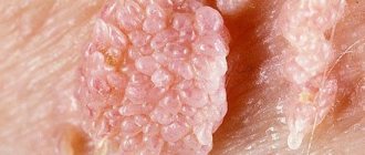
Symptoms
Hemangiomas on the skin initially look like red, uneven spots that protrude slightly upward. It happens that a network of small vessels extends from them. Neoplasms are characterized by intensive growth. They can cover very large areas of the human body.
On this topic
- Other
How to prevent the occurrence of a carbuncle in time
- Inna Viktorovna Zhikhoreva
- August 17, 2020
Hemangiomas of internal organs are asymptomatic. But they can lead to complications:
- nausea;
- vomit;
- feeling of discomfort in the stomach;
- loss of appetite;
- causeless weight loss;
- distension in the abdomen.
The occurrence of such symptoms indicates that you need to be examined by a specialist. He will prescribe an ultrasound to localize the tumor and tomography for a detailed examination.
Possible consequences
When understanding what a hemangioma is, it is important to understand the danger it can pose to humans. So, this is hyperplasia of vascular tissue, which is a benign formation.
Hemangioma is a disease that can affect different layers of the skin.
It does not metastasize, but it can grow. Often such a tumor is located inside the skin for a long time, remaining invisible, and only years later makes itself felt, appearing on the surface.
Important. All hemangiomas are formations that appeared at an early age. This means that the tumor does not form in adulthood, but only appears.
Such hyperplasia often does not pose a fatal health hazard. The bottom line is that the place of its localization in most cases is remote from the internal organs. However, it can grow into the subcutaneous fatty tissue and have a damaging effect on the skin. Sometimes this formation affects bone tissue.
Hemangioma should always be treated
Clearly, this is a problem that cannot be ignored but must be treated.
Diagnostics
Diagnosis of hemangiomas consists of the following mandatory points:
- examination by a specialist;
- instrumental examination;
- laboratory research;
- consultations with various doctors.
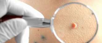
As soon as a red spot of bright red color and a convex shape is detected, growing in a short period of time, you need to consult a doctor as quickly as possible.
He will conduct a survey about how long ago the spot appeared on the body, the rate of its growth, and whether there were similar formations in relatives. The specialist will also examine the tumor under a magnifying glass, examining its structure, applying pressure and searching for other previously unnoticed spots.
Diagnosis of this disease is not particularly difficult and already at the examination stage the doctor can identify cutaneous hemangioma. To detect damage to internal organs, they resort to instrumental studies: thermometry, thermography, ultrasound, MRI, CT, angiography, biopsy.
Laboratory tests are not informative for diagnosing a tumor, but they monitor the occurrence of complications and control over the patient’s well-being. A general blood test is usually prescribed; it shows the platelet count and hemoglobin level. This is how it is determined whether the patient has anemia or bleeding disorders.
Benign breast tumor: treatment
Breast fibroids and other benign tumors can be successfully treated if diagnosed early. Women should pay special attention when choosing a clinic for the treatment of a benign breast tumor.
One of the most important criteria is the openness of the medical staff and the willingness to provide assistance to the patient on any day. Specialists at the Oncology Clinic of the Yusupov Hospital carry out emergency hospitalization of patients in acute conditions.
Hemangioma on the chest and other benign neoplasms are treated at the Yusupov Hospital using the principles of evidence-based medicine. The main principle of the work of specialists at the Yusupov Hospital is to provide high-quality medical care to all patients who apply. However, the patient must take the first step towards treating the disease by making an appointment with oncologists at the Yusupov Hospital.
Treatment
Hemangiomas in adults cannot be treated with medications, so if a tumor is detected, its removal is indicated. If it is localized on the face or genitals, then surgery is performed within three days.
Also, an indication for immediate surgery is the rapid growth of a tumor. Removal can be done by physical and surgical methods.
Physical:
- cryodestruction;
- laser irradiation;
- sclerotherapy;
- electrocoagulation.
Surgical ones are used when organs and tissues inside the body are affected. In the process, the formation itself and a small adjacent area of healthy tissue, about 1-2 millimeters, are removed.
What therapeutic methods are used (3 methods)
Therapy for the treatment of hemangioma on the face in adults or children is used extremely rarely. If it is nevertheless used, then preference is given to compression.
The method applies to formations located in places where it is possible to apply a pressure bandage for a long period. After 2 months, the tumor apparently decreases in size or no trace remains of it.
Another way is to take hormonal drugs. However, the use of the method is strictly limited due to the manifestation of a number of serious associated effects, therefore it is used in rare cases strictly on the recommendation of a specialist.
The dosage of drugs is selected for a specific patient, especially a child.
Another method of treating vascular formations is to take the medications Propranolol and Timolol. The effectiveness of the treatment technique has been proven by experimental studies in Europe and the USA.
Therapy with such drugs is carried out under medical supervision with regular checking of blood pressure and blood glucose levels.
Propranolol or Timolol is prescribed in individual dosages.
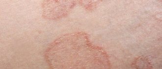
Why do red moles appear on the body?
Red moles are formed by a large number of microscopic blood vessels (hemangiomas) and lymphatic vessels (lymphangiomas). The main reason for the appearance of red moles is dysfunction of the circulatory and lymphatic systems.
The most common causes of angiomas on the body are:
Hormones: expert opinions hormonal changes in women (during pregnancy, taking oral contraceptives);
- skin injuries;
- genetic characteristics of the organism;
- diseases of the gastrointestinal tract, liver and pancreas;
- long exposure to sunlight and frequent visits to the solarium.
Preventing the appearance of red moles
It is impossible to prevent the occurrence of congenital angiomas. A congenital red mole in a child is most often a short-term formation and disappears by the age of 5-7 years. Therefore, if the child does not complain of pain or itching in the area of the angioma, there is no need to worry.
In order to avoid the occurrence of angiomas after birth, it is necessary to follow these recommendations:
- avoid prolonged exposure to direct sunlight and frequent visits to the solarium;
- follow a sleep and nutrition schedule;
- lead a healthy lifestyle.
If you have a red mole, it is important to monitor its development and contact a dermatologist at the first signs of injury or change in size. With timely consultation with a doctor, it is possible to prevent complications of angiomas and, if necessary, begin therapy on time.
Removal of any hemangiomas with laser in Moscow
Removal of hemangioma with a laser in Moscow is performed on the basis of the multidisciplinary medical holding company “SM-Clinic”.
Our specialists have long-term experience, supported by reviews from many satisfied clients. All doctors use only the latest medical equipment, which allows the intervention to be carried out with high precision and without side effects. To make an appointment, you need to call the phone number listed on the website or fill out the online form. The consultation center staff will be happy to answer all your questions and select a convenient time for your visit.
Prices
| Name of service | Price, rub.)* |
| Consultation with a dermatovenerologist (primary) | 1,950 rub. |
| Consultation with a dermatovenerologist (repeated) | 1,700 rub. |
We accept bank cards VISA, MASTERCARD, MAESTRO for payment
The full price list can be found at the reception or by phone.
* The administration of the clinic takes all measures to timely update the price list posted on the website, however, in order to avoid possible misunderstandings, we advise you to check the cost of services at the reception or in the contact center by phone
+7 (495) 292-39-72
.
The posted price is not an offer. Medical services are provided on the basis of a contract.