The best way to prevent skin cancer is to protect its top layer (epidermis) from adverse effects, primarily from ultraviolet radiation. But even a complete refusal to stay in the sun, alas, does not protect against skin cancer 100 percent, but it threatens vitamin D deficiency and some other unpleasant consequences for the body. So, it is impossible to completely protect against melanoma. But skin cancer can be diagnosed early by regularly checking your moles—this greatly increases the chances of successful treatment.
Our expert in this field:
Vertieva Ekaterina Yurievna
Oncologist, dermatovenerologist, cosmetologist
Call the doctor
Local defects
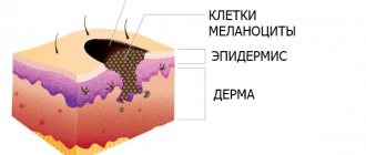
Local developmental defects refer to congenital moles that may appear due to problems with cell division at the final stage of fetal development. Usually such defects are not immediately noticeable at birth. They are first noticed only when the baby turns 3 years old. A variety of injuries play an important role in the appearance of moles: wounds, insect bites, scratches. Immediately after an injury, an inflammatory process occurs that affects different layers of the skin. Because of this inflammation, the tissues activate the production of biologically active substances that stimulate cell growth.
Addresses of clinics in the capital
Where to check moles in Moscow? Best clinics:
Hormonal disorders
Hormonal factors play a major role in the occurrence of moles. Many researchers note that acquired moles often appear in adolescents during puberty. In rare cases, nevi occur in pregnant women, as well as in patients with endocrine disorders. Scientists are also investigating the likelihood of moles due to bacterial and viral infections that affect the skin. The mechanism of formation of moles in these cases is similar to their formation as a result of injuries.
Advantages and features of the study
Examination of moles using a dermatoscope has a number of features and advantages, including the following:
- simplicity, accessibility of diagnostics;
- avoiding injury to tumors during examination;
- no contraindications to the procedure;
- instant receipt of research results;
- lack of special preparatory measures for the procedure.
This research method is at the forefront of preventing the development of melanoma. Due to the effectiveness of the method, it is possible to carry out successful treatment when cancer is detected in the early stages. In addition to skin cancer, dermatoscopy can diagnose other skin pathologies. Particular attention to this research method should be paid to owners of birthmarks of brown, red, black colors of heterogeneous consistency.
Diagnosis of nevi
In the diagnosis of moles, the distinction between benign and malignant neoplasms is of utmost importance. Of course, this is due to the fact that the effectiveness of treatment of a malignant mole depends to a large extent on its timely detection. The following methods are used to diagnose moles:
- Anamnesis collection. In making the correct diagnosis, the main role is played by the accurate collection of patient complaints. It is with the collection of anamnesis that the study of the patient’s disease begins. When interviewing the patient, the doctor pays special attention to clarifying the family history, and is also interested in the presence of factors (external and internal) that could lead to the appearance of a mole.
- Examination of the patient. Different types of nevi differ in specific external manifestations. That is why visual inspection plays a key role in diagnosing moles. The doctor evaluates the neoplasms according to the following criteria: the number of moles, their location, size, consistency, color, time of occurrence. It is also important to record the changes in the mole that have occurred to it since the last examination.
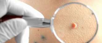
- Dermatoscopy. To make a more accurate diagnosis, the doctor may prescribe a thorough examination of the patient’s skin using a special device that can magnify the image a couple of times. Thanks to this technique, the doctor is able to detect the most minor changes in the patient’s skin. This is a very effective diagnostic method, the use of which allows you to obtain accurate results. The procedure usually lasts no more than 20 minutes.
- Indication by phosphorus isotope. This is a highly sensitive method based on the accumulation of radioactive phosphorus by the tumor. Normally, phosphorus should take part in cell division. If a patient has a malignant neoplasm, phosphorus synthesis increases several times. This process can be detected using an isotope.
- Echography. This technique is considered effective because it involves examining the soft tissue around the mole, which makes it possible to determine its exact size. Many types of moles can grow in the skin. This process is especially characteristic of malignant neoplasms. Therefore, this technique is usually prescribed to prepare the patient for surgery.
- Radiography. To diagnose neoplasms, non-standard equipment is usually used. It is necessary to take a special picture that would enlarge the mole 5-10 times. This technique is used to accurately assess the tissue structure of the mole. For example, a malignant neoplasm usually has a heterogeneous structure with many compactions and small cavities.
- Thermometry. This technique involves measuring the temperature of individual areas of the skin, which is carried out with a special device. The principle of this method is based on studies that indicate an increase in temperature in the event of the appearance of malignant neoplasms. During the procedure, the doctor compares healthy areas of skin and those on which nevi have appeared. This is an accurate, painless and quick method for diagnosing moles, which allows you to effectively monitor changes in their size.
- Biopsy. This is the most reliable method for diagnosing nevi, which allows you to promptly detect any signs of malignancy of a mole. In addition, the accuracy of the biopsy is about 100%. The technique involves taking a small sample of a mole for further investigation of its nature. There are two types of biopsy: puncture (a very small fragment of material is taken for examination) and total (removal of the entire tumor and its subsequent examination).
Degrees of malignancy of cancerous tumors
As the tumor grows in size, its symptoms become more pronounced. It becomes possible to distinguish a mole from a cancerous formation. The degrees of malignancy associated with classic melanoma are as follows.
- First degree. It is characterized by the presence of a malignant formation measuring no more than 1 mm. It acquires a loose and soft consistency, rises above the body with the subsequent formation of pigmented areas.
- Second degree. It is caused by a noticeable increase in the size of the tumor up to 4 mm. Over time, changes are observed in terms of the shape and color characteristics of the growth. The tissues located nearby begin to swell, and ulcers appear on them that bleed.
- Third degree. It is characterized by the fact that the tumor process grows into the deep layers of the skin. The size indicator is more than 4 mm, bleeding, cracking, and pain are observed.
- Fourth degree. The symptoms that make themselves felt are common to the other three types of the disease. There is a general disturbance of well-being due to damage to internal organs. The size indicator can reach 5 mm.
It is important to know! It is necessary to carry out independent periodic checks of moles; any changes occurring in them may indicate pathologies that require immediate treatment.
Ways to remove moles
In most cases, treatment of nevi involves their removal. In this case, surgery can be prescribed either at the request of the patient or according to the doctor’s indications. Typically, the basis for removing a nevus is the risk of its transformation into a malignant tumor. Fortunately, removing skin tumors is not a complicated operation; it is usually performed by dermatologists. However, before removing a nevus, the patient must undergo a thorough diagnostic examination to eliminate the risk of developing melanoma.
Histological analysis
Histological analysis is carried out to diagnose and predict cancer. The principle of histology is the study of biomaterial obtained after operations (biopsy). For example, when cutting out a mole surgically or by biopsy. A piece of skin, having undergone a series of technological treatments, is stained with antigens and examined under a microscope. Histological analysis can take from 10 days and is designed to answer a number of questions:
- from what tissue does the tumor develop?
- stage, term and maturity;
- degree of malignancy;
- nature of occurrence;
- depth of metastases;
- prognosis of treatment outcome.
If a histological analysis is prescribed after removal of a mole, do not despair. Agree immediately, a method of high-quality cancer diagnosis, and eliminates error by almost 100%.
Laser excision of moles
The most common method of removing nevi is laser surgery. The essence of the procedure is that all the liquid is first evaporated from the tissues, which ultimately leads to the death of the cells. This is a very effective procedure, which is convenient to carry out if the patient has a large number of moles. In addition, after their removal, the patient does not have any scars or marks at all. However, large moles should not be removed using laser surgery for the simple reason that it will take a lot of time. In addition, complete removal of such a nevus cannot be guaranteed.
Identifying signs of skin cancer
The task of a dermatologist is to diagnose malignant skin lesions. Unfortunately, skin cancer takes a long time to treat and often leads to death. Through regular monitoring, a doctor can detect a suspicious mole before cancer begins. This means that you can get rid of it in advance.
It is very important that patients monitor their skin for changes at home. You should immediately contact a dermatologist if the mole:
- grows;
- becomes irregular in shape;
- changes along the edges - they rise above the skin;
- changes color - it becomes uneven;
- bleeding;
- inflamed;
- itchy.
If melanoma is diagnosed in time, the cure rate is 90%. The late stage of this cancer is one of the worst cancer diagnoses, often leading to death.
ONLINE REGISTRATION at the DIANA clinic
You can sign up by calling the toll-free phone number 8-800-707-15-60 or filling out the contact form. In this case, we will contact you ourselves.
If you find an error, please select a piece of text and press Ctrl+Enter
Prevention of melanoma development
Some types of moles are very dangerous because they cause cancer. That is why it is necessary to try in every possible way to avoid their occurrence. This can be easily done through various preventive measures. First of all, you need to avoid spending long periods of time outside under the scorching sun. Ultraviolet rays have a detrimental effect on the skin. In particular, it is necessary for people who have many moles to give up tanning. In extreme cases, it would be better to cover the moles with a band-aid.
Doctors also recommend dealing with dry skin, since this is where malignant neoplasms often appear. For this purpose, it is necessary to use special moisturizing cosmetics. Of course, it is necessary to promptly treat all skin diseases. If a rash, itching, peeling, or redness appears, patients with moles should immediately make an appointment with a specialist.
Patients whose moles are on the feet, palms, neck and other places where they may be susceptible to injury need to either protect the nevus or remove it altogether. Constant traumatic impact on a mole can ultimately lead to its inflammation and transformation into a malignant tumor.
It is also necessary to remember that some chemical compounds can provoke cancer. This is why it is extremely important to avoid the negative effects of carcinogens (for example, quit smoking). Research shows that carcinogenic substances are contained even in combustion products of tobacco. It is important for patients who have nevi on the body to undergo regular preventive examinations with a dermatologist. It is advisable to visit a doctor once a year. A dermatologist may prescribe a different schedule of visits if malignancy of moles is suspected. You should also see a doctor if there is a change in the size, color, or consistency of a mole.
Self-diagnosis
At home, it is recommended to carefully monitor the dynamics of changes in the structure of the birthmark. You can independently either check a mole for cancer or draw conclusions about the need to see an oncologist.
Thus, there are five signs on the basis of which a conclusion can be drawn about the degeneration of cells:
- asymmetry of the body of the spot;
- contour violation;
- color change;
- increase in size;
- dynamics of change.
A healthy, harmless nevus is always symmetrical. When cells degenerate, this symmetry is broken. For self-diagnosis, it is recommended to take a photograph and from time to time compare the mole on the body with the photograph taken.
A healthy birthmark or nevus always has a clearly defined, slightly rounded contour. A sudden change in the severity of the contour and its color is a reason to contact the clinic.
Possible consequences
If you do not consult a doctor when the first signs of malignant melanoma appear, this can provoke very unpleasant consequences, even death. Some people consider skin cancer not very dangerous, since they are sure that it is never too late to cut out melanoma. However, this disease is very insidious. It almost always metastasizes, meaning cancer can start anywhere in the body. This is precisely why melanomas are dangerous.
If you want to get reliable results, then you must also undergo examination by an immunologist, endocrinologist and other specialized specialists. Damage to the skin may indicate the development of diabetes. If the tumor is located near the eye, then in this case an ophthalmologist will help you. An immunologist determines how a person’s immune system works. If it is strong enough, it can inhibit the growth of cancer cells for some time.
To avoid various kinds of complications, it is necessary to carry out prevention. To do this, experts recommend removing moles that rise above the skin, as well as those that change color or become damaged.