What is a dysplastic nevus?
Dysplastic nevus is a formation that has a high tendency to acquire malignant properties. The definition of “dysplastic” means the internal and external atypicality of such formations and the difference from ordinary moles. The elements look like moles or pigment spots of different sizes with uneven edges and uneven brown-black color. The surface of the nevus may be completely flat or slightly raised above the skin at the epicenter. Such atypical moles do not have a characteristic localization; the number of spots formed can vary significantly from person to person.
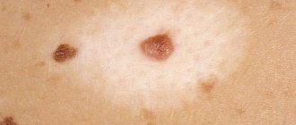
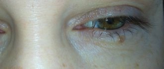
Dysplastic nevus is an irregularly shaped formation of various colors
Dysplastic nevi are clusters of melanocytes that synthesize skin pigment, which enter the epidermis or dermis during fetal development.
Such benign formations occur in approximately 5% of the world's population belonging to the Caucasian race. Atypical nevi can form at any age in people of any gender. A predisposition to the appearance of such moles is inherited.
Dysplastic nevi are also called melanocytic dysplastic nevi, atypical moles, atypical melanocytic nevi, and Clark's nevi by medical professionals.
Classification of formations
Dysplastic melanocytic nevi are divided by dermatologists into two large groups in accordance with the size, shape and degree of risk of malignancy (malignancy):
- sporadic nevi - solitary, a type of acquired neoplasms that are precursors to melanoma;
- familial nevi - multiple atypical formations.
Sporadic nevi
The first type includes:
- A typical shape in which the spot looks like a fried egg - with a raised area in the center. These are formations of various brownish shades and can have different sizes: 1–15 mm - minimal;
- 1.5–10 cm - medium;
- 10–20 cm - large;
- more than 20 cm - gigantic.
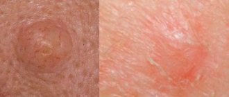
Dysplastic nevi can have different sizes, shapes and shades
Family type of formations
The second, familial, type of dysplastic nevi is a consequence of a genetic predisposition to melanocyte dysplasia under the influence of ultraviolet radiation, while all relatives, even those without age spots, are at risk for developing melanoma - one of the most malignant skin tumors that gives instant metastases.
The familial type of Clark's nevus involves multiple formation of atypical moles on the skin
Based on their tendency to malignant degeneration, oncologists divide Clark’s nevi into 2 forms:
- sporadic (acquired) with a risk of malignancy of the mole 7–70 times: type A - nevus without degeneration into melanoma;
- type C - nevus with malignancy into melanoma;
- type B - mole without transformation into melanoma;
The formation may be stable and not change for a long time, it may spontaneously regress or degenerate into superficial melanoma. It is known that the risk of malignant transformation of a nevus increases in those people whose mole appeared after 40–50 years. Such patients require careful monitoring.
Causes of Clark's nevus
Atypical moles are formed due to local movement of pigment cells under the influence of reasons that are not fully understood. The pigment melanin, which is produced by melanocytes located in different layers of the dermis, determines the shade of the skin and is designed to protect it from the harmful effects of insolation (ultraviolet radiation). In this case, the pigment is found in all skin structures in the same amount. But in people with a predisposition during fetal development or immediately after birth, melanocytes filled with melanin migrate to various areas of the skin. Atypical familial birthmarks are formed - small formations that begin to gradually grow during puberty and become clearly visible.
A nevus is a collection of melanocytes overflowing with melanin.
Sporadic (single) nevi occur in a different scenario. Most researchers believe that ultraviolet radiation and the human papillomavirus play a certain role in the mutation and proliferation (accelerated cell division) of pigment cells. The provocateur of the movement and point accumulation of melanocytes is always sharp hormonal changes - the onset of puberty, pregnancy, etc.
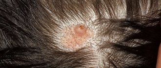
In contrast to the theory that it is insolation that is the main trigger for the development of a sporadic form of pathology, some doctors cite the fact: in many cases, atypical nevi appear in places where direct rays of the sun do not reach, for example, in the perineum, on the genitals or scalp under hair.
It is possible that a significant decrease in the body’s defenses plays some role in the formation of dysplastic nevi, since cases of numerous atypical moles have been reported in connection with skin blistering diseases and some other pathologies that cause immunosuppression (immune suppression).
The risk group for familial dysplastic nevus includes immediate relatives of carriers of atypical moles. It has been proven that the predisposition to the appearance of formations is transmitted in an autosomal dominant manner. Whether a child will develop nevi or not largely depends on the characteristics of his body and the influence of external factors. The closer the relationship with carriers of the pathology, the higher the likelihood of developing multiple nevus syndrome.
The risk of malignancy of Clark's nevus exists due to the persistence of proliferative activity (readiness for increased division) of immature melanocytes in the epidermis and the presence of cell atypia of varying severity.
The danger of Clark's nevus, especially the familial type, is the readiness for malignant transformation into melanoma
Signs of education
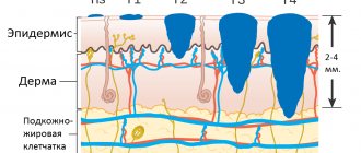
The spot appears on healthy skin. The basis of an atypical mole is an accumulation of pigment in the epidermis and upper layers of the skin. The formation may be the only one, or there may be many pigment spots - more than a hundred.
Table: typical signs of Clark's nevus
| Sign | Feature |
| Amount of elements | From one to several hundred |
| Characteristic location | Arms, chest, buttocks, upper back, less often - on the face, head |
| Color | Shades of brown of varying intensity - from light brown to brown-black. Within one mole there may be several different shades. |
| Magnitude | Initially very large - from 6 mm |
| Surface | May rise slightly above the level of healthy skin. The structure is never smooth or uniform. Usually a coarse or finely lumpy surface, especially in the center of the formation. On the periphery it is smoother. |
| Borders, shape | The shape is always irregular, asymmetrical, the contours are unclear and uneven |
| Other signs |
|
Clark's nevi are initially large in size - always more than 6 mm
Symptoms
Such neoplasms are significantly different from ordinary moles; they are easily recognized by the following signs:
- The spot does not have smooth outlines and is convex towards the center.
- It is large in size.
- Most often they appear on the head, back, and lower back. Sometimes on the palms, feet, chest and other closed places.
- The color of the mole is uneven; it can have several shades at once. The more melanin it contains, the darker it is.
- If you look at a birthmark, you can see many small dots and specks on it that are located around the center.
- The nevus is often covered with hairs.
Diagnostics, difference from other formations
The resulting cutaneous element, one or several, must be shown to a dermatologist. Only a specialist can determine the degree of risk of malignant transformation of the nevus, the need for further observation or removal. To diagnose Clark's nevus, an experienced specialist only needs to conduct a visual examination of the formation.
To exclude malignancy, the patient is prescribed the following diagnostic tests:
- Dermatoscopy.
The procedure involves a thorough examination of the elements using a dermatoscope, which magnifies the image. The purpose of the examination is to detect the slightest changes in the mole. The examination is carried out quickly, painlessly and is highly informative for the patient’s initial and repeated visits. Dermatoscopy does not provide a 100% chance of confirming the diagnosis, but it can tell the specialist which examinations in the future will be the most rational. Recently, digital dermatoscopes have been used. Digital dermatoscopy is performed to visually analyze the nevus - Echography.
It is used infrequently: with large cutaneous elements to exclude malignant growth of a nevus into all layers of the skin or during preoperative preparation to determine the boundaries of a large mole. Ultrasound of the nevus can be performed before surgery - Indication by phosphorus isotope. A highly informative method for diagnosing oncological degeneration of a tumor. Melanoma actively accumulates radioactive phosphorus, since active cell division processes occur in the tumor, in which this element participates. During malignant transformation, the scanner detects intense accumulation of the isotope in the area of formation.
- Local thermometry.
The technique involves measuring the temperature on the surface of the skin using a special device. As the formation grows, active metabolism occurs, accompanied by a slight increase in temperature in this area. During the examination, temperature data obtained from measurements on the surface of the nevus and clean skin are compared. A benign formation will show a difference within 1 °C; with malignant neoplasia, the difference will be higher - up to 2.5 °C. This technique is often used in the follow-up of patients with dysplastic nevi. Using a digital thermometer, local thermometry of nevi is carried out - Biopsy. The most reliable method for diagnosing signs of cancer transformation. A biopsy is the removal of a piece of tissue from a nevus and subsequent microscopic analysis of the sample. A characteristic histological sign of Clark's nevus is atypical proliferation of melanocytes, that is, its chaotic growths in the dermis and epidermis. Two types of examination are used: puncture biopsy, when a sample is obtained with a needle after preliminary anesthesia (the method has limited information content, since cells are taken pointwise and in small quantities);
- a total excisional biopsy, when the entire nevus removed during the operation is examined; if histological signs of oncology are detected, the patient is prescribed chemotherapy.
Nevus biopsy is the most valuable research method from a diagnostic point of view
This method provides information if the element has cracks or pitting. Compared to a biopsy, such an analysis has less diagnostic value. Histological analysis of a smear or scraping from the surface of a mole allows you to exclude or confirm the diagnosis
Blood and urine tests are not used to diagnose atypical nevi, since there are no changes in indicators with such pathology. Clinical and biochemical studies of biological fluids can be carried out before surgical removal of the nevus to assess the functionality of the internal organs.
Differential diagnosis
First of all, a benign Clark nevus is differentiated from the following formations:
- melanoma, which is clinically indistinguishable from a nevus, so a histological examination is necessary;
- Dubreuil's melanosis (lentigo maligna) - a melanoma-dangerous nevus that forms mainly in older people and is a single element with uneven contours located on an open area of the body; the color of the formation can be different - from yellow-brown to black; can reach very large sizes - up to 20 cm;
- acquired nevomelanocytic nevus - a mole with a uniform color of different shades, usually of regular shape, clear boundaries, small in size (up to 6 mm);
- Spitz nevus (benign juvenile melanoma) - a fast-growing birthmark with a smooth (most often) surface, round and convex shape, different colors (pink, yellow, brown) and a fairly large size - up to 2 cm;
- seborrheic keratosis - multiple elements that appear in adults mainly on the back or chest, forearms; keratomas have a round shape, different colors, resemble warts, covered with a thin crust, prone to bleeding; the size of the elements is from 2 to 6 mm, may be accompanied by itching;
- solar lentigo - brownish spots on exposed skin that appear under the influence of ultraviolet radiation in people over 50 years of age (more often in men).
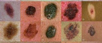
Photo gallery: formations with which dysplastic nevus is differentiated
Melanoma is one of the most malignant skin tumors
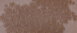
Solar lentigo occurs in older people on exposed skin.
Seborrheic keratosis - multiple elements of brown color
Juvenile nevus, or Spitz nevus, has clear outlines and uniform coloring
Acquired nevomelanocytic nevus is small in size and uniform in color.
Dubreuil's melanosis, or lentigo maligna, is large spots with uneven contours and uneven coloring
Since there is a high probability of Clark’s nevus acquiring malignant properties (especially in the familial, multiple type), you need to know the first signs of malignancy:
- an increase in the size of the nevus - proliferation of the peripheral borders;
- the appearance of a reddish border around the formation;
- thickening of healthy skin around the mole;
- the appearance of itching;
- color change - darkening;
- change in texture (compaction), the appearance of dryness, roughness, cracks on the surface of the nevus.
Anyone who has moles on their body needs to know the signs that may indicate malignant changes
If these signs appear, you should urgently visit a dermatologist.
Video: diagnosis of melanoma-dangerous nevi
Classification
Dysplastic nevi are brown, pink, and in some cases bluish-gray in color. Their classification contains the following forms:
- Typical (“fried egg”) - this nevus is similar to a regular mole, has flat edges, and elevation above the skin is observed only in the middle of the pigment spot.
- Lentiginous is a birthmark that has a slight elevation above the skin and is dark brown in color. Sometimes dysplastic spots are black in color, and their surface is completely flat.
- Keratotic - warty formations that are in some way similar to seborrheic keratosis.
- Erythematous - a pink formation, slightly raised above the skin.
We recommend reading Uterine fibroids - symptoms and signs, treatment
If changes in a dysplastic pigmented nevus begin to appear, then in most cases this indicates its malignancy. Most often, such pigmentation forms in open areas of the body, but can also be located in areas covered by clothing.
Treatment
The only treatment for atypical nevi is radical removal. Moreover, it is irrational to use prophylactic removal of an unchanged dysplastic mole. Formations in which suspicious changes appear, or newly appeared ones with a tendency to grow rapidly, must be removed.
Benign nevi must be observed by a doctor. With single elements (up to 5 formations), clinical examination is carried out 1-2 times a year, with multiple elements (especially if there is a family history of melanoma) - once every 3-6 months.
Dysplastic nevi are removed by excision and a mandatory histological examination of the material is carried out
Since the main indication for surgical removal of nevi is their dynamic changes, which may indicate the manifestation of malignant transformation, the only correct method of surgical intervention is total excision of the nevus. The method involves radical excision of the formation, including an area of healthy skin. To determine the boundaries of removal before surgery, the nevus is examined using a Wood's lamp.
Removal of an atypical nevus is carried out using the method of total excision
The operation is not performed in acute inflammatory diseases, during pregnancy, or in the presence of an infectious pathology.
The intervention is performed under local anesthesia. General anesthesia is used to remove large lesions (especially on the face), as well as in small children.
After excision of the formation, the wound surface is closed with sutures. Healing occurs within 7–10 days. A scar remains at the site of the operation.
After surgery to remove a nevus, a scar remains
Methods of surgical destruction of the formation (cryodestruction, based on freezing cells with their subsequent destruction, and laser removal, which consists of evaporating liquid from tissues) are not used, since after removal of the nevus it is necessary to conduct a histological examination of the material to exclude oncology.
In some cases, prophylactic removal of a benign dysplastic nevus may be used:
- if it is located in a place that is difficult to access for dynamic self-observation;
- the patient expressed such a desire (provided that there are only one or two elements).
In such situations, removal can be performed using a radio wave knife or through electrocoagulation (provided the nevus is small in size).
Multiple atypical moles cannot be removed surgically. For preventive purposes, the patient may be prescribed applications with Fluorouracil, an antitumor drug from the group of antimetabolites, or a course of Interferon therapy. In this case, the patient must regularly undergo mandatory medical examinations.
Fluorouracil is an antitumor drug used prophylactically for multiple atypical nevi
Treatment in children
Indications for surgical treatment of nevus in children are the same as in adults:
- dynamics of education, which indicates possible degeneration;
- large size moles;
- element in a hazardous location.
Prophylactic removal of formations is not performed, since the risk of the appearance of new melanoma-dangerous nevi is not reduced, especially with a family history. In addition, excision of the formation entails the formation of a scar that remains forever.
Radical removal surgery is performed under general anesthesia. The child spends the entire healing period in the hospital.
Video: Doctor Komarovsky about moles in children
Parents of children with dysplastic nevus should pay special attention to prevention:
- protect your child's skin from the sun;
- strengthen the baby's immune system;
- carefully monitor the condition of the mole;
- Visit a dermatologist regularly.
Forecasts
For those who have had a dysplastic nevus removed, the dermatologist will prescribe clinical observation at least once a year for the remaining time. People who have a dysplastic nevus should regularly visit a doctor, monitor the condition of the mole and visit an oncologist if there is the slightest change.